## Distinguishing HSV from VZV Keratitis ### Clinical Presentation Comparison | Feature | HSV Keratitis | VZV Keratitis | |---------|---------------|---------------| | **Ulcer morphology** | Dendritic with terminal bulbs | Pseudodendritic (broader, more amorphous) | | **Terminal bulbs** | Present (pathognomonic) | Absent | | **Anterior uveitis** | Mild to moderate (common) | Severe, granulomatous | | **Skin involvement** | Vesicles on lid margin (may be absent) | Dermatomal vesicular rash (always present) | | **Recurrence** | Frequent (50% within 2 years) | Rare | | **Corneal scarring** | Mild | Severe | **Key Point:** The presence of **terminal bulbs** (small rounded protrusions at the tips of dendritic branches) is the single most reliable morphologic discriminator between HSV and VZV keratitis. ### Pathophysiology **HSV keratitis:** - Dendritic ulcers form due to viral cytolysis and cell-to-cell spread along nerve endings - Terminal bulbs represent accumulation of viral particles and infected epithelial cells at branch tips - Stains with fluorescein (central ulcer) and rose bengal/lissamine green (edges) **VZV keratitis:** - Pseudodendritic ulcers are broader and more irregular - Caused by viral reactivation in ophthalmic division (CN V1) distribution - Always accompanied by dermatomal vesicular rash (distinguishing feature clinically) **High-Yield:** While dermatomal rash is a clinical clue for VZV, it is not always visible at the time of corneal examination. The **terminal bulbs on dendritic ulcers** are the corneal finding that definitively identifies HSV. **Clinical Pearl:** If you see a dendritic ulcer WITHOUT terminal bulbs but with a dermatomal rash and severe anterior uveitis, think VZV. If you see dendritic ulcers WITH terminal bulbs, HSV is the diagnosis regardless of skin findings. [cite:Khurana 6e Ch 5] 
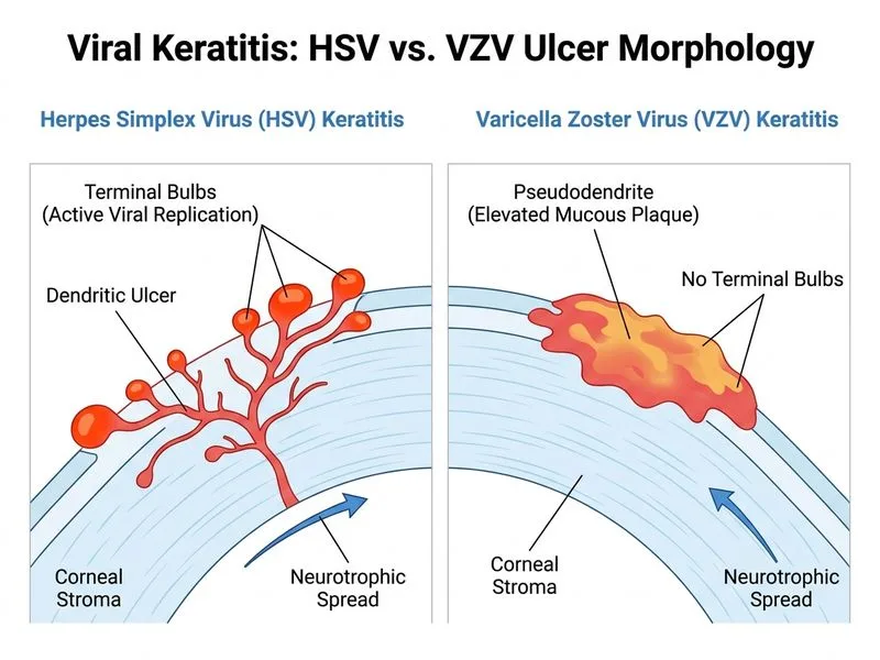
Sign up free to access AI-powered MCQ practice with detailed explanations and adaptive learning.