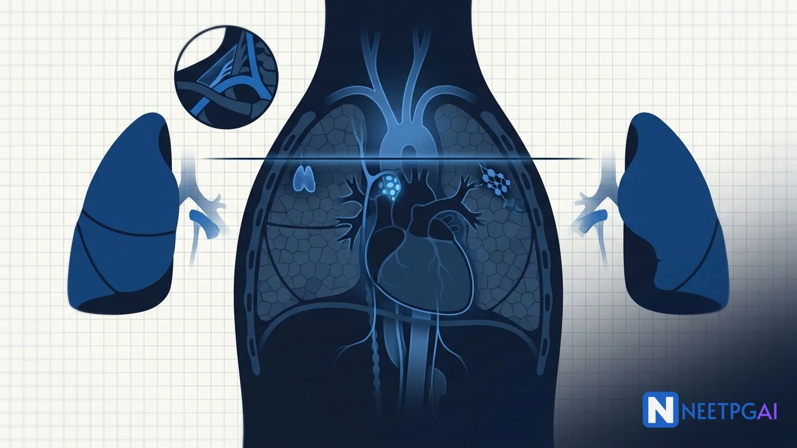
Aortic arch branches — brachiocephalic, left common carotid, left subclavian (the BCS rule).
IASLC nodal map — 14 stations; subcarinal is station 7.
Thoracic outlet — scalene triangle bordered by anterior scalene, middle scalene and first rib.
The mediastinum is the central thoracic compartment between the two pleural sacs and is one of the highest-yield Anatomy topics — almost every clinical vignette featuring a chest X-ray "widened mediastinum", a retrosternal goitre, a posterior mediastinal mass in a young adult, or a phrenic nerve palsy traces back to a compartment-based anatomical reasoning step. NEET PG, NEXT and FMGE all use the IASLC nodal map for lung cancer staging questions and the 4 T's for the anterior mediastinal mass MCQ.
This NEETPGAI deep dive walks through the compartmental anatomy, the cardiac and great-vessel relations, the pleura and lung lobar arrangement, the IASLC lymph node stations, the thoracic outlet, and the neurovascular relations of the phrenic, vagus, recurrent laryngeal and thoracic duct. Pair this with the pulmonary embolism diagnosis guide and the colorectal cancer management deep dive for thoracoabdominal coverage.
Mediastinal compartments
The mediastinum extends from the thoracic inlet superiorly to the diaphragm inferiorly, and from the sternum anteriorly to the vertebral bodies (T1-T12) posteriorly. The transverse thoracic plane of Ludwig — running from the sternal angle (angle of Louis) to the lower border of the T4 vertebra — divides it into superior and inferior. The pericardium then splits the inferior mediastinum into anterior, middle and posterior.
Compartment
Boundaries
Key contents
Superior
Thoracic inlet to T4-T5 disc / sternal angle
Aortic arch and its branches, brachiocephalic veins, SVC origin, trachea, oesophagus, thymus, thoracic duct, vagus, phrenic, left recurrent laryngeal
The sternal angle (T4-T5 level) is the single most useful landmark — it marks the bifurcation of the trachea, the start and end of the aortic arch, the level of the second costal cartilage, the azygos arch over the right main bronchus, and the boundary between superior and inferior mediastinum.
Anterior mediastinal mass — the 4 T's
A widened mediastinum on chest X-ray with the mass projecting anteriorly behind the sternum on lateral view should immediately prompt the 4 T's differential:
Thymoma — most common anterior mediastinal mass in adults; 30 to 50 percent have myasthenia gravis (anti-acetylcholine receptor antibodies). Other associations — pure red cell aplasia, hypogammaglobulinaemia (Good syndrome). Masaoka staging guides treatment.
Teratoma and germ cell tumours — usually in young adults (15 to 35 years); benign mature cystic teratomas, malignant non-seminomatous germ cell tumours (raised AFP, beta-hCG) and pure seminomas.
Thyroid — retrosternal goitre extending downward from the neck through the thoracic inlet; tracheal compression is the clinical concern.
Terrible lymphoma — Hodgkin (nodular sclerosing subtype is classic in young women) and non-Hodgkin lymphoma; bulky anterior mediastinal disease.
Some textbooks add a fifth T — Thoracic aortic aneurysm — which can present as a widened mediastinum mimicking an anterior mass.
Heart in situ and great vessels
The heart sits obliquely in the middle mediastinum, with two-thirds of its bulk on the left side of the midline. Surface markings:
Apex — left fifth intercostal space, mid-clavicular line.
Right border — right margin of sternum, third to sixth costal cartilages (right atrium).
Left border — apex to the left second intercostal space (left ventricle and a bit of left atrium).
Inferior border — xiphisternum to apex (right ventricle).
Superior (base) — second costal cartilages bilaterally (great vessels emerging).
Aortic arch branches
Three branches arise from the convex superior surface in order: brachiocephalic trunk (right), left common carotid, left subclavian (BCS rule). The brachiocephalic divides behind the right sternoclavicular joint into right common carotid and right subclavian. The ligamentum arteriosum (remnant of ductus arteriosus) connects the underside of the arch to the left pulmonary artery; the left recurrent laryngeal nerve hooks under the arch lateral to the ligamentum — a classic site of nerve palsy in aortic aneurysm or left hilar lung cancer.
Venous returns
Superior vena cava — formed by union of right and left brachiocephalic veins at the right first costal cartilage; receives the azygos vein just before entering the right atrium. SVC obstruction (most often from right upper lobe lung cancer or mediastinal lymphoma) causes facial plethora, neck-vein distension and upper limb oedema.
Inferior vena cava — pierces the diaphragm at T8 (caval hiatus) and immediately enters the right atrium.
Azygos system — azygos (right) arches over the right main bronchus at T4 to drain into SVC; hemiazygos and accessory hemiazygos cross the midline behind the aorta at T8-T9 to join the azygos. Provides collateral venous return when IVC or SVC is blocked.
Pulmonary trunk and pulmonary artery
The pulmonary trunk arises from the right ventricle, ascends and bifurcates at the T4-T5 level (sternal angle) under the aortic arch into right and left pulmonary arteries. The right pulmonary artery is longer and passes anterior to the right main bronchus to enter the right hilum. The left pulmonary artery is shorter and arches over the left main bronchus (eparterial bronchus relation differs between sides — see below).
Pleura, lungs and hilar structures
Pleura
Two layers — parietal (lining the thoracic wall, diaphragm, mediastinum and cervical pleura) and visceral (covering the lungs). Parietal pleura is pain-sensitive (somatic innervation — intercostal nerves and phrenic nerve over the diaphragm); visceral pleura is not.
Two key recesses:
Costodiaphragmatic recess — between costal and diaphragmatic pleura; the most dependent pleural space; pleural effusions collect here first. Thoracentesis is performed at the 8th intercostal space in the midaxillary line to enter this recess safely above the diaphragm.
Costomediastinal recess — between costal and mediastinal pleura behind the sternum; deeper on the left side because of the cardiac notch.
Lung lobes and fissures
Lung
Lobes
Fissures
Right
Upper, middle, lower (3 lobes)
Oblique (separates lower from upper/middle) and horizontal (separates upper from middle, runs along 4th rib)
Left
Upper, lower (2 lobes) — lingula is part of upper lobe
Oblique fissure only
The lingula of the left lung is the homologue of the right middle lobe. The cardiac notch of the left upper lobe accommodates the heart.
Hilar structures — the RALS mnemonic
At the hila, the arrangement of pulmonary artery (PA) and bronchus differs:
Right hilum — pulmonary artery is anterior to the right main bronchus (the right main bronchus passes under the azygos arch — eparterial bronchus exists only on the right because the right upper lobe bronchus passes above the right pulmonary artery).
Left hilum — pulmonary artery is superior (Antero-superior) to the left main bronchus (no eparterial bronchus on the left).
RALS mnemonic — Right Anterior, Left Superior. The pulmonary veins are inferior at both hila.
Bronchopulmonary segments
Each lung is divided into bronchopulmonary segments — 10 on the right (3 upper, 2 middle, 5 lower) and 8 to 10 on the left (depending on lingular segment counting). Each segment has its own segmental bronchus, segmental artery, and is drained by intersegmental veins running between segments. Surgical lobectomy and segmentectomy follow these anatomical planes.
Mediastinal lymph node stations — IASLC map
The IASLC (International Association for the Study of Lung Cancer) nodal map standardises 14 stations for lung cancer staging. The midline of the trachea defines right (R) versus left (L):
Station 1 — Low cervical, supraclavicular and sternal notch nodes.
Stations 2R/2L — Upper paratracheal (above the aortic arch).
Station 3 — Prevascular (3a) and retrotracheal (3p).
Stations 4R/4L — Lower paratracheal (below the arch, around the trachea).
Stations 5 and 6 — Aortopulmonary window (5) and para-aortic (6) — left side only.
Station 7 — Subcarinal (between the main bronchi at the carina) — highest-yield MCQ.
Station 8 — Paraoesophageal (below carina).
Station 9 — Pulmonary ligament.
Stations 10 to 14 — Hilar (10), interlobar (11), lobar (12), segmental (13), subsegmental (14) — these are N1 nodes.
Stations 1 to 9 are mediastinal (N2 or N3 depending on side). Stations 10 to 14 are hilar/intrapulmonary (N1). Cervical mediastinoscopy reaches stations 2, 4 and 7; the aortopulmonary window (5, 6) requires Chamberlain procedure (anterior mediastinotomy) or EBUS-TBNA.
Thoracic outlet anatomy
The thoracic outlet (anatomically the superior thoracic aperture) is bounded by the first rib, manubrium, and T1 vertebra. The clinically relevant scalene triangle is bordered by:
Anterior scalene muscle (anteriorly).
Middle scalene muscle (posteriorly).
First rib (inferiorly).
The subclavian artery and lower trunk of the brachial plexus (C8-T1) pass through this triangle. The subclavian vein passes anterior to the anterior scalene (not in the triangle). A cervical rib arising from C7 elevates the lower trunk and subclavian artery against the rib, causing thoracic outlet syndrome — neurogenic (most common, lower trunk distribution, intrinsic hand wasting), arterial (Raynaud-like, post-stenotic dilation, distal emboli), or venous (Paget-Schroetter, effort thrombosis).
Neurovascular relations
Phrenic nerves (C3-C5)
Run anterior to the lung hilum bilaterally, between the mediastinal pleura and pericardium. Carry motor fibres to the diaphragm and sensory fibres from the central diaphragm, pericardium and mediastinal pleura. Right phrenic descends along the right brachiocephalic vein and SVC; left phrenic descends across the aortic arch (lateral to the left vagus and the left recurrent laryngeal hook). Phrenic palsy causes ipsilateral diaphragmatic paralysis (elevated hemidiaphragm on chest X-ray, paradoxical movement on sniff test).
Vagus nerves (CN X)
Run posterior to the lung hilum bilaterally (opposite to phrenic). The right vagus crosses the right subclavian artery and gives off the right recurrent laryngeal nerve which loops under the right subclavian artery and ascends. The left vagus crosses the aortic arch and gives off the left recurrent laryngeal nerve which loops under the aortic arch lateral to the ligamentum arteriosum, then ascends in the tracheo-oesophageal groove. The left recurrent has a much longer thoracic course — making it vulnerable to left hilar masses, aortic aneurysm and mitral stenosis (left atrial enlargement compressing it — Ortner syndrome).
Thoracic duct
Begins at the cisterna chyli (L1-L2), ascends through the aortic hiatus of the diaphragm at T12, runs in the posterior mediastinum between the descending aorta (left) and azygos vein (right), crosses to the left at T5, ascends behind the left subclavian artery and empties into the junction of the left subclavian vein and left internal jugular vein (left venous angle). Injury at any point causes chylothorax (milky pleural fluid with triglyceride greater than 110 mg/dL). The right lymphatic duct drains the right side of the head, neck, upper limb and right thorax into the right venous angle.
NEET PG MCQ traps
Sternal angle at T4-T5 marks the tracheal bifurcation, aortic arch start/end, azygos arch and the boundary of superior and inferior mediastinum.
Posterior mediastinal mass in adult — neurogenic tumours (schwannoma, neurofibroma) most common.
Aortic arch branch order — Brachiocephalic, left Common carotid, left Subclavian (BCS).
Eparterial bronchus only on the right — right upper lobe bronchus passes above the right pulmonary artery.
RALS — Right Anterior, Left Superior — pulmonary artery position relative to bronchus at the hila.
Subcarinal nodes = station 7 — accessible via mediastinoscopy and EBUS-TBNA.
Aortopulmonary window nodes (5, 6) — NOT accessible by standard cervical mediastinoscopy.
Left recurrent laryngeal nerve hooks under the arch lateral to ligamentum arteriosum — vulnerable to left lung cancer and Ortner syndrome (mitral stenosis).
Phrenic anterior, vagus posterior to lung hila.
Thoracic duct enters at left venous angle (junction of left subclavian and left internal jugular).
Costodiaphragmatic recess — site of pleural effusion collection; tap at 8th intercostal space midaxillary line.
Cardiac notch on the left lung — accommodates the heart; the lingula sits below it.
Right lung — 3 lobes, 10 segments; left lung — 2 lobes, 8 to 10 segments.
SVC obstruction — most often from right upper lobe lung cancer.
Azygos arches over right main bronchus at T4 — anatomical landmark on chest CT.
Recent updates and Indian context
IASLC 8th edition TNM (2017) — the current standard for lung cancer staging; nodal map is unchanged from 7th edition but T descriptors were revised. NEET PG continues to test the 14-station map verbatim.
EBUS-TBNA (endobronchial ultrasound) — has replaced cervical mediastinoscopy as the first-line sampling technique for stations 2, 4, 7, 10, 11 across major Indian tertiary centres (AIIMS, PGIMER, Tata Memorial); mediastinoscopy is reserved for EBUS-negative cases with high pre-test probability.
Robotic and VATS thymectomy — increasingly performed for thymoma and myasthenia gravis at AIIMS and Apollo; open trans-sternal approach is still standard for large (greater than 5 cm) or invasive thymomas.
Indian thoracic outlet syndrome epidemiology — overrepresented in young women presenting with intermittent unilateral hand wasting and pain; cervical rib prevalence is around 0.5 to 1 percent.
Mucormycosis post-COVID — has revived interest in invasive mediastinitis pathways from rhino-orbito-cerebral and pulmonary fungal spread along the thoracic inlet planes.
National Cancer Registry data — lung cancer is the leading cancer in Indian men in urban metros; mediastinal nodal staging via EBUS-TBNA is now embedded in the national protocol.
Frequently asked questions
What are the boundaries of the four mediastinal compartments?
The mediastinum sits between the two pleural sacs. The transverse plane of Ludwig (sternal angle to T4-T5 disc) divides it into superior and inferior. The inferior is further split by the pericardium into anterior (between sternum and pericardium), middle (the pericardium and its contents), and posterior (between pericardium and vertebrae T5-T12). Contents differ by compartment and drive the differential diagnosis of mediastinal masses.
What are the 4 T's of an anterior mediastinal mass?
Anterior mediastinal masses are remembered as the 4 T's — Thymoma (most common in adults, associated with myasthenia gravis), Teratoma and other germ cell tumours (young adults, raised AFP and beta-hCG), Thyroid (retrosternal goitre extending down from the neck), and Terrible lymphoma (Hodgkin and non-Hodgkin, often presenting with bulky anterior mediastinal disease). A fifth T — Thoracic aortic aneurysm — is sometimes added.
What is the IASLC lymph node station map?
The IASLC (International Association for the Study of Lung Cancer) map standardises 14 mediastinal and hilar node stations used for lung cancer staging. Stations 1-9 are mediastinal (1 supraclavicular, 2 upper paratracheal, 4 lower paratracheal, 5 aortopulmonary, 6 para-aortic, 7 subcarinal, 8 paraoesophageal, 9 pulmonary ligament). Stations 10-14 are N1 hilar and intrapulmonary nodes. The midline of the trachea separates right from left.
Where does the thoracic duct enter the venous system?
The thoracic duct begins at the cisterna chyli at L1-L2, ascends through the aortic hiatus of the diaphragm at T12, runs in the posterior mediastinum between the aorta and azygos vein, crosses to the left side at T5, and empties into the junction of the left subclavian and left internal jugular veins. Injury during left supraclavicular or thoracic surgery causes chylothorax with milky pleural fluid rich in triglycerides.
What is the difference between the costodiaphragmatic and costomediastinal recesses?
Pleural recesses are potential spaces where parietal pleura reflects without lung filling them, even on full inspiration. The costodiaphragmatic recess is the inferior gutter between costal and diaphragmatic pleura — the dependent space where pleural effusion collects first (best aspirated at the 8th intercostal space in the midaxillary line). The costomediastinal recess lies between costal and mediastinal pleura behind the sternum, deeper on the left because of the cardiac notch of the left lung.
This content is for educational purposes for NEET PG exam preparation. It is not a substitute for professional medical advice, diagnosis, or treatment. Clinical information has been reviewed by qualified medical professionals.
Written by: NEETPGAI Editorial Team
Reviewed by: Pending SME Review
Last reviewed: May 2026