Version 1.0 — Published July 2026
Quick Answer
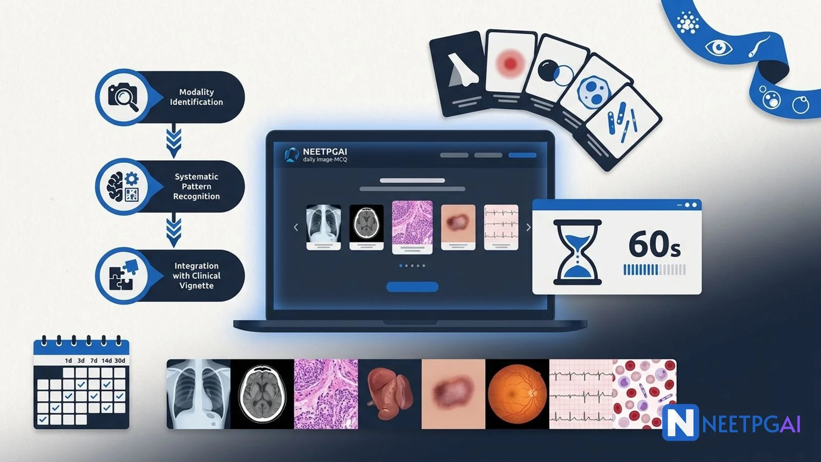
Image-based MCQs (IBQs) now contribute approximately 15-20 percent of NEET PG and 25-30 percent of INI-CET — roughly 27-36 questions on the 180-question NEET PG paper (NEET PG 2027 is expected to follow the revised 2026 pattern until NBEMS publishes the 2027 information bulletin; 50-60 on INI-CET). Building visual pattern recognition through 20-30 image MCQs per day over 8-12 weeks moves accuracy from 40 percent to 80-85 percent, a 40-60 mark swing at the top of the ranking. Use this 10-part playbook:
- Recognise the categories — radiology (X-ray, CT, MRI, USG), pathology (gross, histology, EM), dermatology (clinical photos), ophthalmology (fundus, anterior segment), instruments, ECG, PBS/BM, gross anatomy
- Apply the 3-step approach — modality identification → systematic pattern recognition → integrate with vignette
- Memorise 60-100 Aunt Minnie pearls — a single pathognomonic finding that gives the answer in seconds
- Build a personal image atlas — NEETPGAI daily image MCQ mode, Marrow/PrepLadder atlases, Robbins Atlas, Grant Atlas
- Cap first-pass image MCQ time at 60 seconds — mark and return if stuck
- Master India-specific traps — miliary TB, xerophthalmia, kala-azar PKDL, hydatid cyst, kwashiorkor, pellagra
- Practice 20 image MCQs per day for the last 3 months
- Use spaced repetition — 1d, 3d, 7d, 14d, 30d review of the same weak-cell images
- Subscribe to NEETPGAI's daily image MCQ email for a rolling zero-effort daily drip
- Post-mock review — over-annotate incorrect image MCQs; they are the single highest-leverage revision target
Why image MCQs are exam-favourite in 2027 and beyond
Three forces are pushing NBEMS to test more image MCQs:
Force 1 — Clinical-application testing: the NBEMS 2020 curriculum framework mandates a shift from pure recall to clinical application. Image MCQs — a chest X-ray of miliary TB with a fever-plus-weight-loss vignette, a fundus photo of hypertensive retinopathy with a headache-plus-BP-190/120 vignette — force the candidate to apply knowledge to a real clinical artefact, matching what a first-year resident actually does at the bedside.
Force 2 — Leak-resistance: image MCQs are harder to leak than text MCQs because the visual asset must be reproduced. Bank-based image questions with standardised references (Robbins, Grant, Sabiston) are defensible and hard to game.
Force 3 — Rank discrimination: the top 1 percent of candidates cluster tightly on text-only MCQ accuracy. Image MCQs spread this cluster because visual pattern recognition is a distinct cognitive skill that separates well-trained candidates from rote-memorisers.
Practical implication: if you want to rank in the top 5000 (a competitive PG seat), you must actively drill image MCQs — passive textbook reading is not enough.
Categories of image MCQs
Image MCQs span 8 major categories in NEET PG. Distribution varies by paper; the ranges below reflect 2021-2024 analysis.
| Category | Share | Typical vignette pattern |
|---|
| Radiology (X-ray, CT, MRI, USG) | 40-50 percent | Emergency or ward vignette plus a scan; diagnosis, next step |
| Pathology (gross, histology H&E, EM) | 15-20 percent | Biopsy or specimen vignette plus a slide |
| Dermatology (clinical photos) | 10-15 percent | Rash, ulcer, or lesion plus a colour photo |
| Ophthalmology (fundus, anterior segment) | 5-10 percent | Vision loss, headache, or systemic disease plus a slit-lamp or fundus image |
| Instruments (surgical, dental, ENT, obstetric) | 5-10 percent | Procedure vignette plus an instrument photo; identify the instrument or its use |
| ECG | 5-8 percent | Chest pain, palpitation, or syncope plus a 12-lead ECG |
| PBS / BM smear | 3-5 percent | Anaemia, leucocytosis, or pancytopenia plus a Wright/Giemsa stained smear |
| Gross anatomy specimens | 2-3 percent | Anatomy vignette plus a cadaveric or specimen photo |
Radiology is by far the largest category — invest most of your image-MCQ preparation time here. A quality 4-week radiology image drill can add 15-25 marks alone.
The 3-step approach — the universal reading protocol
Every image MCQ is answered with the same 3-step approach.
Step 1 — Identify modality and tissue first
Before looking at what is in the image, know what kind of image it is. Modality identification triggers the correct mental checklist.
- Chest X-ray — high-contrast black-and-white silhouette; look for airway, breathing, cardiac silhouette, diaphragm, bones, soft tissues
- CT (axial) — slice through body with density in Hounsfield units (bone bright, air black, fat dark grey, soft tissue light grey, contrast bright)
- MRI T1 — fat bright, water dark (opposite in T2)
- MRI T2 / FLAIR — water/CSF bright (dark in FLAIR); oedema bright
- Ultrasound (USG) — anechoic (black — fluid), hypoechoic (dark grey), hyperechoic (bright — bone, gas, calcification)
- Gross pathology — organ specimen, cut surface, colour, texture
- Histology H&E — pink cytoplasm, purple nuclei; low-power scan first (architecture)
- PBS/BM Wright/Giemsa — pink RBCs, purple/dark nuclei, purple cytoplasm
- Fundus — orange-red retinal background, optic disc, macula, vessels
- ECG — 12-lead grid; rate, rhythm, axis, intervals, waves
Trap: a CT scan misread as an X-ray gets density and contrast interpretation entirely wrong. Modality identification is non-negotiable.
Step 2 — Systematic pattern recognition within the modality
Once you know the modality, apply the systematic checklist.
Chest X-ray:
- Airway — trachea central, no deviation
- Breathing — lung fields, pleural line, air bronchograms, opacities
- Cardiac — silhouette shape, size (cardiothoracic ratio under 0.5), position
- Diaphragm — height, contour, costophrenic angles
- Everything else — bones (rib fractures, lytic lesions), soft tissues, foreign bodies, air under diaphragm
CT / MRI:
- Tissue characterisation — hyperdense, hypodense, iso-dense, cystic, solid, enhancing
- Location and organ of origin
- Number of lesions
- Adjacent structures (compression, invasion)
- Contrast enhancement pattern (rim, homogeneous, non-enhancing)
- Specific findings — calcification, fat, blood, gas, necrosis
Pathology histology:
- Low-power — tissue architecture (glandular, papillary, solid sheets, mucin lakes)
- Medium-power — cellular pattern (uniform, pleomorphic, mitoses)
- High-power — specific pathognomonic finding (Reed-Sternberg cell, Auer rods, signet-ring cells, psammoma bodies)
Dermatology:
- Distribution — photodistribution, flexural, palm-sole, mucosal, generalised
- Morphology — macule, papule, vesicle, pustule, plaque, nodule, ulcer
- Colour, texture, scaling
- Associated features — Nikolsky sign, pathergy, mucosal involvement
ECG:
- Rate (60-100)
- Rhythm (sinus, atrial, junctional, ventricular)
- Axis (normal, LAD, RAD)
- Intervals (PR 120-200 ms, QRS under 120, QT under 440)
- P wave, QRS, ST-T changes
- Specific patterns — STEMI, LBBB/RBBB, AF, VT
Step 3 — Integrate with clinical vignette
The vignette narrows the differential to 2-3 likely answers; the image confirms one. Reading the image without the vignette (or vice versa) fails.
Example: a chest X-ray shows a bilateral miliary micronodular pattern. Modality — CXR. Pattern — bilateral micronodular millet-seed distribution. Differential — miliary TB, silicosis, sarcoidosis, pulmonary metastases, hypersensitivity pneumonitis, alveolar microlithiasis. Vignette — 22-year-old immigrant worker with fever, night sweats, weight loss for 3 months, HIV positive. Answer — miliary TB.
Trap: students see a familiar image and answer without reading the vignette. NEET PG frequently tests the same image with different vignettes leading to different answers (a cavitating lung lesion is TB in a poor rural HIV-positive vignette but a septic embolism in an IV drug user vignette).
The Aunt Minnie playbook — 60 high-yield pearls
An Aunt Minnie sign is a single visual finding that is pathognomonic — the moment you see it, you know the diagnosis. Aunt Minnies short-circuit the 3-step approach and give you the answer in seconds. Memorising the top 60-100 Aunt Minnies over 4 weeks pays back 20-30 marks on exam day.
Bone and joint
| Aunt Minnie | Diagnosis |
|---|
| Codman triangle | Osteosarcoma |
| Sunburst periosteal reaction | Osteosarcoma |
| Onion-skin periosteal reaction | Ewing sarcoma |
| Soap-bubble lytic lesion at epiphysis | Giant cell tumour |
| Ground-glass expansion | Fibrous dysplasia |
| Chevron / rugger-jersey spine | Renal osteodystrophy |
| Bamboo spine on X-ray | Ankylosing spondylitis |
| Ivory vertebra | Blastic metastasis, Paget, lymphoma |
| Popcorn calcification | Enchondroma, chondrosarcoma |
Cardiac and vascular
| Aunt Minnie | Diagnosis |
|---|
| Egg-on-side heart on CXR | Transposition of great arteries |
| Boot-shaped heart | Tetralogy of Fallot |
| Figure-of-3 sign on CXR | Coarctation of aorta |
| Snowman heart | Total anomalous pulmonary venous return (supracardiac) |
| Reversed comma pulmonary vessels | Pulmonary hypertension |
| Water-bottle heart | Pericardial effusion |
GI / abdomen
| Aunt Minnie | Diagnosis |
|---|
| Double-bubble on plain X-ray | Duodenal atresia |
| Triple-bubble | Jejunal atresia |
| Bird-beak on barium swallow | Achalasia cardia |
| String sign of Kantor on barium enema | Crohn disease terminal ileitis |
| Lead-pipe colon | Ulcerative colitis |
| Apple-core lesion | Colorectal carcinoma |
| Coffee-bean sign on X-ray | Sigmoid volvulus |
| Whirl sign on CT | Volvulus |
| Target sign on USG | Intussusception |
CNS
| Aunt Minnie | Diagnosis |
|---|
| Cherry-red macula | CRAO, Tay-Sachs, Niemann-Pick |
| Copper-wire / silver-wire arterioles | Hypertensive retinopathy |
| Roth spots on fundus | Infective endocarditis |
| Papilloedema | Raised ICP |
| Kayser-Fleischer ring | Wilson disease |
| Face-of-giant-panda sign on MRI | Wilson disease |
| Eye-of-tiger sign on MRI | Hallervorden-Spatz (NBIA) |
| Empty delta sign on CT venogram | Cerebral venous sinus thrombosis |
| Snake-eye sign on MRI cervical | Anterior spinal cord infarct |
| Ring-enhancing lesion | Neurocysticercosis, TB, toxoplasmosis, abscess, metastasis |
Haematology / PBS / BM
| Aunt Minnie | Diagnosis |
|---|
| Auer rods | AML (M3 = APML) |
| Reed-Sternberg cell | Hodgkin lymphoma |
| Owl-eye giant cell | CMV inclusion body |
| Smudge cells | CLL |
| Rouleaux formation | Multiple myeloma |
| Bite cells / Heinz bodies | G6PD deficiency |
| Howell-Jolly bodies | Post-splenectomy, sickle cell |
| Sickle cells | Sickle cell disease |
| Target cells | Thalassaemia, liver disease |
| Tear-drop cells | Myelofibrosis |
| Hair-on-end skull X-ray | Thalassaemia, sickle cell |
Pathology histology
| Aunt Minnie | Diagnosis |
|---|
| Psammoma bodies | Papillary thyroid carcinoma, meningioma, serous ovarian |
| Signet-ring cells | Gastric adenocarcinoma |
| Rosettes (Homer-Wright, Flexner-Wintersteiner) | Neuroblastoma, retinoblastoma |
| Grenz zone | Leprosy (lepromatous) |
| Non-caseating granulomas | Sarcoidosis, Crohn |
| Caseating granulomas | TB |
| Onion-skinning of arterioles | Malignant hypertension |
| Ferruginous bodies | Asbestosis |
| Councilman body | Yellow fever, viral hepatitis |
| Mallory-Denk hyaline | Alcoholic hepatitis |
Dermatology / STI
| Aunt Minnie | Diagnosis |
|---|
| Christmas-tree distribution | Pityriasis rosea |
| Herald patch | Pityriasis rosea |
| Wickham striae on buccal mucosa | Lichen planus |
| Nikolsky sign | Pemphigus, SJS/TEN |
| Pathergy | Behcet |
| Groove sign inguinal | LGV |
| Painless single indurated ulcer | Primary syphilis (chancre) |
| Multiple painful soft ulcers plus bubo | Chancroid |
| Grouped painful vesicles on erythematous base | Genital HSV |
Ophthalmology
| Aunt Minnie | Diagnosis |
|---|
| Cherry-red spot on fundus | CRAO / storage disorder |
| Bone spicule pigmentation | Retinitis pigmentosa |
| Cotton-wool spots | Diabetic retinopathy, HTN retinopathy |
| Neovascularisation of disc | Proliferative diabetic retinopathy |
| Roth spots | IE |
| Papilloedema | Raised ICP |
| Foster-Kennedy syndrome | Frontal lobe tumour |
Memorising these 60 pearls over 4 weeks (15 per week, 1 pearl per day for spacing) is a 20-30 mark investment.
Building your personal image atlas
You need three parallel image resources.
1. NEETPGAI daily image MCQ mode (free)
The daily image MCQ mode in NEETPGAI's practice interface delivers 5-10 fresh image MCQs per day, tagged by category (radiology, pathology, dermatology, ophthalmology, ECG, PBS, instruments). Each MCQ shows the image inline, offers 4 options, and provides a detailed explanation with the pathognomonic finding highlighted. 20 image MCQs per day over 90 days = 1800 unique image MCQs, covering the majority of PYQ-tested images. The daily image MCQ mode is on the free tier — no Pro subscription needed.
2. Coaching-platform atlases (Marrow, PrepLadder)
Both Marrow and PrepLadder ship image MCQ compilations organised by subject. These are useful as a supplement to a live-solving pipeline, not as a substitute — passive image browsing without answer commitment builds recognition but not recall discrimination.
3. Reference atlases
- Robbins and Cotran Atlas of Pathology — gross and histology
- Grant's Atlas of Anatomy — gross anatomy
- Sabiston Textbook of Surgery — surgical images
- Kanski Clinical Ophthalmology Atlas — fundus, slit-lamp
- Fitzpatrick Atlas of Dermatology — clinical photos
Practical workflow: solve NEETPGAI daily image MCQs → cross-reference the explanation with a coaching-platform image bank if the pearl is unfamiliar → deepen with the reference atlas only for weak subjects.
Timing during the exam
The NEET PG pattern revised in 2026 (NEET PG 2027 is expected to follow it until NBEMS publishes the 2027 information bulletin) allows 210 minutes for 180 questions — approximately 70 seconds per question. The paper is split into 5 sections (Group A–E) of 36 questions with 42 minutes each, and each section locks permanently when its 42-minute timer expires — you cannot go back to an earlier section or jump ahead, and "Mark for Review" works only within the section you are currently in. Image MCQs need slightly more than text-only MCQs.
The 60-second rule: do NOT spend more than 60 seconds on any image MCQ in the first pass. If you cannot identify the finding in 60 seconds, mark for review and move on. Return to it later in the SAME section if time allows — you cannot return once that section has locked, so budget your review pass block by block, not across the whole paper.
Rationale: the compound damage of losing time AND getting the image wrong is far worse than skipping and returning. NEETPGAI mock analytics show that students who consistently cap image-MCQ time at 60 seconds finish each section with buffer for the review pass and reduce rushed errors on text MCQs.
INI-CET: 45 minutes per block of 50 questions = 54 seconds per question, with +1 and -1/3 marking (25 percent negative). The same 60-second cap is even more critical here; leaving hard image MCQs blank is often the right call (expected value calculus: 25 percent chance of correct with random guess = +1 x 0.25 - 0.33 x 0.75 = negative expected value; but with 2-option elimination = +1 x 0.5 - 0.33 x 0.5 = +0.33 positive EV — always guess if you can eliminate 2 options).
India-specific image MCQ traps
Three areas that international textbooks under-emphasise but NEET PG tests heavily.
1. Infections
- Miliary TB on CXR — bilateral micronodular millet-seed pattern; discriminate from silicosis (upper-lobe eggshell hilar calcification), sarcoidosis (bilateral hilar adenopathy plus reticulonodular), metastases (larger, variable size)
- Cavitating pulmonary TB — thick-walled cavities in upper lobes and superior segments of lower lobes
- Meningeal TB on MRI — basal enhancement, tuberculomas, hydrocephalus
- Pott spine — anterior vertebral wedge collapse, gibbus, cold abscess
- Tuberculoma vs neurocysticercosis — TB tuberculoma is a solitary conglomerate ring-enhancing lesion with more surrounding oedema; NCC has multiple lesions at different stages (vesicular, colloidal, granular, calcified) with the "starry-sky" appearance
- Leprosy — hypopigmented anaesthetic patches, madarosis, leonine facies, saddle-nose deformity; slit-skin smear AFB
- Kala-azar and PKDL — post-kala-azar dermal leishmaniasis with hypopigmented macules on face and trunk
- Dengue haemorrhagic rash — petechial, tourniquet-test positive
- Typhoid rose spots — pink papules on trunk
- Malaria PBS — P. falciparum ring forms, banana gametocyte, multiple parasitised RBCs; P. vivax schizonts, Schuffner dots
- Scabies burrows — thread-like burrows on finger web spaces
- Leptospirosis conjunctival suffusion — red eyes without discharge
2. Nutritional deficiencies
- Kwashiorkor — oedema, flag-sign hair (bands of light and dark), dermatosis (flaky-paint), hepatomegaly
- Marasmus — severe wasting, monkey facies, old-man look, no oedema
- Marasmic kwashiorkor — mixed
- Xerophthalmia — Bitot spots (foamy conjunctival triangles), xerosis, keratomalacia, night blindness
- Pellagra — photodistributed 3 Ds (dermatitis in sun-exposed areas — Casal necklace; diarrhoea; dementia)
- Rickets — rachitic rosary, Harrison sulcus, bow legs, splayed epiphyses on X-ray (cupping and fraying)
- Scurvy — perifollicular haemorrhage, corkscrew hair, bleeding gums, subperiosteal haemorrhage on X-ray
- Iodine deficiency goitre — diffuse endemic goitre, cretinism
- B12 / folate deficiency PBS — macro-ovalocytes, hypersegmented neutrophils
3. Tropical parasitic infections
- Filariasis — elephantiasis of leg or scrotum, chylocele, tropical pulmonary eosinophilia
- Amoebic liver abscess — right-lobe subdiaphragmatic hypoechoic lesion on USG; anchovy-sauce pus on aspiration
- Hydatid cyst liver — Gharbi/WHO classification on USG (CE1-CE5); eggshell calcification; daughter cysts (water-lily sign)
- Ascariasis — worm bolus on plain X-ray in bowel obstruction; string-of-pearls in barium meal
- Cysticercosis brain — multiple ring-enhancing lesions at different stages plus scolex (dot sign)
Building an India-specific image atlas alongside the international one is a rank-critical investment — international coaching sources under-emphasise these patterns. NEETPGAI's daily image MCQ deliberately weights India-specific images at approximately 40 percent of the daily set to correct this gap.
Practice cadence — the last 3 months
The high-leverage cadence for the last 90 days before the exam:
- 20 image MCQs per day — 10 fresh + 10 spaced-repetition review
- 90 days x 20 MCQs = 1800 unique + 900 review = 2700 total exposures
- Weekly quiz — 50 mixed-category image MCQs at the end of the week
- Monthly image-only mock — 100 image MCQs in 100 minutes to simulate exam pressure
- Post-mock over-annotation — every incorrect image MCQ gets a 1-line pearl written into a personal notebook or NEETPGAI's saved-questions review deck
Marrow, PrepLadder, and NEETPGAI atlases combined provide sufficient volume; there is no benefit to more than 30 image MCQs per day (attention degrades).
The one thing to over-annotate
Post-mock review is highest-leverage on incorrect image MCQs. For each incorrect image MCQ:
- Screenshot or note the image and vignette
- Write the pathognomonic finding (Aunt Minnie) in one line
- Write the top-2 differentials and how to discriminate
- Add to a personal "trap image" deck for spaced repetition
- Review the deck 1d, 3d, 7d, 14d, 30d post-mock
Students who over-annotate incorrect image MCQs move accuracy 25-35 percent faster than students who only review the correct answer.
How NEET PG tests strategy-of-image-MCQ approach
- Category recognition: modality identification is asked implicitly through image quality
- Pattern recognition: Aunt Minnie signs are tested directly ("What is this finding called and what does it indicate?")
- Integration: vignette-plus-image integration is tested by asking the diagnosis or next step
- Timing: implicitly tested by paper length; students who over-invest in single MCQs run out of time
Key takeaways
- Image MCQs contribute 15-20 percent of NEET PG (27-36/180), 25-30 percent of INI-CET (50-60/200)
- 3-step approach — modality → pattern → integrate with vignette
- Memorise top 60-100 Aunt Minnie pearls (Codman triangle, cherry-red macula, double bubble, Reed-Sternberg, Auer rods, groove sign)
- Build a personal atlas — NEETPGAI daily image MCQ (free) + coaching atlas + reference textbook
- Cap first-pass time at 60 seconds; mark and return
- India-specific traps — miliary TB, xerophthalmia, kala-azar PKDL, hydatid, kwashiorkor
- 20 image MCQs per day for 90 days = 1800 unique exposures
- Post-mock over-annotate incorrect image MCQs — the highest-leverage revision
Frequently Asked Questions
How many image-based MCQs appear in NEET PG and INI-CET and why are they growing?
Image-based MCQs (IBQs) now contribute approximately 15-20 percent of the NEET PG paper (27-36 of 180 questions under the 2026 pattern) and approximately 25-30 percent of INI-CET (50-60 of 200 questions), with a clear rising trend over the last 5 years. The growth is driven by three factors — (1) the NBEMS shift toward clinical-application testing rather than pure recall, matching the international USMLE-style trend; (2) the availability of high-quality medical image banks and standardised references (Robbins Atlas, Grant Atlas, Sabiston Atlas) that make image questions defensible and difficult-to-leak; (3) the discriminatory value of image MCQs — students who have only mugged textbook facts often struggle with visual pattern recognition, which separates ranks at the top. Image MCQ categories span radiology (X-ray, CT, MRI, USG — most numerous), pathology (gross, histology, EM), dermatology (clinical photos), ophthalmology (fundus, anterior segment), instruments (surgical, dental, ENT), ECG, peripheral blood smear and bone marrow smear, and gross anatomy specimens. Building visual pattern recognition through 20-30 image MCQs per day over 8-12 weeks moves accuracy from 40 percent to 80-85 percent — a 40-60 mark swing at the top of the ranking.
What is the 3-step approach to answering an image MCQ correctly?
The 3-step approach to any image MCQ is (1) identify modality and tissue first — before looking at the image content, know what kind of image you are reading: X-ray or CT, gross specimen or histology H&E, ECG or peripheral blood smear. Modality identification triggers the correct mental checklist and prevents catastrophic misreads (a CT scan misread as an X-ray gets the density and contrast interpretation entirely wrong). (2) Systematic pattern recognition within the modality — for radiology, use the ABCDE approach for chest X-ray (airway, breathing, cardiac silhouette, diaphragm, everything else); for CT/MRI, tissue characterisation (hyperdense, hypodense, enhancing, cystic, solid, calcification, fat, blood, gas); for pathology histology, low-power then high-power scan (architecture then cellular detail then specific pathognomonic finding); for dermatology, distribution (photodistribution, flexural, palm-sole, mucosal) plus morphology (macule, papule, vesicle, pustule, plaque). (3) Integrate with clinical vignette — the vignette narrows the differential to 2-3 likely answers; the image confirms one. Reading the image without the vignette (or vice versa) fails; both must inform each other. Common trap — students see a familiar image and answer without reading the vignette, missing that the vignette specifies a paediatric patient when the image would suggest an adult diagnosis. Practising the 3-step approach on 20 image MCQs per day for 8 weeks locks the reflex.
What is an Aunt Minnie sign and which 15-20 pearls are the highest-yield?
An Aunt Minnie sign is a single visual finding that is pathognomonic — the moment you see it, you know the diagnosis (like recognising your Aunt Minnie in a crowd). Aunt Minnies short-circuit the 3-step approach and give you the answer in seconds. The top 15-20 high-yield Aunt Minnies for NEET PG are (1) Codman triangle on X-ray = osteosarcoma; (2) sunburst periosteal reaction = osteosarcoma; (3) onion-skin periosteal reaction = Ewing sarcoma; (4) soap-bubble lytic lesion of the epiphysis = giant cell tumour; (5) ground-glass expansion = fibrous dysplasia; (6) cherry-red macula on fundus = CRAO or Tay-Sachs / Niemann-Pick storage disorder; (7) copper wire and silver wire arterioles = hypertensive retinopathy; (8) roth spots = infective endocarditis; (9) Kayser-Fleischer ring = Wilson disease; (10) target lesion on CT liver = colorectal metastasis or abscess; (11) egg-on-side heart on CXR = transposition of great arteries; (12) boot-shaped heart = tetralogy of Fallot; (13) figure-of-3 sign = coarctation of aorta; (14) double bubble on abdominal X-ray = duodenal atresia; (15) Bird's beak on barium swallow = achalasia cardia; (16) String sign of Kantor on barium enema = Crohn ileitis; (17) lead pipe colon = ulcerative colitis; (18) Owl-eye giant cell = CMV inclusion body; (19) Reed-Sternberg cell = Hodgkin lymphoma; (20) Auer rods on PBS = AML M3 (APML). Memorising the top 60-100 Aunt Minnies over 4 weeks pays back 20-30 marks on exam day.
How should image MCQ timing be managed during the NEET PG exam?
The NEET PG pattern revised in 2026 (NEET PG 2027 is expected to follow it until NBEMS publishes the 2027 information bulletin) allows 210 minutes for 180 questions — approximately 70 seconds per question on average. The paper is now split into 5 sections (Group A–E) of 36 questions with 42 minutes each, and each section locks permanently when its timer expires, so 'return in the second pass' means returning within the SAME active section, never to an earlier locked one. Image MCQs require slightly longer than text-only MCQs (they involve modality identification, pattern recognition, and integration with vignette). The rule is DO NOT spend more than 60 seconds on any image MCQ in the first pass — if you cannot identify the finding in 60 seconds, mark it for review and move on; return to it later in the same section if time allows. Doing this preserves buffer for other MCQs and prevents the compound damage of losing time AND getting the image wrong. If you are still stuck, use the process of elimination on the options plus your vignette clues to make an educated best guess — never leave an image MCQ blank because although NEET PG uses negative marking (+4 correct, -1 wrong), a correct answer is worth four times the penalty, so even a random 4-option guess carries positive expected value. On INI-CET where the paper has 4 blocks of 50 questions each with 45 minutes per block (54 seconds per question) and marking is +1/-1/3 (25 percent negative), the tighter time pressure means the same 60-second cap is even more critical. NEETPGAI's mock test analytics show that students who consistently cap image-MCQ time at 60 seconds on the first pass finish each section with buffer for the review pass.
What are the India-specific image MCQ traps every NEET PG aspirant must know?
India-specific image MCQ traps cluster into three areas that international textbooks under-emphasise. (1) Infections — miliary TB on chest X-ray (bilateral micronodular millet-seed pattern; discriminate from silicosis, sarcoidosis, metastases), cavitating TB (thick-walled cavities in upper lobes), meningeal TB with basal enhancement on MRI, spinal TB (Pott spine with anterior wedge collapse and gibbus), abdominal TB (bowel wall thickening, mesenteric adenopathy, doughy abdomen), tuberculoma (ring-enhancing lesion with surrounding oedema on MRI brain — differential with neurocysticercosis); leprosy skin (hypopigmented anaesthetic patches, madarosis, leonine facies); kala-azar (post-kala-azar dermal leishmaniasis PKDL with hypopigmented macules, papules); dengue fever haemorrhage rash; typhoid rose spots; malaria peripheral blood smear (P. falciparum ring forms, P. vivax schizonts); scabies burrows; leprosy AFB slit-skin smear; leptospirosis conjunctival suffusion. (2) Nutritional deficiencies — kwashiorkor (edema, flag sign hair, dermatosis, hepatomegaly), marasmus (severe wasting, monkey facies, old-man look), xerophthalmia (Bitot spots, xerosis, keratomalacia), pellagra (photodistributed 3 Ds — dermatitis, diarrhoea, dementia), rickets (rachitic rosary, harrison sulcus, bow legs on X-ray), scurvy (perifollicular haemorrhage, corkscrew hair), iodine deficiency goitre. (3) Tropical parasitic infections — filariasis (elephantiasis, hydrocele), schistosomiasis (rare in India but pyelogram findings tested), amoebic liver abscess (right-lobe subdiaphragmatic hypoechoic lesion on USG), hydatid cyst liver (Ghosh classification on USG/CT, egg-shell calcification), ascariasis (worm bolus on barium meal in bowel obstruction), roundworm on plain X-ray. NEET PG tests these India-specific patterns more heavily than international boards do — build a dedicated India-specific image atlas alongside the international one.
This content is for educational purposes for NEET PG exam preparation. It is not a substitute for professional medical advice, diagnosis, or treatment. Clinical information has been reviewed by qualified medical professionals.
Written by: NEETPGAI Editorial Team
Reviewed by: Pending SME Review
Last reviewed: July 2026