Version 1.0 — Published May 2026
Quick Answer
Acute cholecystitis is one of the most testable surgical emergencies on NEET PG — the Tokyo Guidelines 2018 (TG18) framework, early laparoscopic cholecystectomy timing, and complication recognition appear in nearly every paper. A 65-year-old obese woman with right-upper-quadrant pain after a fatty meal, fever, and a positive Murphy's sign needs the following 8-step workflow:
- Apply TG18 diagnostic criteria — Cluster A (local inflammation: Murphy's sign or RUQ pain/mass/tenderness) plus Cluster B (systemic inflammation: fever, raised CRP, raised WBC) plus Cluster C (characteristic imaging) for a definite diagnosis
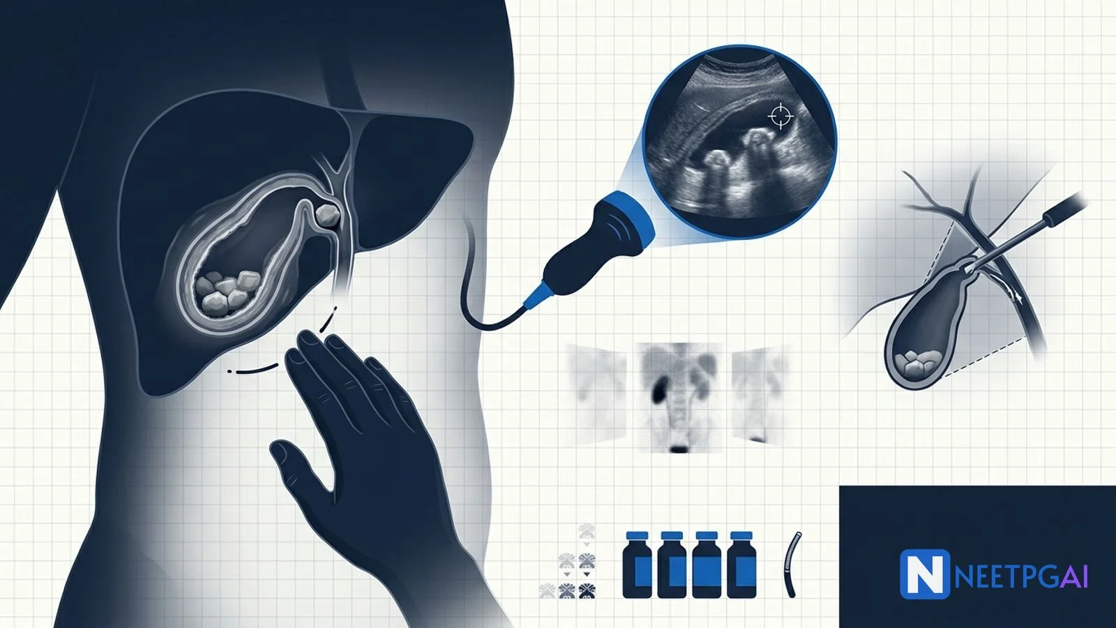
- First-line imaging — abdominal ultrasound — gallbladder wall over 3 mm, pericholecystic fluid, sonographic Murphy's sign, stones with posterior acoustic shadowing
- Second-line — HIDA scan or MRCP — when ultrasound is equivocal; HIDA shows non-visualisation of the gallbladder at 60 minutes
- Grade severity TG18 I/II/III — based on local inflammation and organ dysfunction
- Resuscitation and pregnancy-safe broad-spectrum antibiotics — piperacillin-tazobactam OR ceftriaxone plus metronidazole
- Differentials — acute hepatitis, acute pancreatitis, peptic ulcer perforation, inferior myocardial infarction, right basal pneumonia, hepatic abscess
- Definitive treatment — early laparoscopic cholecystectomy within 72 hours for Grade I and II; percutaneous cholecystostomy bridge for Grade III with delayed interval cholecystectomy at 6-8 weeks
- Watch for complications — gangrene, perforation, Mirizzi syndrome, gallstone ileus, emphysematous cholecystitis
The case
A 65-year-old woman walks into the emergency department of a tertiary hospital in Lucknow at 2 AM with severe upper-abdominal pain that began approximately 8 hours earlier. She had a heavy fried-food dinner at a family wedding the previous evening (mutton biryani, fried snacks, paneer dishes, sweets). The pain woke her from sleep at midnight. It started as a vague upper-abdominal discomfort and progressively localised to the right upper quadrant, with radiation to the right shoulder blade. She rates it 8 of 10 — constant, sharp, worse on inspiration and movement. Multiple antacid tablets at home gave no relief.
Associated features — nausea since the onset, two episodes of bilious vomiting in the last 3 hours. Subjective fever and chills since 4 AM. No haematemesis, no melaena, no chest pain, no shortness of breath. Bowel habit unchanged. Urine slightly darker than usual. No haematuria. No yellowish discolouration of eyes noticed by the family, but she lives alone since her husband's death 2 years ago.
Past medical history — Type 2 diabetes mellitus diagnosed 12 years ago on metformin 1 g twice daily and glimepiride 2 mg daily; HbA1c 7.8 percent at last check 3 months ago. Hypertension on amlodipine 5 mg and telmisartan 40 mg, well controlled. Dyslipidaemia on atorvastatin 20 mg. Known to have asymptomatic gallstones — ultrasound 2 years ago showed multiple gallstones in a normal gallbladder; she was advised elective cholecystectomy but declined due to fear and family commitments. Two previous episodes of self-limited biliary colic in the past year managed conservatively at home. No history of jaundice. Two normal vaginal deliveries 35 and 32 years ago. Postmenopausal for 15 years, no hormone replacement therapy.
Surgical history — appendicectomy at age 22. Otherwise no surgery.
Drug allergies — penicillin (mild rash, no anaphylaxis — documented in records).
Family and social history — non-smoker, non-alcoholic, vegetarian Indian diet with frequent ghee-rich cooking, sedentary lifestyle, BMI 31 (Asian-Indian obesity), lives alone, daughter visits weekly from Delhi.
On examination — alert but uncomfortable, in distress from pain. Temperature 38.5 degrees Celsius. Pulse 108/min regular. Blood pressure 132/84. Respiratory rate 22/min. Oxygen saturation 96 percent on room air. Mild scleral icterus on close inspection of the sclerae under good light. Pallor mild. No lymphadenopathy. Hydration mild dehydration.
Cardiovascular and respiratory examinations unremarkable apart from raised pulse. Abdomen — obese pendulous abdomen, no scars apart from the right iliac fossa appendicectomy scar. Tenderness maximal at the right upper quadrant, with localised guarding. Murphy's sign POSITIVE — sudden inspiratory arrest when the examiner palpates deep beneath the right costal margin during inspiration. A palpable tender mass is appreciated in the right upper quadrant approximately 3 cm below the costal margin, consistent with an inflamed gallbladder or pericholecystic phlegmon. No rebound elsewhere. Bowel sounds present, reduced. Liver edge cannot be reliably defined due to obesity and tenderness. Murphy's sign on inspiration with sonographic confirmation is planned. Per-rectal examination unremarkable, stools normal coloured on glove. Genitourinary examination unremarkable.
The emergency physician initiates immediate work-up and consults the on-call general surgery team with a working impression of acute cholecystitis in a 65-year-old obese diabetic woman with known gallstones until proven otherwise.
ABCD assessment and initial investigations
This is a time-critical surgical emergency — elderly diabetics with acute cholecystitis are high-risk for gangrenous or emphysematous cholecystitis with sepsis. Delay raises perforation and mortality.
A — Airway: patent, no concerns.
B — Breathing: RR 22/min mildly raised consistent with pain and fever; SpO2 96 percent on room air.
C — Circulation: Pulse 108/min, BP 132/84. Mild tachycardia from pain and dehydration; no haemodynamic instability. Two large-bore IV lines, IV Ringer lactate 1 L over 30 minutes, then maintenance.
D — Disability: GCS 15, oriented, capillary blood glucose 168 mg/dL on arrival (held off oral hypoglycaemics, started variable-rate insulin infusion).
E — Exposure and Environment: Temperature 38.5; mild scleral icterus; no rash; no flank discolouration (Grey-Turner or Cullen sign absent — rules against haemorrhagic pancreatitis).
Initial investigations
- CBC — Hb 11.4 g/dL, WBC 18,400/microL (markedly raised), platelets 285,000, neutrophils 88 percent with band forms (left shift)
- CRP — 124 mg/L (very high)
- Urea, creatinine, electrolytes — urea 42 mg/dL, creatinine 1.1 mg/dL (baseline), Na 138, K 4.1
- LFTs — total bilirubin 2.4 mg/dL (mildly raised — direct 1.6, indirect 0.8 suggesting cholestasis), AST 78 U/L, ALT 92 U/L, ALP 240 U/L (raised), GGT 180 U/L (raised) — picture of mild cholestasis, possibly suggesting Mirizzi syndrome or common bile duct stone needing exclusion
- Serum lipase — 80 U/L (normal — rules out concurrent acute pancreatitis); serum amylase 110 U/L
- HbA1c — 8.2 percent; random blood glucose 168 mg/dL
- Coagulation profile — INR 1.1, PT 13 sec, aPTT 32 sec
- Lactate — 2.4 mmol/L (mildly raised — early sepsis or hypoperfusion)
- ECG — sinus tachycardia, no acute ischaemic changes (important — RUQ pain in elderly can be referred from inferior MI; troponin sent at 0 and 3 hours both negative)
- Chest X-ray — clear lung fields, no free air under diaphragm, no right basal pneumonia
- Urine routine — trace ketones (fasting), no leucocytes, no nitrites
- Blood culture — sent before antibiotics
Imaging
- Abdominal ultrasound — performed at bedside within 1 hour. Gallbladder is distended (long axis 11 cm, short axis 5.2 cm), wall thickening 6 mm (over 3 mm), pericholecystic fluid present, multiple stones in the body and fundus, a 14 mm stone impacted in the Hartmann pouch (concerning for Mirizzi syndrome). Sonographic Murphy's sign POSITIVE — maximal tenderness over the visualised gallbladder. Common bile duct diameter 7 mm (upper limit of normal for age — over 6 mm is suspicious for obstruction). Intrahepatic biliary radicles not dilated. No free fluid in the peritoneal cavity. No air in the gallbladder wall (rules out emphysematous cholecystitis).
- MRCP — performed urgently given raised bilirubin, raised ALP/GGT, and impacted Hartmann-pouch stone. Confirms acute cholecystitis with multiple gallstones, a 14 mm stone in the Hartmann pouch compressing the common hepatic duct (Mirizzi type I — extrinsic compression without fistula), mild intrahepatic biliary radicle dilatation, and a 6 mm distal common bile duct stone. No pancreatic duct dilatation, no acute pancreatitis features. Liver normal.
- CT abdomen with contrast — not done initially (MRCP gave the necessary biliary tree detail), but a low-threshold for CT remains if perforation, emphysematous cholecystitis, or gangrene is suspected later.
Investigations confirm acute cholecystitis with Mirizzi syndrome type I and a coexisting CBD stone — a complex biliary emergency in an elderly diabetic.
TG18 diagnostic application
Cluster A (local inflammation): Murphy's sign positive AND RUQ tender mass. PRESENT.
Cluster B (systemic inflammation): fever 38.5, WBC 18,400, CRP 124. PRESENT.
Cluster C (imaging): gallbladder wall over 3 mm, pericholecystic fluid, sonographic Murphy's sign, stones, distended gallbladder. PRESENT.
Diagnosis — DEFINITE acute cholecystitis (TG18).
Severity grading
- WBC over 18,000 — YES (Grade II criterion)
- Palpable RUQ mass — YES (Grade II criterion)
- Duration over 72 hours — NO (8 hours)
- Marked local inflammation features (gangrene, abscess, peritonitis, emphysematous) — NOT seen on imaging
- Organ dysfunction (cardiovascular, neurological, respiratory, renal, hepatic, haematological) — NONE (vitals stable, GCS 15, creatinine normal, INR normal, platelets normal)
Grade II (moderate) acute cholecystitis. Plus coexisting Mirizzi syndrome type I and a 6 mm CBD stone.
The diagnostic algorithm — confirming cholecystitis and excluding mimics
NEET PG tests the RUQ-pain differential extensively. Memorise the 8-cause list.
Differential diagnosis of RUQ pain with fever in an elderly woman
| Diagnosis | Distinguishing features | First-line investigation |
|---|
| Acute cholecystitis | RUQ pain after fatty meal, Murphy's sign, fever, leukocytosis with left shift, raised CRP, gallstones on USG | Abdominal ultrasound; MRCP if jaundice |
| Acute hepatitis (viral or drug-induced) | Prodrome of malaise/anorexia, raised AST/ALT in thousands, viral markers positive | LFTs, viral serology (hepatitis A, B, C, E IgM) |
| Acute pancreatitis | Epigastric pain radiating to back, raised lipase greater than 3x normal, history of gallstones or alcohol | Serum lipase, CT abdomen with contrast |
| Peptic ulcer perforation | Sudden severe upper abdominal pain, board-like rigidity, free air under diaphragm on erect chest X-ray | Erect chest X-ray, CT abdomen |
| Inferior myocardial infarction | Substernal or epigastric pain, diaphoresis, autonomic features, ST changes inferior leads | ECG, troponin I/T |
| Right basal pneumonia | Cough, fever, breathlessness, dullness and crackles right base, consolidation on chest X-ray | Chest X-ray, sputum |
| Hepatic abscess (amoebic or pyogenic) | Tender hepatomegaly, fever, swinging temperature, RUQ pain referred to right shoulder, raised ALP, history of dysentery (amoebic) or biliary disease (pyogenic) | Abdominal ultrasound or CT; aspiration; amoebic serology |
| Mirizzi syndrome | Cholecystitis features plus obstructive jaundice from impacted Hartmann stone compressing CBD | MRCP |
| Acute appendicitis (high-riding appendix in pregnancy or anatomic variant) | Migratory pain, less commonly localised in RUQ in non-pregnant adults | Clinical, USG, CT |
Why our patient is acute cholecystitis with Mirizzi syndrome
Eight features confirm it: (1) classic post-fatty-meal RUQ pain with right scapular radiation, (2) Murphy's sign positive on examination AND on ultrasound, (3) fever 38.5 with WBC 18,400 and CRP 124, (4) ultrasound demonstrates wall thickening, pericholecystic fluid, gallstones with an impacted Hartmann stone, (5) raised ALP/GGT and direct hyperbilirubinaemia consistent with biliary obstruction, (6) MRCP confirms cholecystitis plus Mirizzi type I plus a 6 mm CBD stone, (7) ECG and troponin rule out MI, (8) no features of pancreatitis, perforation, or hepatic abscess.
Diagnosis
Acute calculous cholecystitis (TG18 Grade II) with Mirizzi syndrome type I and a 6 mm common bile duct stone, in a 65-year-old obese type-2 diabetic woman, currently haemodynamically stable, planned for ERCP-stone extraction followed by laparoscopic cholecystectomy.
Management — biliary clearance and source control
The two parallel goals are (1) biliary tree clearance (extraction of the CBD stone and management of Mirizzi compression) and (2) source control of the inflamed gallbladder by cholecystectomy.
Pre-operative preparation
- IV crystalloid resuscitation — Ringer lactate to maintain urine output 0.5 mL/kg/hr
- Broad-spectrum antibiotics within 1 hour — given the penicillin allergy (mild rash, no anaphylaxis), the surgical team opts for IV ceftriaxone 2 g daily plus metronidazole 500 mg IV every 8 hours rather than piperacillin-tazobactam. Cross-reactivity with cephalosporins after a non-anaphylactic penicillin reaction is low (under 1 percent for third-generation cephalosporins). Blood culture sent before first dose.
- Variable-rate insulin infusion for glycaemic control (target 140-180 mg/dL); metformin and glimepiride held; HbA1c noted for endocrine follow-up
- Anti-emetic — ondansetron 4 mg IV
- Analgesia — paracetamol 1 g IV regular; tramadol IV for breakthrough; opioids preferred over NSAIDs given mild renal risk and concurrent diabetes
- VTE prophylaxis — graduated compression stockings; LMWH (enoxaparin 40 mg subcutaneous daily) started 6 hours post-procedure given high-risk profile (elderly, obese, immobile, inflammatory state)
- Anaesthetic assessment — pre-operative ECG, echo (LVEF assessment in elderly diabetic), spirometry, ASA grade assignment (ASA III given diabetes, hypertension, obesity, acute illness)
- Consent — staged biliary clearance: ERCP with stone extraction first, then laparoscopic cholecystectomy; risks include conversion to open (10-20 percent in Mirizzi syndrome due to obscured anatomy), bile duct injury (under 0.5 percent in expert hands but higher in inflammatory states), bleeding, infection, retained stone
Definitive treatment strategy — staged approach
In our patient with Mirizzi syndrome type I plus a CBD stone, a two-stage approach is chosen:
Stage 1 — ERCP with sphincterotomy, CBD stone extraction, and biliary stent placement within 24 hours. The 6 mm distal CBD stone is removed using a Dormia basket after endoscopic sphincterotomy. A 7 Fr plastic biliary stent is placed across the Mirizzi compression to ensure biliary drainage.
Stage 2 — Laparoscopic cholecystectomy within 48-72 hours of ERCP (still within the early window). The critical view of safety (CVS) — clearing the hepatocystic triangle, visualising only two structures (cystic duct and cystic artery) entering the gallbladder, and exposing the cystic plate — is the universal safety standard. In Mirizzi syndrome, however, dense inflammation may obscure the anatomy; the surgical team has a low threshold for conversion to open if the CVS cannot be safely achieved, or for subtotal cholecystectomy (leaving a small cuff of gallbladder near the impacted stone) to avoid bile duct injury.
Surgical approach — laparoscopic with CVS dissection
Laparoscopic cholecystectomy (preferred):
- Patient position — supine, reverse Trendelenburg with left lateral tilt (American technique) OR French position (between legs)
- Port placement — standard 4-port technique (umbilical 10 mm camera, epigastric 10 mm, two right subcostal 5 mm working ports); for obese patients port positions may shift slightly inferior
- Pneumoperitoneum — 12-14 mmHg
- Critical view of safety dissection — clear the hepatocystic triangle of fat and fibrous tissue, expose the lower one-third of the gallbladder off the cystic plate, identify only two structures entering the gallbladder (cystic duct and cystic artery); both must be visualised before any clip or division
- Cystic artery and duct — clipped and divided; intraoperative cholangiogram (IOC) considered in Mirizzi syndrome to confirm biliary anatomy and exclude bile duct injury
- Specimen retrieval — through the umbilical port in a retrieval bag; sent for histopathology to rule out incidental gallbladder carcinoma (1-3 percent incidence in elderly Indian women with chronic gallstones)
- Drain — a closed-suction drain (typically 14 Fr Robinson or Jackson-Pratt) placed in the gallbladder fossa, especially when subtotal cholecystectomy or Mirizzi dissection has been performed
- Operating time — typically 45-90 minutes; longer in Mirizzi and elderly diabetic patients
Conversion to open is indicated by: dense adhesions obscuring the CVS, suspected bile duct injury, uncontrolled bleeding, suspected gallbladder cancer, or anatomic uncertainty. Conversion rates are 5-10 percent in routine acute cholecystitis but 15-30 percent in Mirizzi syndrome — conversion is a sign of good surgical judgement, not failure.
Percutaneous cholecystostomy — when to use
Percutaneous cholecystostomy (PC) is a percutaneous transhepatic drainage of the gallbladder under ultrasound or CT guidance, used as a bridge in patients unfit for immediate surgery. Indications:
- TG18 Grade III acute cholecystitis with organ dysfunction
- Patients with severe comorbidities (ASA IV-V), severe heart failure, terminal illness
- Patients on anticoagulation that cannot be reversed safely
- Failure of medical management at 24-48 hours
After PC, interval laparoscopic cholecystectomy is planned 6-8 weeks later once inflammation settles. In Indian district hospitals where interventional radiology is unavailable, surgical cholecystostomy (open or laparoscopic placement of a drain) is the alternative.
Intra-operative findings and post-operative care
In our patient — ERCP performed at 18 hours from admission. CBD stone retrieved, sphincterotomy completed, 7 Fr biliary stent placed. Laparoscopic cholecystectomy performed at 60 hours from admission (within the 72-hour window). Intra-operative findings — distended oedematous gallbladder, dense pericholecystic inflammation, impacted Hartmann stone visible. CVS achieved with patient dissection. Cystic duct found short and oedematous; intraoperative cholangiogram performed to confirm anatomy and stent position. Cholecystectomy completed in 105 minutes. Estimated blood loss 80 mL. No bile leak, no bowel injury. Specimen sent for histopathology — acute on chronic cholecystitis, no malignancy. Drain placed in gallbladder fossa.
Post-operative care:
- Day 0 — IV ceftriaxone + metronidazole continued for 24 hours, then orally for total 5-7 days (Grade II)
- Day 1 — clear oral fluids, ambulation, deep breathing exercises, insulin infusion transitioned to subcutaneous regimen
- Day 2-3 — soft diet, drain removed if low output (under 30 mL per day, non-bilious), oral metformin resumed once eating normally
- Day 3-4 — discharge if afebrile, eating, pain controlled, drain removed
- Follow-up — outpatient review at 2 weeks for wound check and histology report; ERCP biliary stent removal at 4-6 weeks via repeat ERCP; long-term diabetic and lipid follow-up
Complications — local and systemic
Local
- Gangrenous cholecystitis — 15-25 percent of acute cases in elderly diabetics; transmural necrosis; needs urgent surgery
- Gallbladder perforation — 10 percent in delayed presentations; free intraperitoneal (generalised biliary peritonitis), localised (pericholecystic abscess), or fistulised (cholecystoenteric)
- Emphysematous cholecystitis — Clostridium and E coli gas-formers; classic in elderly diabetic males; 50 percent gangrenous, 15-20 percent mortality; gas in gallbladder wall on CT or X-ray; needs urgent surgery
- Mirizzi syndrome — type I (extrinsic compression), type II-V (cholecystocholedochal fistula with progressive bile duct involvement); needs MRCP characterisation; subtotal cholecystectomy with bile duct repair for higher types
- Gallstone ileus — large stone (over 2.5 cm) eroding into duodenum via cholecystoenteric fistula; classic Rigler triad on plain X-ray (pneumobilia, ectopic gallstone in pelvis or RLQ, small bowel obstruction); needs enterolithotomy with delayed cholecystectomy
- Bouveret syndrome — gallstone ileus variant with stone impaction in duodenum causing gastric outlet obstruction
- Cholecystoenteric fistula — chronic erosion into duodenum, colon, or stomach; needs fistula closure plus cholecystectomy
- Hepatic abscess — pyogenic spread from severe cholecystitis; needs drainage plus antibiotics
Systemic
- Sepsis and septic shock — high in elderly diabetics; needs urgent source control and broad-spectrum antibiotics
- ARDS, AKI, multi-organ dysfunction — in Grade III disease
- DVT/PE — pregnancy plus surgery doubles risk; obese elderly diabetic at very high risk; LMWH prophylaxis essential
- Wound infection, intra-abdominal abscess — 2-5 percent post-laparoscopic, 5-15 percent post-open
- Bile duct injury — under 0.5 percent in expert hands, higher in Mirizzi (2-5 percent); needs hepatobiliary referral for Roux-en-Y hepaticojejunostomy if major injury
India-specific considerations
- Northern Indo-Gangetic belt has the highest gallstone prevalence in India (15-22 percent in women over 40 in published Lucknow, Kanpur, and Varanasi series) vs 5-7 percent in southern India — high dietary fat, parity, obesity, and genetic predisposition
- Gallbladder cancer prevalence is also highest in this belt (Varanasi, Allahabad, Lucknow have some of the world's highest age-adjusted rates); always send gallbladder specimens for histopathology and have a low threshold for further imaging in incidentalomas, polyps over 1 cm, porcelain gallbladder, and chronic typhoid carriers
- Diagnostic delay is common in rural and Tier-2/3 areas — average symptom-to-presentation time in published Indian series is 48-72 hours vs 12-24 hours in urban tertiary centres
- Limited MRI/ERCP access outside metropolitan tertiary hospitals — many district hospitals rely on USG only; the threshold for transfer to a centre with ERCP and laparoscopic expertise should be low in suspected Mirizzi syndrome or CBD stones
- PMJAY (Pradhan Mantri Jan Arogya Yojana) covers emergency biliary surgery including laparoscopic cholecystectomy and ERCP for eligible families
- Cost considerations — ceftriaxone plus metronidazole remains the dominant antibiotic combination in Indian district hospitals due to availability and cost (~Rs 300/day) vs piperacillin-tazobactam (~Rs 1500/day); both are TG18-compliant
- Stigma against surgery in the elderly — families may delay consent fearing surgical risk; clear communication that delay raises perforation and mortality is essential
How NEET PG tests acute cholecystitis
Seven recurring patterns.
Pattern 1 — The TG18 diagnostic-criteria question: Vignette gives the patient's symptoms, signs, and imaging; asks which is the most appropriate diagnostic framework. Tokyo Guidelines 2018 (TG18) — clusters A, B, C. A definite diagnosis needs one from each cluster.
Pattern 2 — The severity-grading question: Vignette gives WBC count, organ dysfunction parameters; asks the TG18 grade. Grade I (mild) — no organ dysfunction, mild gallbladder inflammation. Grade II (moderate) — any of WBC over 18,000, RUQ mass, duration over 72 hours, gangrene, abscess, biliary peritonitis, emphysematous. Grade III (severe) — organ dysfunction.
Pattern 3 — The early vs delayed cholecystectomy question: Best surgical timing for Grade I/II acute cholecystitis? Early laparoscopic cholecystectomy within 72 hours of symptom onset. Why? Shorter total hospital stay, no recurrence risk, similar bile duct injury rate, lower cost. Delayed interval cholecystectomy at 6-8 weeks now restricted to Grade III with PC bridge and patients unfit for immediate surgery.
Pattern 4 — The imaging question: First-line imaging in suspected acute cholecystitis? Abdominal ultrasound. Findings? Gallbladder wall over 3 mm, pericholecystic fluid, sonographic Murphy's sign, gallstones with posterior acoustic shadowing, gallbladder distension over 8 cm long axis or 4 cm short axis. Second-line when ultrasound equivocal? HIDA scan (cholescintigraphy) — non-visualisation of gallbladder 60 minutes after tracer suggests cystic duct obstruction.
Pattern 5 — The complication question: Elderly diabetic male with gas in gallbladder wall on CT? Emphysematous cholecystitis (Clostridium and E coli, 15-20 percent mortality, needs urgent surgery). Stone in Hartmann pouch compressing CHD with obstructive jaundice? Mirizzi syndrome type I. Plain abdominal X-ray with pneumobilia plus ectopic stone plus small bowel obstruction? Gallstone ileus (Rigler triad).
Pattern 6 — The antibiotic question: Most appropriate empirical antibiotic in community-acquired Grade I/II acute cholecystitis? Piperacillin-tazobactam OR ceftriaxone plus metronidazole. Duration after source control? 24 hours for Grade I, 4-7 days for Grade II. Avoid in penicillin-allergic with anaphylaxis? All beta-lactams; use carbapenem (with cross-reactivity caution) or fluoroquinolone plus metronidazole.
Pattern 7 — The critical view of safety question: Universal safety standard during laparoscopic cholecystectomy? Critical view of safety (CVS) — three criteria: hepatocystic triangle cleared of fat and fibrous tissue, lower one-third of gallbladder separated from cystic plate, only two structures (cystic duct and cystic artery) seen entering the gallbladder. Indication for intraoperative cholangiogram? Suspected Mirizzi, unclear anatomy, suspected CBD stone, intraoperative bile duct injury.
High-yield one-liners:
- Acute cholecystitis is one of the commonest non-trauma surgical emergencies in India, especially the northern belt
- TG18 — three clusters (A local, B systemic, C imaging); definite diagnosis needs one from each
- Severity Grade I/II/III — operate within 72 hours for I/II; PC bridge plus delayed interval cholecystectomy at 6-8 weeks for III
- Murphy's sign — sudden inspiratory arrest on deep RUQ palpation
- Sonographic Murphy's sign is MORE specific than clinical Murphy's
- Gallbladder wall over 3 mm, pericholecystic fluid, sonographic Murphy's, gallstones — diagnostic ultrasound tetrad
- HIDA scan — non-visualisation of gallbladder at 60 minutes confirms cystic duct obstruction
- Elderly diabetic with gas in gallbladder wall — emphysematous cholecystitis (urgent surgery)
- Hartmann-pouch stone compressing CHD — Mirizzi syndrome (MRCP for typing)
- Rigler triad (pneumobilia, ectopic stone, SBO) — gallstone ileus
- Pre-operative antibiotics — piperacillin-tazobactam OR ceftriaxone plus metronidazole
- Critical view of safety (CVS) — universal safety standard
- Convert to open if CVS cannot be safely achieved
- Send gallbladder specimens for histopathology — gallbladder cancer in 1-3 percent elderly Indian women
Frequently Asked Questions
What are the Tokyo Guidelines 2018 (TG18) diagnostic criteria for acute cholecystitis?
TG18 uses three clusters. A — local signs of inflammation: Murphy's sign positive OR right upper quadrant pain, mass, or tenderness. B — systemic signs of inflammation: fever, raised CRP, or raised WBC. C — imaging findings characteristic of acute cholecystitis on ultrasound, CT, or MRI (gallbladder wall thickening over 3 mm, pericholecystic fluid, sonographic Murphy's sign, gallbladder enlargement over 8 cm long axis or 4 cm short axis, gallstone or sludge). A suspected diagnosis requires one item from A plus one from B. A definite diagnosis requires one from A plus one from B plus C. NEET PG tests these criteria as a structured algorithm — the older textbook combination of Murphy's sign and gallstones on ultrasound alone is now refined into this three-cluster framework.
How is the severity of acute cholecystitis graded under TG18 and why does it matter?
TG18 severity is graded I to III based on organ dysfunction and local inflammation. Grade I (mild) — acute cholecystitis without organ dysfunction and only mild gallbladder inflammation; suitable for early laparoscopic cholecystectomy within 72 hours. Grade II (moderate) — any of WBC over 18,000 per microL, palpable tender mass in right upper quadrant, duration over 72 hours, marked local inflammation (gangrene, pericholecystic abscess, hepatic abscess, biliary peritonitis, emphysematous cholecystitis); requires urgent surgery or percutaneous cholecystostomy bridge if patient unfit. Grade III (severe) — organ dysfunction in cardiovascular (hypotension requiring vasopressors), neurological (decreased consciousness), respiratory (PaO2/FiO2 ratio under 300), renal (creatinine over 2 mg/dL or oliguria), hepatic (INR over 1.5), or haematological (platelets under 100,000); resuscitation, broad-spectrum antibiotics, and percutaneous cholecystostomy first, with delayed interval cholecystectomy in 6-8 weeks. Grading drives the operate-now versus drain-first decision and is heavily tested.
Why is early laparoscopic cholecystectomy within 72 hours preferred over delayed interval cholecystectomy?
Multiple randomised trials and meta-analyses (the ACDC trial 2013, CHOCOLATE 2018, the 2021 Cochrane review) consistently show early laparoscopic cholecystectomy within 72 hours of symptom onset is superior to delayed interval surgery for Grade I and Grade II disease. Benefits include shorter total hospital stay (5-7 days vs 12-15 days when readmission for recurrent attacks is counted), lower overall cost, lower conversion-to-open rates (4-10 percent in fresh inflammation due to oedematous tissue planes), no risk of recurrent attacks during the wait (20-30 percent rate over 6 weeks), and similar bile duct injury rates when performed by trained laparoscopic surgeons. Delayed interval cholecystectomy at 6-8 weeks is now reserved for Grade III disease bridged by percutaneous cholecystostomy, patients unfit for immediate surgery, or those presenting beyond 7-10 days when dense adhesions raise complication risk. NEET PG repeatedly tests the 72-hour window as the upper limit for safe early laparoscopic cholecystectomy.
What are the major complications of acute cholecystitis and which patient populations are at highest risk?
Complications cluster into local and systemic. Local — gangrenous cholecystitis (15-25 percent of acute cases in elderly diabetics, transmural necrosis), gallbladder perforation (10 percent — free into peritoneum causing generalised biliary peritonitis, localised pericholecystic abscess, or fistulisation), emphysematous cholecystitis (gas-forming organisms Clostridium and E coli, characteristic gas in gallbladder wall on CT, 50 percent gangrenous, 15-20 percent mortality, classic in elderly diabetic males), Mirizzi syndrome (impacted stone in Hartmann pouch compressing common hepatic duct causing obstructive jaundice), cholecystoenteric fistula and gallstone ileus (large stone eroding into duodenum, classic Rigler triad on abdominal X-ray — pneumobilia, ectopic gallstone, small bowel obstruction). Systemic — sepsis, ARDS, multi-organ dysfunction. Highest-risk groups are elderly (over 65), diabetics, immunocompromised, chronic steroid users, and patients with delayed presentation. India-specific risk factors include high gallstone prevalence in the northern Indo-Gangetic belt, dietary fat, and delayed referral from primary care.
Which antibiotic regimens are recommended for acute cholecystitis under Indian and TG18 guidelines?
TG18 antibiotic choice is risk-stratified. Community-acquired Grade I or II in low-risk patients — single agent with biliary penetration: piperacillin-tazobactam 4.5 g IV every 8 hours, OR a third-generation cephalosporin (ceftriaxone 2 g IV daily) plus metronidazole 500 mg IV every 8 hours to cover anaerobes. Grade III or high-risk patients (recent hospitalisation, immunosuppression, comorbid sepsis) — broader-spectrum cover with carbapenem (meropenem 1 g IV every 8 hours) or piperacillin-tazobactam plus vancomycin if MRSA suspected. Antibiotic duration after surgical source control is 24 hours for Grade I, 4-7 days for Grade II, and longer (10-14 days) for Grade III or perforation with peritonitis. Common Indian gallbladder bile cultures grow Escherichia coli (the dominant organism), Klebsiella, Enterococcus, Enterobacter, and Pseudomonas; anaerobes are uncommon in community-acquired cases. In rural Indian district hospitals, ceftriaxone plus metronidazole remains the most widely used regimen due to cost and availability. Aminoglycosides are added only for severe sepsis with renal function permitting.
This content is for educational purposes for NEET PG exam preparation. It is not a substitute for professional medical advice, diagnosis, or treatment. Clinical information has been reviewed by qualified medical professionals.
Written by: NEETPGAI Editorial Team
Reviewed by: Pending SME Review
Last reviewed: May 2026