image mcq
radiology
chest x ray
pneumonia
pneumothorax
pulmonary edema
miliary TB
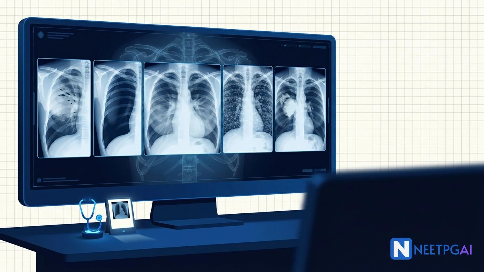
5 high-yield CXR image MCQs for NEET PG: lobar pneumonia, tension/deep-sulcus pneumothorax, batwing pulmonary edema, miliary TB, lung cancer with hilar lymphadenopathy.
Version 1.0 — Published April 2026
Chest X-ray image MCQs contribute 4-5 questions per NEET PG paper across pulmonology, internal medicine, and radiology. Five CXR patterns recur reliably: lobar pneumonia (homogeneous lobar opacity with air bronchograms), pneumothorax (visceral pleural line with absent peripheral markings; deep sulcus sign in supine; tension shows mediastinal shift), pulmonary edema (batwing perihilar haze with Kerley B lines and cardiomegaly), miliary TB (diffuse millet-seed nodules), and lung cancer with hilar lymphadenopathy (asymmetric hilar mass with or without mediastinal widening).
The pattern-to-diagnosis mapping plus knowledge of the silhouette sign and lobar collapse patterns covers nearly every CXR NEET PG question.
Chest X-ray remains the single most-tested radiology image in NEET PG, and the patterns are highly stereotyped — pattern recognition is the single most cost-effective preparation strategy. Indian medical education retains heavy emphasis on CXR interpretation in clinical and PG entrance exams, and infectious causes (pneumonia, TB) are over-represented relative to Western boards.
Five patterns recur in nearly every paper: lobar pneumonia, pneumothorax (deep sulcus and tension), cardiogenic pulmonary edema, miliary TB, and lung cancer with hilar nodes. Drill these five patterns plus 5-10 PYQ CXR images daily for 2 weeks, and your accuracy will move from 40 to 85 percent on radiology image MCQs.
A radiopacity in anatomic contact with a normally air-bordered structure obliterates that border:
| Border lost | Lobe / segment involved |
|---|---|
| Right heart border | Right middle lobe |
| Left heart border (lingula) | Lingular segment of LUL |
| Right hemidiaphragm | Right lower lobe |
| Left hemidiaphragm | Left lower lobe |
| Aortic knob | Apicoposterior segment of LUL |
| Descending aorta | Posterior basal segments of LLL |
| Heart border preserved + opacity behind heart | Posterior basal LLL behind the heart |
This content is for educational purposes for NEET PG exam preparation. It is not a substitute for professional medical advice, diagnosis, or treatment. Clinical information has been reviewed by qualified medical professionals.
Start practicing NEET PG MCQs with AI-powered explanations.
Start Free PracticeMaster GI secretions, digestion, absorption transporters, motility patterns, and gut hormones with high-yield NEET PG 2026 traps and India-context examples.
Master labor stages, Friedman vs Zhang curves, WHO partograph, AMTSL, episiotomy and India JSY/LaQshya policies for NEET PG 2026 OBG MCQs.
5 anterior segment ophthalmology image MCQs for NEET PG: hypopyon and Behcet, Kayser-Fleischer ring in Wilson, Brushfield spots in Down, corneal arcus, and pterygium vs pinguecula.
Daily MCQs, study tips, and topper strategies on Telegram.
Join on Telegram →| Lobe |
|---|
| Collapse pattern |
|---|
| RUL | Up-and-medial; raised right hilum, S-sign of Golden if mass at hilum (suggests obstructing cancer) |
| RML | Loss of right heart border; sail sign or thin triangular opacity on lateral |
| RLL | Down and posterior; preserved right heart border, loss of right hemidiaphragm, increased retrocardiac opacity |
| LUL (including lingula) | Up and anterior; veil-like haze across entire left hemithorax with preserved cardiac silhouette through it; luftsichel sign (hyperlucent crescent) |
| LLL | Down and posterior; loss of left hemidiaphragm, retrocardiac triangular opacity (sail sign) |
Image description: [PA chest radiograph of an adult patient. There is a homogeneous, dense opacity occupying the right upper zone, sharply demarcated inferiorly by the horizontal fissure and laterally by the chest wall. Within the opacity, branching tubular lucencies (air bronchograms) are clearly visible. The right hilum is at normal height. The right heart border is preserved. The right hemidiaphragm is sharp. There is no pleural effusion, no mediastinal shift. The contralateral lung is clear. The cardiothoracic ratio is normal.]
Clinical vignette: A 64-year-old retired farmer presents with 4 days of productive cough with rust-colored sputum, fever 39.4 C, pleuritic right-sided chest pain, and dyspnea. He is a non-smoker with no chronic illness. Examination reveals dullness to percussion, bronchial breathing, increased vocal fremitus and crepitations over the right upper zone. WBC is 18,400 with neutrophilia.
Options:
Correct answer: (a) Right upper lobe lobar pneumonia
Reasoning: Homogeneous lobar opacity sharply demarcated by the horizontal fissure with prominent air bronchograms plus rust-coloured sputum, high fever, neutrophilia, and bronchial breathing on examination is textbook lobar pneumonia, most often pneumococcal in a community-acquired setting. Air bronchograms confirm an alveolar filling process with patent bronchi (cancer would obstruct the bronchus and abolish bronchograms).
RUL collapse from an obstructing tumour would show volume loss (raised right hilum, tracheal deviation, S-sign of Golden at the hilum) and absent air bronchograms. Apical TB with cavitation typically shows patchy upper-zone heterogeneous opacity with cavities and fibrosis, often with hilar lymphadenopathy. A right pleural effusion would produce a meniscus-shaped opacity at the costophrenic angle with blunting, not a lobar opacity respecting the fissure.
Teaching pearl: Lobar consolidation differential — Streptococcus pneumoniae (rust sputum, classic), Klebsiella pneumoniae (currant-jelly sputum, bulging fissure sign especially RUL in alcoholics and diabetics), Legionella (extrapulmonary features — confusion, hyponatremia, diarrhea, hepatic dysfunction), staphylococcal pneumonia (post-influenza, abscesses, pneumatoceles).
CURB-65 / qSOFA for severity assessment determines outpatient vs inpatient vs ICU. Empirical therapy in India: outpatient — amoxicillin or doxycycline; inpatient ward — ceftriaxone + azithromycin; ICU — ceftriaxone + azithromycin or beta-lactam + respiratory fluoroquinolone with anti-pseudomonal cover if risk factors. Always send sputum Gram stain and culture, blood cultures, urinary antigen for pneumococcus and Legionella in severe disease.
Image description: [PA chest radiograph of a young, thin male. The right hemithorax shows a thin curvilinear visceral pleural line running parallel to the chest wall, with absent lung markings beyond the line and increased lucency. The right lung is partially collapsed toward the hilum. The mediastinum is in the midline. The right hemidiaphragm is at normal height. Trachea is central. The contralateral left lung is clear. No rib fractures, no surgical emphysema.]
Clinical vignette: A 25-year-old tall thin male medical student presents with sudden onset right-sided pleuritic chest pain and mild dyspnea while studying. He is a non-smoker with no prior episodes. Vitals: pulse 96/min, BP 122/76, respiratory rate 22, SpO2 96 percent on room air. Examination shows reduced breath sounds and hyperresonance on the right.
Options:
Correct answer: (a) Primary spontaneous pneumothorax
Reasoning: Visceral pleural line with absent lung markings beyond it and partial lung collapse, in a tall thin young male non-smoker, with stable vitals and central mediastinum is primary spontaneous pneumothorax — the classic patient profile is a tall thin male 18-35 years old, often from rupture of apical subpleural blebs.
Tension pneumothorax produces complete lung collapse against the hilum, contralateral mediastinal shift, ipsilateral hemidiaphragm depression/inversion, and is a clinical emergency with hypotension, distended neck veins, and severe distress — none present here. Pulmonary embolism rarely changes CXR substantially; classic but rare signs include Westermark sign (oligaemia distal to embolus), Hampton hump (peripheral wedge-shaped opacity), and Fleischner sign (enlarged proximal pulmonary artery). Pleural effusion produces a homogeneous opacity with meniscus and often shifts the mediastinum away if large.
Teaching pearl: Pneumothorax classification:
| Type | Setting |
|---|---|
| Primary spontaneous | Tall thin young males, smokers, apical bleb rupture |
| Secondary spontaneous | Underlying lung disease — COPD, TB, cystic fibrosis, Marfan, LAM, Birt-Hogg-Dubé |
| Traumatic | Blunt or penetrating trauma; often associated with rib fractures, hemothorax |
| Iatrogenic | Central line insertion, thoracentesis, mechanical ventilation barotrauma, transbronchial biopsy |
| Tension | Any of the above with one-way valve mechanism — clinical emergency |
| Catamenial | Recurrent pneumothorax in women during menstruation; thoracic endometriosis |
Management of primary spontaneous pneumothorax (BTS guidelines):
Image description: [PA chest radiograph of an elderly woman. Both lungs show symmetrical bilateral perihilar haziness with a central distribution radiating outward — the so-called batwing or butterfly pattern. Multiple short horizontal lines (Kerley B lines) are visible at both costophrenic angles. There is peribronchial cuffing visible at the hila. Pulmonary vessels are dilated in the upper zones (cephalization). Both costophrenic angles are blunted with small bilateral pleural effusions, right slightly more than left. The cardiac silhouette is enlarged with a cardiothoracic ratio of approximately 0.6.]
Clinical vignette: A 78-year-old woman with hypertension, type 2 diabetes, and known ischemic heart disease presents to the emergency room with sudden onset severe dyspnea, orthopnea, and a productive cough with pink frothy sputum starting 4 hours ago. She has a 2-pillow orthopnea and recent ankle swelling. Examination: SpO2 88 percent on room air, BP 168/96, pulse 124/min, raised JVP, S3 gallop, and bilateral basal crackles up to mid-zones.
Options:
Correct answer: (c) Cardiogenic pulmonary edema
Reasoning: Symmetrical bilateral perihilar batwing opacities, Kerley B lines, peribronchial cuffing, cephalization, bilateral pleural effusions, and cardiomegaly in an elderly hypertensive diabetic with orthopnea, raised JVP, S3 gallop, and pink frothy sputum is the canonical CXR of cardiogenic pulmonary edema secondary to acute decompensated heart failure.
Bilateral lobar pneumonia would be unusual to present this symmetrically and would not show Kerley lines or cephalization; cardiomegaly would be incidental rather than central to the picture. ARDS shows patchy bilateral peripheral or diffuse opacities with normal cardiac silhouette, no Kerley lines, no significant pleural effusions, and refractory hypoxemia despite high FiO2. Pulmonary alveolar hemorrhage shows patchy bilateral alveolar opacities with hemoptysis or anemia and is a different clinical context (vasculitides, anti-GBM disease, mitral stenosis).
Teaching pearl: Cardiogenic vs non-cardiogenic pulmonary edema CXR features:
| Feature | Cardiogenic | Non-cardiogenic (ARDS) |
|---|---|---|
| Distribution | Central (perihilar batwing) | Peripheral or patchy diffuse |
| Kerley B lines | Common | Rare |
| Pleural effusion | Often, bilateral | Rare unless coexisting pathology |
| Cardiomegaly | Common | Uncommon |
| Vascular pedicle width | Wide (>70 mm) | Normal |
| Air bronchograms | Less prominent | Prominent |
Kerley lines are pleural-based linear opacities representing thickened interlobular septa from interstitial edema:
Management of acute cardiogenic pulmonary edema (LMNOP):
Treat the underlying cause: ACS, hypertensive crisis, valvular disease, arrhythmia, ischemic cardiomyopathy. Echocardiography urgently to assess EF and structural lesions.
Image description: [PA chest radiograph of a young adult male. Both lungs show innumerable, small (1-3 mm), uniformly sized, well-defined nodules distributed homogeneously throughout all zones, including the apices and bases — without zonal predominance. The hila are mildly prominent with possible bilateral hilar lymphadenopathy. The cardiac silhouette is normal. There are no cavities, no pleural effusion, no consolidation, no significant fibrosis.]
Clinical vignette: A 32-year-old male, HIV-positive on irregular ART for 4 years, presents with 6 weeks of low-grade evening fever, drenching night sweats, 8 kg weight loss, anorexia, and progressive dry cough. CD4 count is 110/μL. There is no productive sputum. Examination reveals cervical lymphadenopathy, oral candidiasis, and bilateral fine crepitations.
Options:
Correct answer: (b) Miliary tuberculosis
Reasoning: Innumerable uniformly sized 1-3 mm nodules distributed homogeneously throughout both lungs (millet-seed pattern) in an immunocompromised patient with constitutional symptoms is miliary tuberculosis. India is endemic, HIV co-infection markedly raises the risk, and the diagnosis is presumptive until proven by sputum AFB, GeneXpert, or biopsy.
Pneumocystis jirovecii pneumonia (PCP) typically shows bilateral perihilar reticular or ground-glass opacities (not discrete miliary nodules) and a more rapid clinical course; LDH is markedly elevated; treatment is high-dose cotrimoxazole. Pulmonary metastases ("cannonball") are typically larger (≥1 cm), variable in size, and fewer in number — classic primaries are renal cell carcinoma, choriocarcinoma, and sarcomas. Sarcoidosis shows bilateral hilar lymphadenopathy with reticulonodular interstitial opacities (often upper-zone predominant) but rarely the uniform miliary pattern.
Teaching pearl: Miliary pattern differential (NEET PG list):
| Disease | Distinguishing features |
|---|---|
| Miliary TB | Endemic, immunocompromise, fever, weight loss, prototype |
| Miliary fungal | Histoplasmosis, coccidioidomycosis, blastomycosis (geography) |
| Hematogenous metastases | Thyroid, RCC, melanoma, choriocarcinoma; size variable |
| Sarcoidosis | BHL, upper-zone reticulonodular, rarely truly miliary |
| Pneumoconiosis | Silicosis, coal-worker — upper-zone, occupational history |
| Hypersensitivity pneumonitis | Acute/subacute, exposure history, ground-glass micronodules |
| Viral pneumonias (rare) | Varicella pneumonia produces nodules in adults |
Diagnosis of miliary TB:
Treatment under RNTCP/NTEP: standard 4-drug regimen (HRZE) for 2 months intensive phase, then 4 months continuation phase (HR) for pulmonary disease without CNS involvement. Extend to 9-12 months for TB meningitis with adjunctive corticosteroids (dexamethasone or prednisolone tapered over 6-8 weeks; mortality benefit). ART continued or initiated within 2-8 weeks of TB therapy in HIV co-infection (earlier in CD4 <50 to reduce mortality, balanced against IRIS risk).
Image description: [PA chest radiograph of an elderly male. There is a well-defined, large (5-6 cm) rounded opacity at the right hilum, with associated widening of the right hilum and mediastinum from probable lymphadenopathy. The mass appears to be partially obstructing the right upper lobe bronchus, with subtle volume loss in the RUL (right hilum elevated, slight tracheal shift to the right). The contralateral lung is clear. No pleural effusion, no rib destruction visible. The cardiothoracic ratio is normal. A small ipsilateral hilar lymph node mass produces an additional bulge below the right hilum.]
Clinical vignette: A 68-year-old male, 50 pack-year smoker, presents with 3 months of cough, intermittent hemoptysis (streaks of blood), 10 kg unintentional weight loss, anorexia, and worsening dyspnea over 6 weeks. He has clubbing of his fingers and a recent diagnosis of hypercalcemia (corrected calcium 12.4 mg/dL) without obvious cause. He has no known TB contact, no fever, no night sweats.
Options:
Correct answer: (b) Lung cancer with hilar lymphadenopathy (likely squamous cell carcinoma)
Reasoning: A large hilar mass with associated hilar lymphadenopathy and possible RUL volume loss in an elderly heavy smoker with hemoptysis, weight loss, clubbing, and hypercalcemia (paraneoplastic, often PTHrP from squamous cell carcinoma) points strongly to non-small cell lung cancer. Squamous cell carcinoma is the histologic subtype most associated with central airway location, hemoptysis from cavitation, and PTHrP-mediated hypercalcemia.
TB with hilar adenopathy is more common in children (primary TB) and the mass would more typically be parenchymal (Ghon focus) with smaller hilar nodes. Sarcoidosis Stage I shows bilateral hilar lymphadenopathy (BHL) without a parenchymal mass and is uncommon at age 68. Lymphoma typically presents with bilateral and/or anterior mediastinal lymphadenopathy with constitutional B-symptoms — a unilateral central mass with ipsilateral hilar nodes is less typical.
Teaching pearl: Lung cancer histologic subtypes — high-yield associations:
| Subtype | Location | Smoking | Notable features |
|---|---|---|---|
| Squamous cell carcinoma | Central | Strong | Cavitates; PTHrP → hypercalcemia |
| Adenocarcinoma | Peripheral | Less strong (commonest in non-smokers and women) | EGFR, ALK, ROS1, KRAS mutations; targetable in non-smokers |
| Large-cell carcinoma | Peripheral | Strong | Aggressive; least common NSCLC subtype |
| Small-cell carcinoma | Central | Very strong | Limited/extensive stage; SIADH (ADH), Cushing (ACTH), Lambert-Eaton; chemo and radiotherapy primary; not surgical |
| Carcinoid tumor | Central | Not strongly associated | Younger patients; carcinoid syndrome rare unless metastatic |
| Mesothelioma | Pleural | Asbestos | Pleural plaques and effusion |
Paraneoplastic syndromes (NEET PG cluster):
Workup of suspected lung cancer:
Stage and histology drive management — surgery for early-stage NSCLC, concurrent chemoradiotherapy for stage III, targeted therapy or immunotherapy for stage IV NSCLC, etoposide-platinum chemotherapy + thoracic RT for limited-stage SCLC.
A skin fold can mimic a pneumothorax line on supine films but lung markings extend BEYOND the skin fold (whereas pneumothorax has absent markings beyond the visceral pleural line). Bullae produce hyperlucency without a visible visceral pleural line and have thin curvilinear walls — distinguishing emphysematous bullae from pneumothorax is critical because a chest tube placed in a giant bulla is catastrophic. CT confirms when uncertain.
Both can produce a sharply demarcated opacity, but lobar pneumonia preserves volume (no shift, no raised hilum) while collapse loses volume (hilum displaced toward the opacity, mediastinal shift toward the opacity, narrowed intercostal spaces, raised hemidiaphragm). The S-sign of Golden (an obstructing central mass causing collapse with a concave-then-convex inferior border) suggests cancer. Air bronchograms are present in pneumonia and absent in obstructive collapse.
Pulmonary edema is central, symmetric, with Kerley lines, cardiomegaly, pleural effusions; ARDS is patchy/peripheral, no Kerley lines, normal heart size; diffuse pneumonia (PCP, viral) is often bilateral with ground-glass and/or interstitial pattern without cardiomegaly. Clinical context (BNP, ejection fraction, hypoxemia severity) and the trajectory matter as much as the image.
Miliary TB has uniform 1-3 mm nodules throughout all zones; PCP has bilateral perihilar ground-glass and interstitial opacities (less commonly cysts); cannonball metastases are larger, variable in size, and fewer in number. HRCT clarifies: random nodule distribution (TB, metastases), centrilobular (HP, infectious bronchiolitis), perilymphatic (sarcoidosis, lymphangitis carcinomatosa).
Young patient with bilateral symmetric hilar lymphadenopathy and no parenchymal mass — sarcoidosis Stage I, lymphoma, infection (TB, fungal). Elderly smoker with unilateral hilar mass and parenchymal opacity — lung cancer until proven otherwise. Mediastinal mass differential is age- and location-specific (4 Ts of anterior mediastinum: Thymoma, Teratoma, Thyroid, Terrible lymphoma).
The silhouette sign, described by Felson, states that an intrathoracic radiopacity in anatomic contact with a normally air-bordered structure (heart, aorta, diaphragm) will obliterate that border on a frontal chest X-ray. Loss of the right heart border localizes pathology to the right middle lobe (because the medial RML is in contact with the right atrium); loss of the left heart border localizes to the lingula; loss of the right hemidiaphragm to the right lower lobe; loss of the left hemidiaphragm to the left lower lobe; loss of the aortic knob silhouette to the apicoposterior segment of the left upper lobe. Preserved heart borders with retrocardiac opacity suggest a posterior basal segment lower-lobe lesion behind the heart.
Tension pneumothorax produces hyperlucent hemithorax, complete collapse of the ipsilateral lung against the hilum, depression or inversion of the ipsilateral hemidiaphragm, widening of the ipsilateral intercostal spaces, and contralateral mediastinal shift away from the affected side — signs of accumulating pleural pressure compressing mediastinal structures and impairing venous return. However, tension pneumothorax is a CLINICAL diagnosis: severe respiratory distress, hypotension, tachycardia, distended neck veins, tracheal deviation, ipsilateral hyperresonance, and absent breath sounds mandate immediate needle thoracostomy in the second intercostal space mid-clavicular line (or fourth-fifth ICS in the safe triangle) WITHOUT waiting for the X-ray. Imaging confirms after decompression.
The deep sulcus sign appears on a supine chest X-ray when free pleural air rises to the most non-dependent location (anteriorly and inferiorly in supine patients) rather than the apex (where it collects in upright films). It produces an abnormally deepened, hyperlucent costophrenic sulcus on the side of the pneumothorax. The deep sulcus sign is critical in trauma and ICU patients who cannot sit up, where standard upright signs of pneumothorax (visceral pleural line, absent lung markings beyond the line) may be absent. Adjuncts include the double diaphragm sign (anterior and posterior diaphragm both visible), abnormal hyperlucency over the upper abdomen, and a sharply defined cardiac border. Bedside lung ultrasound (absent lung sliding, lung point sign) is now often more sensitive than supine CXR.
Cardiogenic pulmonary edema typically shows symmetrical bilateral perihilar (batwing or butterfly) opacities, Kerley B lines (short horizontal lines at the costophrenic angles, 1-2 cm long, perpendicular to the pleura, representing thickened interlobular septa from interstitial edema), Kerley A lines (longer, at the upper zones, radiating from the hila), peribronchial cuffing, vascular redistribution to upper zones (cephalization), bilateral pleural effusions (often right greater than left), and an enlarged cardiac silhouette (CTR over 0.5). ARDS produces patchy bilateral peripheral or diffuse opacities, normal cardiac silhouette, no Kerley lines, no significant pleural effusions, and a clinical context of hypoxemia disproportionate to imaging. Pneumonia is typically focal, lobar or segmental, may show air bronchograms, and lacks the symmetric perihilar pattern.
The millet seed (miliary) pattern on chest X-ray is a diffuse, fine, uniform 1-3 mm nodular pattern distributed throughout both lung fields without zonal predominance, classically described as resembling millet seeds (the small grain). It signifies hematogenous dissemination of an infection or process. The differential includes miliary tuberculosis (the prototype, especially in immunocompromised patients, children, and post-partum), miliary fungal infections (histoplasmosis, coccidioidomycosis, blastomycosis), metastatic disease (especially thyroid, renal cell, melanoma, choriocarcinoma), sarcoidosis (less uniform, more reticulonodular), pneumoconioses (silicosis, coal-worker lung — typically upper-zone predominant), and hypersensitivity pneumonitis. In India and other endemic areas, miliary TB is the working diagnosis until proven otherwise; sputum AFB, GeneXpert, and IGRA testing follow.
This content is for educational purposes for NEET PG exam preparation. It is not a substitute for professional medical advice, diagnosis, or treatment. Clinical information has been reviewed by qualified medical professionals.
Written by: NEETPGAI Editorial Team Reviewed by: Pending SME Review Last reviewed: April 2026