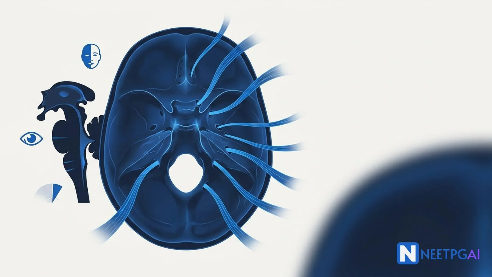
cranial nerves
anatomy NEET PG
brainstem syndromes
skull foramina
Bell's palsy
Wallenberg syndrome
NEET PG 2026
Master 12 cranial nerves, nuclei, foramina of exit, brainstem syndromes and clinical lesions for NEET PG 2026 with mnemonics and exam traps.
Cranial nerves contribute 4–6 NEET PG questions per paper across Anatomy, Medicine and ENT. The exam-ready framework:
The cranial nerves are NEET PG examiner gold — every nerve has a clean anatomical path, a predictable lesion picture, and a foramen. The trick is to combine three perspectives: brainstem nucleus location (vertical level), foraminal exit (skull base anatomy), and clinical lesion vignette (medicine and surgery). When you can switch between these layers fluently, every cranial nerve MCQ becomes solvable in seconds.
This NEETPGAI deep dive covers all 12 cranial nerves, their nuclei, foramina, branches, common lesions, brainstem syndromes and the exam-favourite clinical tests. Pair this with the brachial plexus complete guide and the common anatomy mistakes primer.
Mnemonic for names: Oh Oh Oh To Touch And Feel Very Good Velvet, Ah Heaven (Olfactory, Optic, Oculomotor, Trochlear, Trigeminal, Abducens, Facial, Vestibulocochlear, Glossopharyngeal, Vagus, Accessory, Hypoglossal).
Mnemonic for modality (Sensory/Motor/Both): Some Say Marry Money But My Brother Says Big Brains Matter More.
| CN | Name | Modality | Function |
|---|---|---|---|
| I | Olfactory | Sensory | Smell |
| II | Optic | Sensory | Vision |
Start practicing NEET PG MCQs with AI-powered explanations.
Start Free PracticeMaster GI secretions, digestion, absorption transporters, motility patterns, and gut hormones with high-yield NEET PG 2026 traps and India-context examples.
Master labor stages, Friedman vs Zhang curves, WHO partograph, AMTSL, episiotomy and India JSY/LaQshya policies for NEET PG 2026 OBG MCQs.
5 anterior segment ophthalmology image MCQs for NEET PG: hypopyon and Behcet, Kayser-Fleischer ring in Wilson, Brushfield spots in Down, corneal arcus, and pterygium vs pinguecula.
Daily MCQs, study tips, and topper strategies on Telegram.
Join on Telegram →| III |
| Oculomotor |
| Motor (+ parasympathetic) |
| All extraocular muscles except SO and LR; levator palpebrae; pupil constriction; accommodation |
| IV | Trochlear | Motor | Superior oblique (depression in adduction, intorsion) |
| V | Trigeminal | Both | V1 ophthalmic (sensory), V2 maxillary (sensory), V3 mandibular (sensory + muscles of mastication) |
| VI | Abducens | Motor | Lateral rectus |
| VII | Facial | Both | Muscles of facial expression; taste anterior 2/3 tongue (chorda tympani); lacrimation; submandibular/sublingual salivation; stapedius |
| VIII | Vestibulocochlear | Sensory | Hearing (cochlear); balance (vestibular) |
| IX | Glossopharyngeal | Both | Taste posterior 1/3 tongue; carotid body; stylopharyngeus; parotid salivation |
| X | Vagus | Both | Pharynx, larynx, thoracic and abdominal viscera; baroreceptor reflex |
| XI | Accessory | Motor | Sternocleidomastoid, trapezius |
| XII | Hypoglossal | Motor | All intrinsic + extrinsic tongue muscles except palatoglossus (CN X) |
| Foramen | Cranial fossa | Contents |
|---|---|---|
| Cribriform plate (ethmoid) | Anterior | CN I (olfactory filaments) |
| Optic canal (lesser wing of sphenoid) | Middle | CN II + ophthalmic artery |
| Superior orbital fissure (SOF) | Middle | CN III, IV, V1, VI; superior ophthalmic vein |
| Foramen rotundum | Middle | CN V2 (maxillary) |
| Foramen ovale | Middle | CN V3 (mandibular); accessory meningeal artery; lesser petrosal nerve |
| Foramen spinosum | Middle | Middle meningeal artery; meningeal branch of V3 |
| Foramen lacerum | Middle | Internal carotid (passes over, not through) |
| Internal acoustic meatus (IAM) | Posterior | CN VII, VIII; labyrinthine artery |
| Jugular foramen | Posterior | CN IX, X, XI; sigmoid sinus → IJV |
| Hypoglossal canal | Posterior | CN XII |
| Foramen magnum | Posterior | Medulla, vertebral arteries, spinal portion of CN XI |
SOF mnemonic: "Lazy French Tart Lying Flat In Anticipation" (Lacrimal, Frontal, Trochlear, Lateral SOV, Frontal nerve... use whichever sequence you prefer; the key NEET PG mnemonic for SOF contents is simply III, IV, V1, VI + SOV).
| Syndrome | Site | Vessel | Features |
|---|---|---|---|
| Weber | Ventral midbrain | Paramedian branches of PCA | Ipsilateral CN III palsy + contralateral hemiplegia (corticospinal tract) |
| Benedikt | Midbrain tegmentum | PCA | Ipsilateral CN III palsy + contralateral involuntary movements (red nucleus) |
| Claude | Dorsal midbrain | PCA | Ipsilateral CN III + contralateral cerebellar ataxia |
| Parinaud | Dorsal midbrain | Pineal lesion | Vertical gaze palsy + light-near dissociation + lid retraction (Collier sign) |
| Millard-Gubler | Ventral pons | Pontine branches of basilar | Ipsilateral CN VI + CN VII + contralateral hemiplegia |
| Foville | Dorsal pons | Pontine branches | Lateral gaze palsy + ipsilateral CN VII + contralateral hemiplegia |
| Wallenberg (lateral medullary) | Lateral medulla | PICA / vertebral | Ipsilateral V (face), Horner, dysphagia (IX/X), ataxia + contralateral body spinothalamic loss |
| Medial medullary (Dejerine) | Medial medulla | Anterior spinal artery | Ipsilateral CN XII (tongue) + contralateral hemiplegia + contralateral medial lemniscus loss |
| Locked-in syndrome | Bilateral ventral pons | Basilar artery | Quadriplegia, anarthria; only vertical eye movement and blinking preserved |
NEET PG memory rule: in brainstem strokes, the cranial nerve sign is ipsilateral to the lesion, and the long-tract signs (motor, sensory) are contralateral.
Master NEET PG with AI-powered practice — adaptive MCQs with instant explanations.
Start Free Practice →CN I — cribriform plate; CN II — optic canal; CN III, IV, V1, VI — superior orbital fissure; CN V2 — foramen rotundum; CN V3 — foramen ovale; CN VII, VIII — internal acoustic meatus; CN IX, X, XI — jugular foramen; CN XII — hypoglossal canal. The middle meningeal artery (not a cranial nerve) passes through foramen spinosum.
UMN (supranuclear, e.g., stroke) facial palsy spares the forehead because the upper face has bilateral cortical innervation. Bell's palsy is a peripheral LMN lesion of CN VII — the entire ipsilateral half of the face, including the forehead, is paralysed. Bell's palsy may also include hyperacusis, taste loss in the anterior 2/3 of the tongue, and reduced lacrimation depending on lesion site.
Wallenberg (lateral medullary) syndrome is caused by occlusion of the posterior inferior cerebellar artery (PICA) or vertebral artery. Features: ipsilateral facial pain/temperature loss (CN V spinal nucleus), Horner's syndrome (descending sympathetic), dysphagia and hoarseness (CN IX, X — nucleus ambiguus), vertigo and nystagmus (vestibular), ataxia (inferior cerebellar peduncle), and contralateral body pain/temperature loss (spinothalamic tract).
Vestibular schwannoma (acoustic neuroma) arises from the vestibular division of CN VIII at the cerebellopontine angle. It causes progressive sensorineural hearing loss, tinnitus and disequilibrium. Larger tumours compress CN V (facial numbness, loss of corneal reflex) and CN VII (facial weakness — late). Bilateral schwannomas are pathognomonic of neurofibromatosis type 2.
A 'down and out' eye with a fixed dilated pupil and ptosis suggests a compressive CN III palsy — the parasympathetic fibres travel on the outside of the nerve and are affected first. Common causes are posterior communicating artery aneurysm and uncal herniation. A pupil-sparing CN III palsy (motor weakness without pupil involvement) suggests ischaemic causes such as diabetes mellitus.
This content is for educational purposes for NEET PG exam preparation. It is not a substitute for professional medical advice, diagnosis, or treatment. Clinical information has been reviewed by qualified medical professionals.
Written by: NEETPGAI Editorial Team Reviewed by: Pending SME Review Last reviewed: May 2026