Version 1.0 — Published April 2026
Quick Answer
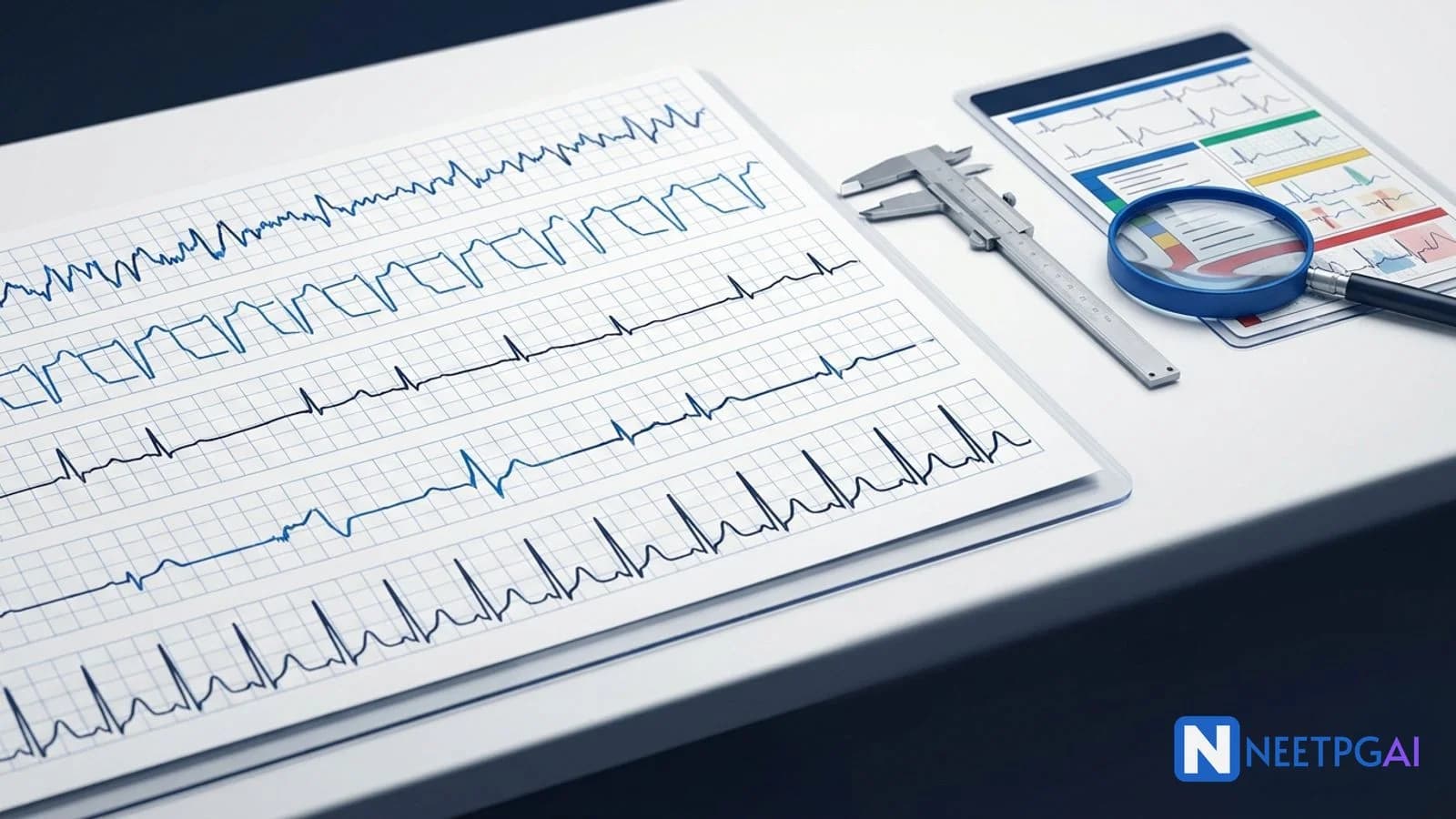
ECG interpretation contributes 4-6 questions per NEET PG paper. Five image-based arrhythmia patterns recur reliably: atrial fibrillation, ventricular tachycardia, complete heart block, Wolff-Parkinson-White syndrome, and hyperkalemia ECG changes. Master the systematic approach: rate, rhythm, axis, intervals (PR, QRS, QT), P-wave morphology, QRS morphology, ST/T changes, U waves. The three highest-yield diagnostic clues: AV dissociation = VT until proven otherwise; sawtooth waves = flutter; peaked T + wide QRS = hyperkalemia (give IV calcium NOW).
Why ECG image MCQs are high-yield in NEET PG
NEET PG cardiology contributes 5-7 questions; of these, 2-3 are ECG-image-based. Unlike pure recall, ECG MCQs reward pattern recognition: a 5-second glance often gives the diagnosis if you have practiced. The five patterns below cover almost every recent NEET PG ECG question (2019-2024 papers). For each, we walk through the image description, four answer options, the correct answer with reasoning, and a teaching pearl.
Pair this with a daily 10-strip practice habit for 2 weeks and your ECG accuracy will jump from 40 to 85 percent.
MCQ 1: Irregular tachycardia in a 70-year-old man
Image description: [ECG showing an irregularly irregular rhythm at approximately 130 bpm with no discernible P waves; instead a fibrillatory undulating baseline. QRS complexes are narrow (90 ms). RR intervals vary widely.]
Clinical vignette: A 70-year-old man with hypertension and CKD presents with palpitations and dyspnea for 2 days. BP 132/86, pulse 130/min irregular.
Options:
- (a) Multifocal atrial tachycardia
- (b) Atrial flutter with variable AV block
- (c) Atrial fibrillation
- (d) Sinus tachycardia with frequent PACs
Correct answer: (c) Atrial fibrillation
Reasoning: Three diagnostic features of AF — (1) irregularly irregular rhythm with no pattern, (2) absent P waves (replaced by fibrillatory waves at 350-600 bpm), (3) narrow QRS unless concomitant bundle branch block. Multifocal atrial tachycardia (MAT) shows at least 3 distinct P-wave morphologies (often in COPD); flutter shows sawtooth waves typically in II, III, aVF; sinus tachycardia with frequent PACs has visible P waves with intermittent ectopics.
Teaching pearl: Once AF is identified, the immediate workflow is CHA2DS2-VASc for stroke risk and HAS-BLED for bleeding risk to decide on anticoagulation. CHA2DS2-VASc components: CHF (1), HT (1), Age >=75 (2), DM (1), prior Stroke/TIA (2), Vascular disease (1), Age 65-74 (1), Sex female (1). Score >=2 in men or >=3 in women = anticoagulation indicated (DOAC preferred over warfarin in non-valvular AF). Rate control with metoprolol or diltiazem is first-line; rhythm control (cardioversion or ablation) for symptomatic patients despite rate control.
MCQ 2: Wide-complex tachycardia in a 65-year-old with prior MI
Image description: [ECG showing a regular wide-complex tachycardia at 170 bpm. QRS width 160 ms. Independent P waves visible marching through QRS at a slower rate (AV dissociation). Extreme left axis deviation. Concordance — all precordial QRS complexes are negative.]
Clinical vignette: A 65-year-old man with prior anterior MI 2 years ago presents with sudden palpitations, lightheadedness, BP 96/60.
Options:
- (a) Supraventricular tachycardia with aberrancy
- (b) Atrial flutter with 1:1 conduction
- (c) Monomorphic ventricular tachycardia
- (d) Pre-excited atrial fibrillation
Correct answer: (c) Monomorphic ventricular tachycardia
Reasoning: The five VT clues are visible: AV dissociation (independent P waves), QRS >140 ms in this RBBB-pattern lead, extreme axis (northwest), negative concordance in precordial leads, and clinical context (prior MI = high pretest probability of VT). Brugada criteria: any one positive criterion = VT. The Vereckei aVR algorithm focuses on initial R wave or rapid downstroke in aVR.
Teaching pearl: In a wide-complex tachycardia, assume VT until proven otherwise, particularly in patients with structural heart disease. Treatment of stable VT: IV amiodarone 150 mg over 10 minutes, then 1 mg/min infusion. Unstable VT (hypotension, altered mental status, chest pain): synchronized DC cardioversion 100 J biphasic. Pulseless VT or VF: defibrillate at 200 J biphasic, CPR, IV adrenaline 1 mg every 3-5 minutes, IV amiodarone 300 mg bolus. Never give AV nodal blockers (verapamil, adenosine) for unknown wide-complex tachycardia — if it is actually VT, they cause hemodynamic collapse.
MCQ 3: Bradycardia in an 80-year-old with syncope
Image description: [ECG showing a ventricular rate of 38 bpm with regular QRS complexes (width 150 ms, RBBB-like morphology). P waves are visible at a regular rate of 90 bpm but bear no consistent relationship to the QRS — PR intervals vary randomly. AV dissociation with atrial rate faster than ventricular rate.]
Clinical vignette: An 80-year-old woman is brought after a syncopal episode. BP 110/68, pulse 38, mild confusion. ECG above.
Options:
- (a) Sinus bradycardia
- (b) Mobitz type I (Wenckebach) second-degree AV block
- (c) Mobitz type II second-degree AV block
- (d) Third-degree (complete) AV block
Correct answer: (d) Third-degree (complete) AV block
Reasoning: Complete AV dissociation with the atrial rate faster than the ventricular rate, and a wide QRS at a slow rate (escape rhythm from below the bundle of His), confirms third-degree heart block. In Mobitz I, PR prolongs progressively; in Mobitz II, PR is constant with sudden dropped beats but most QRS still conduct. In CHB, no atrial impulse conducts. The escape rhythm width tells you the level: narrow QRS = junctional escape (40-60 bpm); wide QRS = ventricular escape (20-40 bpm) — much less reliable.
Teaching pearl: Complete heart block management: stabilize with transcutaneous pacing at the bedside; bridge with IV atropine 0.5-1 mg (rarely effective in CHB) or IV adrenaline/dopamine infusion; arrange urgent transvenous pacing by cardiology; definitive treatment is permanent pacemaker (dual-chamber DDD if intact AV nodal function below the block, otherwise single-chamber VVI). Reversible causes to exclude: drugs (beta-blockers, CCBs, digoxin toxicity), inferior MI (often resolves), Lyme disease, sarcoidosis. Indications for PPM: any symptomatic AV block, asymptomatic Mobitz II, asymptomatic complete AV block.
MCQ 4: Palpitations in a 19-year-old college student
Image description: [ECG showing sinus rhythm at 78 bpm with PR interval 100 ms (short), QRS width 130 ms (wide), and a slurred upstroke at the start of QRS in leads V1-V3 (delta wave). Delta wave is positive in V1.]
Clinical vignette: A 19-year-old college student reports occasional palpitations. ECG done as part of pre-employment screening shows the above findings. He is asymptomatic today.
Options:
- (a) Left bundle branch block
- (b) Wolff-Parkinson-White syndrome (Type A)
- (c) Right bundle branch block with first-degree AV block
- (d) Lown-Ganong-Levine syndrome
Correct answer: (b) Wolff-Parkinson-White syndrome (Type A)
Reasoning: WPW classic triad: short PR (<120 ms), wide QRS (>120 ms), and delta wave (slurred upstroke). Positive delta in V1 indicates a left-sided accessory pathway = Type A. Type B has a negative delta in V1 (right-sided pathway). LBBB has wide QRS but no short PR or delta wave. RBBB with first-degree block has wide QRS and long PR — opposite of WPW. Lown-Ganong-Levine is short PR with normal QRS and no delta wave (a less well-defined entity).
Teaching pearl: WPW danger is pre-excited atrial fibrillation — an accessory pathway with a short refractory period can conduct AF impulses at >250 bpm and degenerate to VF, causing sudden cardiac death. Never give AV nodal blockers (adenosine, verapamil, digoxin, beta-blockers) in pre-excited AF — blocking the AV node forces all conduction down the accessory pathway, accelerating ventricular rate. Correct treatment of pre-excited AF: synchronized DC cardioversion if unstable, or IV procainamide/ibutilide if stable. Definitive treatment of symptomatic WPW: catheter ablation of the accessory pathway (success rate >95 percent). Asymptomatic WPW in low-risk patients can be observed; high-risk features (short refractory period <240 ms, multiple pathways, family history of SCD) warrant ablation.
MCQ 5: ESRD patient who missed dialysis — ECG findings
Image description: [ECG showing sinus rhythm at 92 bpm with markedly tall, peaked, narrow-based T waves (tented appearance) in V2-V5. QRS width 130 ms (widened). PR interval 230 ms (prolonged). P waves are flattened. Lab: serum K+ 7.4 mEq/L.]
Clinical vignette: A 55-year-old man on hemodialysis for 4 years missed his last 2 sessions. He presents with weakness and palpitations. ECG above; serum K+ 7.4 mEq/L.
Options:
- (a) Hypocalcemia
- (b) Hyperkalemia
- (c) Hypothermia
- (d) Acute pericarditis
Correct answer: (b) Hyperkalemia
Reasoning: Sequential ECG changes of hyperkalemia: (1) K+ 5.5-6.5 mEq/L — peaked T waves with narrow base ("tented T waves," best seen in V2-V4); (2) K+ 6.5-7.5 — widened QRS, prolonged PR; (3) K+ 7.5-8.5 — flattened/absent P waves; (4) K+ >8.5 — sine wave pattern (merged QRS-T), asystole/VF. This patient has changes at all four stages, consistent with K+ around 7.4. Hypocalcemia prolongs QT but does not produce peaked T waves. Hypothermia causes Osborn (J) waves at the J point. Acute pericarditis causes diffuse concave-up ST elevation with PR depression.
Teaching pearl: Hyperkalemia treatment is staged by ECG severity:
| Stage | Treatment | Onset |
|---|
| Membrane stabilization | IV calcium gluconate 10 mL of 10% solution OR calcium chloride (central line) | 1-3 minutes; lasts 30-60 minutes |
| Intracellular shift | IV insulin 10 units + 25 g dextrose IV; nebulized salbutamol 10-20 mg | 15-30 minutes |
| Removal from body | Oral patiromer or sodium zirconium cyclosilicate; loop diuretics if euvolemic; dialysis definitive in ESRD | 1-3 hours |
Critical teaching point: Calcium does NOT lower potassium — it stabilizes the cardiac membrane against arrhythmia while potassium-lowering measures take effect. Always start with calcium when ECG changes are present. Sodium bicarbonate has fallen out of favor (limited efficacy outside acidosis); kayexalate has GI necrosis risk and is being replaced by patiromer/zirconium cyclosilicate.
Common pitfalls in ECG MCQs
Pitfall 1: Confusing AF with MAT. AF has no organized P waves; MAT has at least 3 distinct P morphologies. MAT is associated with COPD; AF with structural heart disease.
Pitfall 2: Calling VT "SVT with aberrancy." When in doubt, treat as VT — SVT will tolerate amiodarone/cardioversion, but VT can degenerate during inappropriate adenosine.
Pitfall 3: Missing AV dissociation in CHB. Look for P waves marching at a different rate from QRS. Use calipers (or the corner of an answer sheet) to compare PP and RR intervals.
Pitfall 4: Diagnosing Mobitz I as Mobitz II. Mobitz I has progressively prolonging PR; Mobitz II has constant PR with sudden dropped beats. Mobitz II usually has a wide QRS (infranodal) and is dangerous.
Pitfall 5: Treating WPW + AF with adenosine. Memorize: pre-excited AF requires DC cardioversion or procainamide, NEVER AV nodal blockers.
Pitfall 6: Confusing hyperkalemia peaked T with hyperacute T waves of STEMI. Hyperkalemia T waves are narrow-based and tented, in multiple precordial leads. STEMI hyperacute T waves are broad-based, regional (one coronary territory), and accompanied by ST changes.
Pitfall 7: Reading ECG without knowing the rate calculator. 1500 / number of small squares between QRS = rate (regular rhythms). For irregular rhythms, count QRS complexes in a 6-second strip and multiply by 10.
Systematic ECG approach (the 8-step checklist)
Apply this to every NEET PG ECG MCQ:
- Rate — atrial and ventricular separately if they differ
- Rhythm — regular or irregular; if irregular, regularly irregular or irregularly irregular
- Axis — normal, left, right, extreme/northwest
- PR interval — 120-200 ms; short = pre-excitation; long = first-degree block
- QRS width — <120 ms narrow; >120 ms wide (BBB, VT, pre-excitation)
- QT interval — corrected with Bazett formula; long QT >460 ms in women, >450 ms in men
- P-wave morphology — present? upright in II? bifid (left atrial enlargement)? peaked (right atrial enlargement)?
- ST/T changes — elevation, depression, inversion, peaking, U waves
How NEET PG tests ECG arrhythmias
NEET PG cardiology carries 5-7 questions per paper, with 2-3 ECG-image questions. Six dominant patterns:
- Pattern 1 — AF identification + CHA2DS2-VASc: anticoagulation decision based on score
- Pattern 2 — Wide-complex tachycardia: VT versus SVT with aberrancy; assume VT
- Pattern 3 — Heart block grading: Mobitz I, II, III differentiation
- Pattern 4 — WPW recognition: delta wave, short PR; pre-excited AF management
- Pattern 5 — Electrolyte ECG changes: hyperkalemia (peaked T), hypokalemia (U waves), hypocalcemia (long QT), hypercalcemia (short QT)
- Pattern 6 — STEMI localization: anterior (V1-V4), inferior (II, III, aVF), lateral (I, aVL, V5-V6), posterior (tall R in V1)
High-yield one-liners for last-day revision:
- AF = irregularly irregular, no P waves
- Sawtooth = atrial flutter (II, III, aVF)
- VT = wide regular + AV dissociation; assume VT in structural heart disease
- CHB = AV dissociation with atrial rate > ventricular rate
- WPW = short PR + wide QRS + delta wave
- Pre-excited AF: never give adenosine/verapamil/digoxin
- Hyperkalemia: peaked T → wide QRS → flat P → sine wave
- IV calcium gluconate FIRST in hyperkalemia with ECG changes
- Mobitz II usually has wide QRS and progresses to CHB — needs PPM
- Long QT: torsades risk; treat with IV magnesium sulfate 2 g
Frequently Asked Questions
How do you differentiate VT from SVT with aberrancy on ECG?
Five clues favor VT over SVT with aberrancy: (1) AV dissociation (P waves marching independently through QRS), (2) capture or fusion beats, (3) QRS width above 140 ms in RBBB pattern or above 160 ms in LBBB pattern, (4) extreme axis deviation (northwest axis), (5) concordance of QRS in all precordial leads (all positive or all negative). Brugada criteria and Vereckei aVR criteria formalize this. Importantly, when in doubt assume VT — it is more dangerous and SVT will tolerate VT treatment but not vice versa.
What are the classic ECG findings of WPW syndrome?
WPW (Wolff-Parkinson-White) shows the triad: short PR interval (less than 120 ms), wide QRS (more than 120 ms), and a delta wave (slurred upstroke at the start of QRS). The delta wave represents pre-excitation through an accessory pathway (bundle of Kent) bypassing the AV node. Classification: Type A (positive delta in V1, suggesting left-sided pathway) and Type B (negative delta in V1, right-sided pathway). The danger: AF in WPW can conduct rapidly via the accessory pathway and degenerate to VF — never give AV nodal blockers (digoxin, verapamil, adenosine) in pre-excited AF.
How do hyperkalemia ECG changes progress with rising serum potassium?
Sequential progression: K+ 5.5-6.5 mEq/L causes peaked, narrow-based T waves (tented T waves, often best seen in precordial leads). K+ 6.5-7.5 widens QRS and prolongs PR interval. K+ 7.5-8.5 flattens or eliminates P waves. K+ above 8.5 produces a sine wave pattern (merged QRS-T) and asystole/VF. Treatment is staged: IV calcium gluconate 1 g (membrane stabilization, 1-3 min onset, lasts 30-60 min), insulin-dextrose, beta-2 agonists, kayexalate or patiromer, dialysis for severe/refractory.
What is the difference between Mobitz I and Mobitz II second-degree AV block?
Mobitz I (Wenckebach) shows progressive PR prolongation until a P wave fails to conduct (dropped QRS), then the cycle repeats. Block is at the AV node, usually benign, often vagal. Treatment is rarely needed unless symptomatic. Mobitz II shows constant PR intervals with sudden non-conducted P waves. Block is below the AV node (His-Purkinje), often progresses to complete heart block, and requires permanent pacemaker. The clue: Mobitz II often has a wide QRS (infranodal), while Mobitz I usually has a narrow QRS.
How is atrial fibrillation differentiated from atrial flutter on ECG?
Atrial fibrillation: irregularly irregular rhythm, no discernible P waves (replaced by fibrillatory baseline at 350-600 bpm), variable ventricular response. Atrial flutter: regular sawtooth flutter waves (best seen in II, III, aVF) at 250-350 bpm, often with 2:1 or 4:1 AV block giving a regular ventricular response (commonly 150 bpm with 2:1 block). Vagal maneuvers or adenosine can transiently reveal flutter waves by slowing ventricular response. AF management focuses on rate control plus anticoagulation by CHA2DS2-VASc score; flutter often responds to cavotricuspid isthmus ablation.
This content is for educational purposes for NEET PG exam preparation. It is not a substitute for professional medical advice, diagnosis, or treatment. Clinical information has been reviewed by qualified medical professionals.
Written by: NEETPGAI Editorial Team
Reviewed by: Pending SME Review
Last reviewed: April 2026