Complete Guide to NEET PG Surgery High-Yield Topics | NEETPGAI
surgeryneet pghigh yieldsurgical anatomy
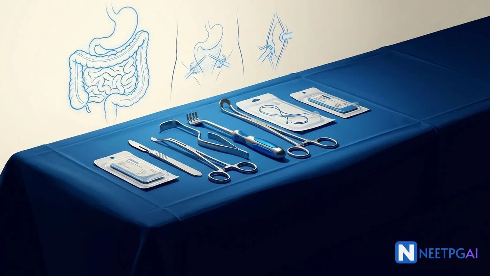
Complete Guide to NEET PG Surgery High-Yield Topics
Master every high-yield surgery topic for NEET PG: surgical anatomy, GI surgery, trauma, hernias, breast, thyroid, urology, and vascular surgery with real exam facts and study strategies.
NEETPGAI EditorialReviewed by SME Agent
Published 16 Nov 2025
32 min read
Version 1.1 — Updated April 2026 (added organization protocol compliance, TOC, references, author attribution)
Quick Answer
Surgery contributes 25–30 questions to NEET PG and rewards candidates who master a focused set of high-yield topics rather than attempting encyclopedic coverage. The eight topics that return with the highest frequency are:
Surgical anatomy — inguinal canal, Hesselbach triangle, triangle of Calot, and named incisions
GI surgery — appendicitis, intestinal obstruction, colorectal cancer, peptic ulcer complications, and the Whipple procedure
Hernias — direct versus indirect differentiation, Shouldice and Mayo repairs, and sliding hernias
Breast surgery — TNM staging, modified radical mastectomy versus breast-conserving surgery, and sentinel lymph node biopsy
Thyroid surgery — nerve injury, parathyroid preservation, and surgical indications
Trauma — ATLS primary survey, lethal six of chest trauma, FAST scan, and Burns management
Urology — renal calculi, BPH, and urological emergencies
Vascular surgery — varicose veins, peripheral arterial disease, and aneurysms
This guide covers each topic with the clinical facts that NBE tests, the incisions and anatomical landmarks you must know cold, and a practical study strategy to lock in 20+ marks from Surgery alone.
Surgery is the subject where anatomical precision meets clinical judgment. Unlike Pharmacology, where mechanism chains give you predictive power across drug classes, Surgery demands that you know specific facts — which nerve is at risk during thyroidectomy, which incision is used for appendicectomy, which layer is weakest in a direct hernia. There is no way to derive McBurney's point from first principles under exam pressure. You either know it or you do not.
That specificity is what makes Surgery both dangerous and rewarding. Dangerous because the knowledge base is wide and a scattered approach wastes time. Rewarding because NBE returns to the same landmarks, incisions, staging systems, and operative principles year after year with only minor rephrasing. A candidate who has drilled the eight high-yield areas in this guide will recognize more than 80% of Surgery stems on sight.
This guide is structured around those eight areas. Each section gives you the clinical facts that NBE tests, the anatomical detail that distinguishes correct from close-but-wrong options, and the common trap patterns that cost marks. Pair it with the full Surgery subject hub and daily MCQ practice to convert reading into retrievable exam knowledge.
Surgical anatomy: the foundation every question rests on
Surgical anatomy is not a standalone topic — it is the substrate of every surgery question. A hernia question is really an inguinal canal anatomy question. A cholecystectomy complication question is really a triangle of Calot question. A thyroidectomy nerve injury question is really a neck anatomy question. Master the anatomy, and the clinical questions answer themselves.
Share this article
This content is for educational purposes for NEET PG exam preparation. It is not a substitute for professional medical advice, diagnosis, or treatment. Clinical information has been reviewed by qualified medical professionals.
Ready to put this into practice?
Start practicing NEET PG MCQs with AI-powered explanations.
The inguinal canal is the single most tested anatomical structure in NEET PG Surgery. You need to know the boundaries cold:
Anterior wall — external oblique aponeurosis for the entire length; internal oblique reinforces the lateral third
Posterior wall — transversalis fascia for the entire length; conjoint tendon (fused internal oblique and transversus abdominis aponeurosis) reinforces the medial third
Roof — arching fibers of internal oblique and transversus abdominis
Floor — inguinal ligament (Poupart ligament) and lacunar ligament medially
The deep inguinal ring lies at the midpoint of the inguinal ligament (midway between the anterior superior iliac spine and the pubic tubercle), lateral to the inferior epigastric artery. The superficial inguinal ring is a triangular opening in the external oblique aponeurosis, superomedial to the pubic tubercle. The contents differ by sex: spermatic cord in males (with vas deferens, testicular artery, pampiniform plexus, cremasteric artery and vein, genital branch of the genitofemoral nerve, ilioinguinal nerve on the cord surface), round ligament of the uterus in females.
NBE frequently tests the relationship of the inferior epigastric artery to hernias, so burn this into memory: indirect hernias are lateral to the inferior epigastric vessels; direct hernias are medial.
Hesselbach triangle
Hesselbach triangle defines the anatomical region of direct inguinal hernias:
Medial boundary — lateral border of the rectus abdominis muscle
Lateral boundary — inferior epigastric artery
Inferior boundary — inguinal ligament
A hernia that emerges through this triangle is a direct inguinal hernia by definition. It does not traverse the inguinal canal and does not descend into the scrotum (in the vast majority of cases). This is a favorite single-best-answer target.
Triangle of Calot (cystohepatic triangle)
This triangle is the critical surgical landmark during cholecystectomy:
Medial boundary — common hepatic duct
Inferior boundary — cystic duct
Superior boundary — inferior surface of the liver (segment V)
The cystic artery (usually a branch of the right hepatic artery) runs within or near this triangle. Safe identification of the cystic artery and cystic duct within the triangle of Calot is the central safety step of laparoscopic cholecystectomy. The "critical view of safety" technique requires clearing the triangle until only the cystic duct and cystic artery are seen entering the gallbladder. NBE tests this in the context of iatrogenic bile duct injury prevention.
The anatomical snuffbox
Bounded by the tendons of the extensor pollicis longus (ulnar border) and the abductor pollicis longus and extensor pollicis brevis (radial border). The radial artery crosses its floor. Its clinical importance: tenderness in the anatomical snuffbox after a fall on an outstretched hand suggests a scaphoid fracture, even if the initial radiograph is normal. This is a classic NEET PG one-liner.
Named incisions you must know
Incision
Location
Use
Key Fact
McBurney (grid-iron)
Right iliac fossa, at McBurney point (junction of lateral 1/3 and medial 2/3 of the line from ASIS to umbilicus)
Open appendicectomy
Muscle-splitting, not muscle-cutting
Lanz
Transverse incision in the right iliac fossa, at the same level as McBurney
Appendicectomy (cosmetically superior)
Along Langer lines; better scar
Kocher (subcostal)
Right subcostal, 2.5 cm below the costal margin
Open cholecystectomy
Risk of intercostal nerve injury
Rooftop (chevron/double Kocher)
Bilateral subcostal
Hepatic, biliary, or pancreatic surgery
Excellent upper abdominal access
Pfannenstiel
Transverse suprapubic
Pelvic surgery, cesarean section
Low transverse; cosmetically favorable
Midline laparotomy
Vertical through linea alba
Emergency abdominal exploration
Fastest access; no muscle cutting
Battle incision
Paramedian vertical
Rarely used now; historical
Through rectus sheath, displacing rectus
GI surgery: the largest single block of Surgery marks
GI surgery consistently accounts for 8–10 questions in NEET PG. The topics repeat: appendicitis, intestinal obstruction, colorectal cancer, peptic ulcer surgery, and hepatobiliary procedures. Mastering this section alone can secure a third of your Surgery marks.
Appendicitis
Appendicitis is the most common surgical emergency and a perennial NEET PG topic.
McBurney point is the surface marking of the base of the appendix — the junction of the lateral one-third and medial two-thirds of a line drawn from the right anterior superior iliac spine (ASIS) to the umbilicus. This is tested so frequently that getting it wrong is an unforced error.
Alvarado score (MANTRELS): Migration of pain to RIF, Anorexia, Nausea/vomiting, Tenderness in RIF, Rebound tenderness, Elevated temperature, Leukocytosis, Shift to the left. A score of 7–10 strongly suggests appendicitis. NBE often presents a clinical scenario and asks you to calculate or interpret this score.
Surgical approach: The traditional open approach uses a McBurney (grid-iron) or Lanz incision. Laparoscopic appendicectomy is now standard in many centers and is preferred in women of reproductive age (to exclude gynecological pathology), in obese patients, and when diagnosis is uncertain. The mesoappendix is divided, the appendicular artery (branch of the ileocolic artery, which is a branch of the superior mesenteric artery) is ligated, and the appendix base is crushed, ligated, and divided.
Positions of the appendix: retrocaecal (65–70%, the most common), pelvic (30%), subcaecal, preileal, and postileal. The retrocaecal position explains why some patients present with loin pain mimicking ureteric colic — a favorite misleading stem in NEET PG.
Intestinal obstruction
The distinction between small bowel obstruction (SBO) and large bowel obstruction (LBO) is a high-yield clinical discrimination:
Multiple air-fluid levels; valvulae conniventes visible (full width of bowel)
Haustra visible (partway across bowel)
Perforation risk
Lower
Caecum in closed-loop obstruction (competent ileocaecal valve)
Sigmoid volvulus is the most common type of volvulus in adults and produces a characteristic "coffee bean sign" on abdominal X-ray. Initial decompression is attempted with a flatus tube via sigmoidoscopy. Caecal volvulus produces the "kidney bean sign."
Colorectal cancer
Dukes staging is still tested despite being largely replaced by TNM in clinical practice:
Stage
Extent
Lymph nodes
Notes
Dukes A
Confined to bowel wall (mucosa and submucosa)
No
Best prognosis
Dukes B
Through muscularis propria
No
Adjuvant chemo debated
Dukes C
Any depth
Yes (C1: apical negative; C2: apical positive)
Adjuvant chemo standard
Dukes D
Any depth
Any
Distant metastases
CEA (carcinoembryonic antigen) is not a screening marker — it is used for post-operative surveillance. A rising CEA after resection suggests recurrence. NBE loves the "what is the role of CEA" question because most students incorrectly mark it as a diagnostic tool.
Surgical margins: for rectal cancer, a distal margin of at least 2 cm (1 cm for low rectal with neoadjuvant therapy) and a circumferential resection margin (CRM) of more than 1 mm are required for curative resection. Total mesorectal excision (TME) is the standard for mid and low rectal cancers.
Peptic ulcer surgery
While the incidence of elective ulcer surgery has declined with proton pump inhibitors and H. pylori eradication, NBE still tests the operative principles because they test applied anatomy and physiology.
Indications for surgery: perforation, hemorrhage not controlled by endoscopy, obstruction (gastric outlet), and malignancy suspicion.
Perforation management: Graham omental patch repair (an omental plug sutured over the perforation) is the standard for perforated duodenal ulcer. The perforation site is most commonly the anterior wall of the first part of the duodenum.
Posterior duodenal ulcer erodes into the gastroduodenal artery, causing massive hemorrhage — a classic NEET PG association.
Truncal vagotomy reduces acid secretion by denervating the parietal cell mass but also denervates the pylorus, requiring a drainage procedure (pyloroplasty or gastrojejunostomy). Highly selective vagotomy (HSV) preserves the nerve of Latarjet (anterior and posterior nerves of the lesser curvature) and denervates only the parietal cell mass, avoiding the need for a drainage procedure. HSV has the lowest recurrence rate and the fewest side effects — a frequently tested comparison.
The Whipple procedure (pancreaticoduodenectomy)
Whipple procedure is the standard operation for periampullary carcinoma and carcinoma of the head of the pancreas.
Structures resected: head of the pancreas, entire duodenum, distal common bile duct, gallbladder, and antrum of the stomach (in the classic Whipple; the pylorus-preserving variant retains the stomach and proximal duodenum).
Three anastomoses (in order from proximal to distal):
Pancreaticojejunostomy — the most dangerous anastomosis; pancreatic fistula is the most feared post-operative complication
Hepaticojejunostomy — biliary drainage
Gastrojejunostomy (or duodenojejunostomy in pylorus-preserving Whipple)
Courvoisier law often appears in the stem: a palpable, non-tender gallbladder in the presence of jaundice is unlikely to be due to gallstones (the chronically inflamed gallbladder from stones is fibrosed and cannot distend). This suggests malignant obstruction — periampullary carcinoma until proven otherwise.
Hernias: anatomy that directly determines the answer
Hernia questions are anatomy questions in disguise. NBE tests classification, anatomical relations, repair techniques, and complications. This section recurs every single year.
Inguinal hernias: direct versus indirect
The single most important distinction is the relationship to the inferior epigastric artery:
Indirect inguinal hernia: enters the deep inguinal ring lateral to the inferior epigastric artery, traverses the inguinal canal, and can descend into the scrotum. More common in young males. Congenital in origin (patent processus vaginalis).
Direct inguinal hernia: pushes through the posterior wall of the inguinal canal (Hesselbach triangle) medial to the inferior epigastric artery. More common in older males. Acquired, due to weakness of the transversalis fascia. Rarely descends into the scrotum.
Pantaloon hernia (saddlebag hernia): both direct and indirect components, straddling the inferior epigastric vessels.
Femoral hernia
Passes through the femoral canal, below and lateral to the pubic tubercle (inguinal hernias are above and medial to the pubic tubercle — this is a favorite discriminator). More common in females, but inguinal hernias are still the most common hernia in females overall. Femoral hernias have the highest risk of strangulation among groin hernias due to the rigid boundaries of the femoral ring (inguinal ligament anteriorly, pectineal ligament posteriorly, lacunar ligament medially, femoral vein laterally).
Hernia repair techniques
Repair
Type
Key Feature
Shouldice
Tissue repair (inguinal)
Four-layer continuous suture repair of the posterior wall; the gold standard tissue repair with the lowest recurrence (less than 1% in expert hands)
Lichtenstein
Tension-free mesh (inguinal)
Polypropylene mesh placed over the posterior wall; most widely performed open repair worldwide
Mayo
Overlap repair (umbilical)
Overlapping of fascial flaps in a "vest-over-pants" fashion; for umbilical and paraumbilical hernias
Bassini
Tissue repair (inguinal)
Suturing of the conjoint tendon to the inguinal ligament behind the cord; historical importance
Laparoscopic TEP/TAPP
Mesh (inguinal)
Totally extraperitoneal (TEP) or transabdominal preperitoneal (TAPP); lower post-operative pain, faster return to work
Shouldice repair is the benchmark tissue repair. It involves opening the transversalis fascia, overlapping it in four layers with continuous non-absorbable suture, and reinforcing the posterior wall. The recurrence rate is under 1% in the Shouldice Hospital series — the lowest reported for any tissue repair — but this result is operator-dependent and not universally reproducible, which is why mesh repair (Lichtenstein) has become the global default.
Complications of hernia
Irreducibility — hernia contents cannot be returned to the abdomen
Obstruction — bowel within the hernia becomes obstructed (symptoms of intestinal obstruction)
Strangulation — blood supply to the herniated contents is compromised; surgical emergency. The earliest feature is pain; the latest feature is absent cough impulse
Richter hernia — only part of the circumference of the bowel wall is trapped (antimesenteric border); can strangulate without obstruction — a classic NBE trap because students expect obstruction before strangulation
Littre hernia — Meckel diverticulum within the hernia sac
Maydl hernia (hernia-en-W) — two loops of bowel within the sac with the intervening loop inside the abdomen at risk of strangulation
Breast surgery: staging and operative decisions
Breast pathology generates 3–5 questions in most NEET PG papers. NBE tests TNM staging, operative choices, and the clinical differentiation of benign from malignant lesions.
TNM staging essentials
The critical T-stage cutoffs for surgical decisions:
Stage
Size / Extent
Key features
T1
Up to 2 cm
BCS usually feasible
T2
2–5 cm
BCS if tumor-to-breast ratio favorable
T3
Greater than 5 cm
Mastectomy usually indicated
T4a
Any size, chest wall extension
Locally advanced
T4b
Any size, skin involvement
Edema, ulceration, satellite nodules
T4c
Both T4a + T4b
T4d
Inflammatory carcinoma
Peau d'orange; neoadjuvant chemo before surgery
Inflammatory breast carcinoma (T4d) is a clinical diagnosis — peau d'orange appearance, erythema, and warmth over at least one-third of the breast. It is not a surgical candidate upfront; neoadjuvant chemotherapy is mandatory before considering surgery. This is a frequently tested management sequence.
Modified radical mastectomy (MRM) versus breast-conserving surgery (BCS)
Modified radical mastectomy (Patey/Auchincloss):
Removes the entire breast, nipple-areola complex, and axillary lymph nodes (levels I and II)
Preserves the pectoralis major muscle (this is the key distinction from the Halsted radical mastectomy, which removes both pectoralis major and minor)
Patey modification: removes pectoralis minor to access level III nodes
Auchincloss modification: preserves pectoralis minor, clears only levels I and II
Breast-conserving surgery (BCS/wide local excision/lumpectomy):
Removes the tumor with a margin of normal tissue (at least 1 mm for invasive carcinoma, 2 mm for DCIS is the current consensus)
Must be followed by whole-breast radiotherapy — BCS without radiotherapy has unacceptably high local recurrence
Survival equivalence to mastectomy was established by the NSABP B-06 trial and the Milan trial
Contraindications to BCS: multicentric disease, large tumor relative to breast size (unfavorable tumor-to-breast ratio), prior chest wall radiation, first and second trimester pregnancy (radiation contraindicated), and diffuse suspicious microcalcifications on mammography.
Halsted principles and radical mastectomy
William Stewart Halsted introduced the radical mastectomy based on the principle that breast cancer spreads in an orderly, centrifugal fashion through lymphatics. His operation removed the breast, both pectoral muscles, and all three levels of axillary lymph nodes en bloc. The Halsted radical mastectomy is now almost never performed because the Fisher hypothesis (breast cancer is a systemic disease from inception) and survival data showed that less extensive surgery with adjuvant therapy achieves equivalent outcomes.
However, NBE still tests the historical distinction — radical mastectomy removes pectoralis major and minor; modified radical mastectomy preserves pectoralis major.
Sentinel lymph node biopsy (SLNB)
The sentinel lymph node is the first lymph node to receive lymphatic drainage from the tumor. It is identified using blue dye (patent blue V or isosulfan blue) or radioactive technetium-99m sulfur colloid (or both — the dual technique). If the sentinel node is negative on histopathology, full axillary dissection is avoided, reducing the risk of lymphedema. The false-negative rate is approximately 5–10%.
NBE tests the principle: "If the sentinel lymph node is negative, what is the next step?" The answer is observation/no further axillary surgery.
Benign breast disease
Fibroadenoma — most common benign tumor in young women (15–35 years). Firm, mobile ("breast mouse"), well-circumscribed. Excision is indicated if growing, greater than 3 cm, or causing anxiety. FNAC shows clusters of epithelial cells in a clean background.
Phyllodes tumor — fibroepithelial tumor that can be benign, borderline, or malignant. Distinguished from fibroadenoma by rapid growth, larger size, and leaf-like stromal pattern on histology. Treatment is wide local excision with at least 1 cm margin (not enucleation). Axillary dissection is not indicated because phyllodes spreads hematogenously, not lymphatically.
Breast cyst — most common cause of a breast lump in women aged 35–55. Aspiration yields straw-colored fluid; if fluid is bloodstained, send for cytology.
Thyroid surgery: nerves, glands, and complications
Thyroid surgery questions test two things: the indications for surgery and the anatomical hazards during thyroidectomy. The complications are entirely predictable from the anatomy, which is why this section belongs in any serious Surgery prep plan.
Indications for thyroidectomy
Malignancy — confirmed or suspected thyroid carcinoma
Compression symptoms — retrosternal goitre causing tracheal or esophageal compression
Thyrotoxicosis — failed medical management, patient preference, or contraindication to radioiodine (pregnancy, large goitre, suspected malignancy)
Cosmetic — large visible goitre
Surgical anatomy of the thyroid
Blood supply: Superior thyroid artery (first branch of the external carotid artery) and inferior thyroid artery (branch of the thyrocervical trunk from the subclavian artery). The thyroidea ima artery, when present, arises from the brachiocephalic trunk or the aortic arch and supplies the isthmus — it is a cause of unexpected bleeding during tracheostomy.
Venous drainage: Superior and middle thyroid veins drain into the internal jugular vein. The inferior thyroid veins drain into the brachiocephalic veins.
The recurrent laryngeal nerve (RLN) is the most important structure at risk during thyroidectomy:
On the right, it hooks around the right subclavian artery
On the left, it hooks around the arch of the aorta (longer course, more vulnerable)
It runs in the tracheoesophageal groove (Berry ligament area is the danger zone) and enters the larynx behind the cricothyroid joint
Unilateral RLN injury causes hoarseness (ipsilateral vocal cord palsy in the paramedian/cadaveric position)
Bilateral RLN injury causes stridor and airway obstruction — a surgical emergency requiring immediate reintubation or tracheostomy
The external branch of the superior laryngeal nerve (EBSLN) runs with the superior thyroid artery and is at risk during ligation of the superior pole vessels. Injury causes loss of the cricothyroid muscle function — the patient cannot tense the vocal cord, resulting in a monotonous voice and inability to sing high notes ("the nerve of opera singers" or Amelita Galli-Curci nerve). This is a classic NEET PG association.
Post-thyroidectomy complications
Complication
Cause
Clinical Feature
Hypocalcemia
Inadvertent removal or devascularization of parathyroid glands
Tingling, Chvostek sign, Trousseau sign; most common early complication
Recurrent laryngeal nerve injury
Stretching, cutting, or diathermy injury to RLN
Hoarseness (unilateral), stridor (bilateral)
Hemorrhage/hematoma
Reactive bleeding from thyroid bed
Neck swelling, dyspnea; open wound at bedside if airway compromised
Thyroid storm
Inadequate preoperative preparation of thyrotoxic patient
Hyperpyrexia, tachycardia, altered sensorium; medical emergency
Wound infection
Standard surgical risk
Rare in clean thyroid surgery
Chvostek sign (tapping the facial nerve anterior to the ear causes twitching of the facial muscles) and Trousseau sign (inflating a blood pressure cuff above systolic for 3 minutes causes carpal spasm) are the bedside tests for hypocalcemia. Both are tested as post-thyroidectomy complications.
Trauma surgery: ATLS and emergency protocols
Trauma is tested as clinical scenarios. NBE gives you a patient with injuries and expects you to apply the ATLS framework, identify the life-threatening condition, and choose the correct intervention. Pure recall of protocols is necessary but insufficient — you must apply them to the stem.
The ATLS primary survey: ABCDE
The sequence is non-negotiable and is itself tested:
A — Airway with cervical spine protection. Establish a patent airway. Jaw thrust (not head tilt) in suspected cervical spine injury. Definitive airway if needed.
B — Breathing and ventilation. Look, listen, and feel. Identify and treat tension pneumothorax (needle decompression at the 2nd intercostal space, midclavicular line, followed by chest tube), open pneumothorax (three-sided occlusive dressing), and flail chest (paradoxical movement; underlying pulmonary contusion is the main cause of hypoxia, not the flail segment itself).
C — Circulation with hemorrhage control. Two large-bore IV cannulae, initiate crystalloid resuscitation. Identify and control external hemorrhage. Assess for internal hemorrhage (chest, abdomen, pelvis, long bones, "the floor" — external). Class III and IV hemorrhagic shock (greater than 30% blood loss) require blood transfusion.
D — Disability (neurological status). Glasgow Coma Scale, pupillary response. GCS of 8 or below mandates intubation for airway protection.
E — Exposure and environment. Fully undress the patient to identify all injuries; prevent hypothermia.
The lethal six of chest trauma
These are the six immediately life-threatening thoracic injuries identified during the primary survey:
Tension pneumothorax — clinical diagnosis (do not wait for X-ray). Tracheal deviation away from the affected side, absent breath sounds, hyperresonance, hypotension. Treatment: needle decompression followed by tube thoracostomy.
Open pneumothorax (sucking chest wound) — seal with a three-sided occlusive dressing (the unsealed side acts as a flutter valve), then chest tube.
Massive hemothorax — greater than 1500 mL blood in the chest. Chest tube drainage and autotransfusion; if output exceeds 200 mL/hour for 2–4 hours, thoracotomy is indicated.
Flail chest — two or more ribs fractured in two or more places creating a free-floating segment. The underlying pulmonary contusion, not the mechanical flail, is what kills. Management is analgesic (epidural or intercostal nerve block), chest physiotherapy, and ventilatory support if needed.
Cardiac tamponade — Beck triad: hypotension, distended neck veins, muffled heart sounds. Pulsus paradoxus. Emergency pericardiocentesis via the subxiphoid approach. Definitive treatment is surgical (pericardial window or thoracotomy).
Tracheobronchial injury — persistent air leak after chest tube placement, massive subcutaneous emphysema. Requires surgical repair.
FAST scan (Focused Assessment with Sonography for Trauma)
The FAST scan is a bedside ultrasound looking for free fluid in four areas:
Hepatorenal space (Morrison pouch) — the most dependent part of the peritoneal cavity in a supine patient; fluid collects here first
Splenorenal space
Pelvis (pouch of Douglas/rectovesical pouch)
Pericardial space (subxiphoid view)
A positive FAST in a hemodynamically unstable patient is an indication for emergency laparotomy.
Burns
Rule of nines (Wallace) for estimating total body surface area (TBSA) burns in adults: head 9%, each upper limb 9%, anterior trunk 18%, posterior trunk 18%, each lower limb 18%, perineum 1%.
Parkland formula for fluid resuscitation: 4 mL x body weight (kg) x %TBSA burn. Half is given in the first 8 hours from the time of burn (not from the time of arrival), and the remaining half over the next 16 hours. Use Ringer lactate. This calculation appears in NEET PG with near-annual regularity.
Burn depth classification: Superficial (erythema only, first degree), superficial partial thickness (blisters, painful, second degree), deep partial thickness (white/mottled, diminished sensation), and full thickness (leathery, painless, third degree).
Urology: stones, obstruction, and emergencies
Urology contributes 2–4 questions per NEET PG paper. The yield is concentrated in renal calculi, benign prostatic hyperplasia, and urological emergencies.
AR cystinuria; positive cyanide-nitroprusside test
Staghorn calculus fills the renal pelvis and calyces, forming a branching "antler" shape. Most commonly struvite. Treatment is percutaneous nephrolithotomy (PCNL) for stones greater than 2 cm, extracorporeal shock wave lithotripsy (ESWL) for stones less than 2 cm in the renal pelvis.
Benign prostatic hyperplasia (BPH)
BPH involves hyperplasia of the periurethral (transition) zone of the prostate — a frequently tested distinction from prostate carcinoma, which arises from the peripheral zone.
Medical management: alpha-1 blockers (tamsulosin, prazosin) for symptomatic relief; 5-alpha reductase inhibitors (finasteride, dutasteride) to reduce prostate volume (effective only in large prostates, takes 3–6 months).
Surgical management: Transurethral resection of the prostate (TURP) is the gold standard. Complication to know: TUR syndrome — dilutional hyponatremia from absorption of hypotonic glycine irrigation fluid, causing confusion, nausea, visual disturbance, and seizures.
Urological emergencies
Testicular torsion — peak incidence in adolescence. Sudden onset scrotal pain, high-riding testis, absent cremasteric reflex, bell-clapper deformity. Surgical emergency — do not delay for imaging. Surgical exploration and orchidopexy within 6 hours for best salvage rate. Bilateral orchidopexy is performed because the contralateral testis may have the same predisposing anatomy.
Paraphimosis — the retracted foreskin cannot be returned to its normal position, causing venous congestion and edema of the glans. Manual reduction (compressing the glans and sliding the foreskin forward) is the first step. Dorsal slit if reduction fails.
Acute urinary retention — most commonly caused by BPH in older males. Immediate catheterization. Monitor for post-obstructive diuresis.
Vascular surgery: veins, arteries, and aneurysms
Vascular surgery appears as 2–3 questions, focused on varicose veins, peripheral arterial disease, and aortic aneurysms.
Varicose veins
Varicose veins of the lower limb result from incompetent valves in the superficial venous system (great saphenous vein or small saphenous vein) or at the saphenofemoral/saphenopopliteal junction.
Clinical tests:
Trendelenburg test — tests saphenofemoral junction competence. Patient lies supine, leg elevated to empty veins, pressure applied at the SFJ, patient stands. Rapid filling on release indicates SFJ incompetence.
Brodie-Trendelenburg test — modified version with a tourniquet
Perthes test — tests deep vein patency. Tourniquet applied, patient walks. If varicosities persist or worsen, deep vein obstruction is present and superficial stripping is contraindicated.
Schwartz test (tap test) — tapping the proximal varicosity transmits an impulse distally if valves are incompetent
Complications of varicose veins: edema, pigmentation (hemosiderin deposition), lipodermatosclerosis, venous eczema, venous ulceration (typically at the medial malleolus, above the ankle — the "gaiter area"), and thrombophlebitis.
Venous ulcer versus arterial ulcer: Venous ulcers are shallow, irregular, painless (or mildly painful), located at the medial malleolus, with surrounding pigmentation and lipodermatosclerosis. Arterial ulcers are deep, punched-out, painful, located at the tips of toes or pressure points, with absent pulses and cold limb. This differentiation is a favorite MCQ stem.
Peripheral arterial disease
Leriche syndrome — aortoiliac occlusive disease presenting with the triad of intermittent claudication in the buttocks and thighs, absent femoral pulses, and erectile dysfunction in males.
Buerger disease (thromboangiitis obliterans) — inflammatory vasculitis of small and medium arteries and veins. Affects young male smokers. Presents with rest pain, digital gangrene, superficial migratory thrombophlebitis, and Raynaud phenomenon. Pathology shows organized thrombus with intact vessel wall. Complete smoking cessation is the most important management step.
Fontaine classification of chronic limb ischemia:
Stage
Presentation
Management
I
Asymptomatic
Risk factor modification
IIa
Claudication >200 m
Exercise therapy, risk factor modification
IIb
Claudication <200 m
Consider revascularization
III
Rest pain
Urgent revascularization
IV
Gangrene or ulceration
Revascularization or amputation
Abdominal aortic aneurysm (AAA)
An AAA is defined as a focal dilatation of the abdominal aorta to greater than 3 cm (normal diameter is approximately 2 cm). Most are infrarenal.
Risk factors: smoking (strongest modifiable risk factor), age greater than 65, male sex, hypertension, family history.
Screening: single ultrasound screening for men aged 65–75 who have ever smoked (USPSTF recommendation, frequently tested).
Management by size:
Less than 5.5 cm — surveillance with serial ultrasound (every 6–12 months based on size)
Greater than or equal to 5.5 cm — elective repair (open or endovascular/EVAR)
Symptomatic at any size — urgent repair
Ruptured — emergency repair. The classic presentation of ruptured AAA is sudden severe back/abdominal pain, hypotension, and a pulsatile abdominal mass. Do not delay for CT if the patient is hemodynamically unstable.
Study strategy: converting knowledge into exam marks
Knowing the content is necessary but not sufficient. Surgery in NEET PG rewards candidates who can retrieve specific facts under time pressure and apply them to clinical scenarios. The strategy below is designed for exactly that conversion.
Phase 1: Foundation reading (2 weeks)
Cover the eight high-yield areas in this guide using SRB Manual of Surgery or Bailey and Love (selectively). Spend two days on each major topic. For each topic, build a one-page summary with the key anatomy, named procedures, and complications. Do not take extensive notes — the one-page summary is your revision tool.
Solve 15 surgery MCQs daily on the topic you studied that day. Mark every question you get wrong and note the specific fact you were missing.
Phase 2: MCQ drilling (2 weeks)
Increase to 30–40 surgery MCQs daily. Mix topics — do not cluster by subtopic. Use previous year question banks and simulated clinical vignettes. For each wrong answer, trace the error to one of three categories:
Anatomy gap — you did not know the anatomical relationship
Procedure confusion — you mixed up two operations or their indications
Staging/classification error — you misremembered a staging criterion
The category distribution tells you where to reinvest study time.
Read our spaced repetition guide for NEET PG and build a deck of the named incisions, anatomical landmarks, and staging systems from this guide. Review the deck daily — ten minutes is enough if the deck is well-curated.
Phase 3: Revision and mocks (1 week)
In the final week, solve one full-length surgery mock under timed conditions. Review the entire mock with explanations. Revise your one-page summaries in reverse order (the earliest topics have decayed most). On the day before the exam, review only three things: the named incisions table, the TNM staging cutoffs, and the ATLS ABCDE sequence.
For targeted MCQ practice on operative procedures and clinical scenarios, use the Pharmacology MCQ strategy guide — its five-step solve flow for clinical vignettes applies equally to Surgery stems.
Sources and references
S. Das, A Manual on Clinical Surgery, 14th Edition, 2022 — standard Indian clinical surgery reference for NEET PG.
Bailey & Love, Short Practice of Surgery, 28th Edition (Williams et al., 2023) — gold-standard surgical textbook for principles and operative detail.
Sabiston, Textbook of Surgery, 21st Edition (Townsend et al., 2021) — comprehensive operative and pathophysiological reference.
SRB's Manual of Surgery, 6th Edition (Sriram Bhat M., 2019) — widely used concise reference for Indian PG entrance exams.
American College of Surgeons, ATLS Student Course Manual, 10th Edition, 2018 — definitive trauma management protocol.
AJCC Cancer Staging Manual, 8th Edition (Amin et al., 2017) — current TNM staging reference for breast, colorectal, and thyroid carcinoma.
Frequently asked questions
How many surgery questions appear in NEET PG?
Surgery typically contributes 25–30 questions in NEET PG, making it the second-highest-weighted clinical subject after Medicine. Of these, GI surgery alone accounts for 8–10 questions most years. Surgical anatomy, breast, thyroid, and trauma questions appear with near-annual regularity. The effective yield is higher because orthopedic, ENT, and ophthalmology questions often test surgical principles taught in General Surgery.
Which surgery topics are tested most frequently in NEET PG?
GI surgery dominates with appendicitis, intestinal obstruction, colorectal cancer, and peptic ulcer complications. Hernias appear almost every year — inguinal anatomy, direct versus indirect differentiation, and repair techniques. Breast surgery TNM staging and surgical options, thyroid surgery with nerve injury complications, and trauma ATLS protocols are the next tier. Surgical anatomy questions on the inguinal canal, triangle of Calot, and Hesselbach triangle are perennial.
Should I read Bailey and Love cover-to-cover for NEET PG surgery?
No. Bailey and Love is an excellent reference but reading it cover-to-cover in exam prep is a poor use of time. Use it selectively for conceptual depth on high-yield topics — hernia anatomy, breast staging, thyroid nerve relations, and GI surgical principles. For day-to-day prep, use SRB Manual of Surgery or a concise high-yield guide and reserve Bailey for topics where mechanism-level understanding is weak.
How do I differentiate direct from indirect inguinal hernia in MCQs?
The key anatomical landmark is Hesselbach triangle, bounded by the inferior epigastric artery laterally, the rectus sheath medially, and the inguinal ligament inferiorly. A direct hernia pushes through Hesselbach triangle medial to the inferior epigastric vessels. An indirect hernia enters the deep inguinal ring lateral to the inferior epigastric vessels and traverses the inguinal canal. NBE commonly tests this with a stem describing vessel relations or the ring of entry.
What are the most commonly tested incisions in NEET PG surgery?
The perennial favorites are Lanz incision for appendicectomy (transverse, cosmetically superior, in the right iliac fossa along Langer lines), Kocher subcostal incision for open cholecystectomy, Pfannenstiel incision for pelvic surgery, midline laparotomy for emergency access, and McBurney (grid-iron) incision at McBurney point for traditional appendicectomy. Know the anatomical basis, layers traversed, and clinical advantage of each.
How should I study trauma surgery for NEET PG?
Focus on ATLS protocols — the primary survey ABCDE sequence, the lethal six of chest trauma (tension pneumothorax, open pneumothorax, flail chest, massive hemothorax, cardiac tamponade, and tracheobronchial injury), and the FAST scan for abdominal trauma. Understand damage control surgery principles, Glasgow Coma Scale scoring, and Burns classification including the Parkland formula for fluid resuscitation. These are tested as clinical scenarios, not as factual recall.
Is the Whipple procedure tested in NEET PG, and what should I know about it?
Yes, pancreaticoduodenectomy (Whipple procedure) is a recurring topic. Know the indication (periampullary carcinoma, head of pancreas carcinoma), what is resected (head of pancreas, duodenum, distal common bile duct, gallbladder, and the antrum of the stomach in a classic Whipple), and the three anastomoses performed (pancreaticojejunostomy, hepaticojejunostomy, gastrojejunostomy). The most dangerous post-operative complication is pancreatic fistula.
What is the best strategy for last-minute surgery revision before NEET PG?
In the final two weeks, stop reading new material. Revise high-yield tables — TNM staging for breast and colorectal cancer, hernia classification and repair types, thyroid surgery complications, and ATLS protocols. Solve 30–40 surgery MCQs daily with timed conditions. Use spaced repetition for surgical anatomy landmarks and named incisions. On exam day, review only your self-made one-page summaries of the eight core topics covered in this guide.
Start your surgery prep today. Open the Surgery subject page and solve your first 15 MCQs — the anatomy you drill now is the anatomy you will retrieve on exam day. Want unlimited AI-powered surgery MCQs with detailed explanations? Explore NEETPGAI Pro.
Written by: NEETPGAI Editorial Team
Reviewed by: Dr. SME Agent, NEETPGAI Medical Advisory Board
Last reviewed: April 2026
This article is reviewed by qualified medical professionals for clinical accuracy and exam relevance. For corrections or updates, contact the editorial team.