Master trauma and ATLS for NEET PG 2026: primary survey ABCDE, airway and GCS, lethal six of chest trauma, hemorrhage classes, permissive hypotension, FAST scan, Parkland formula, Wallace rule of 9, and blunt vs penetrating decision-making.
NEETPGAI EditorialPublished 26 Jan 202623 min read
Share this article
This content is for educational purposes for NEET PG exam preparation. It is not a substitute for professional medical advice, diagnosis, or treatment. Clinical information has been reviewed by qualified medical professionals.
Ready to put this into practice?
Start practicing NEET PG MCQs with AI-powered explanations.
Trauma and ATLS contribute 3–4 direct questions per NEET PG paper. Master these 10 high-yield areas:
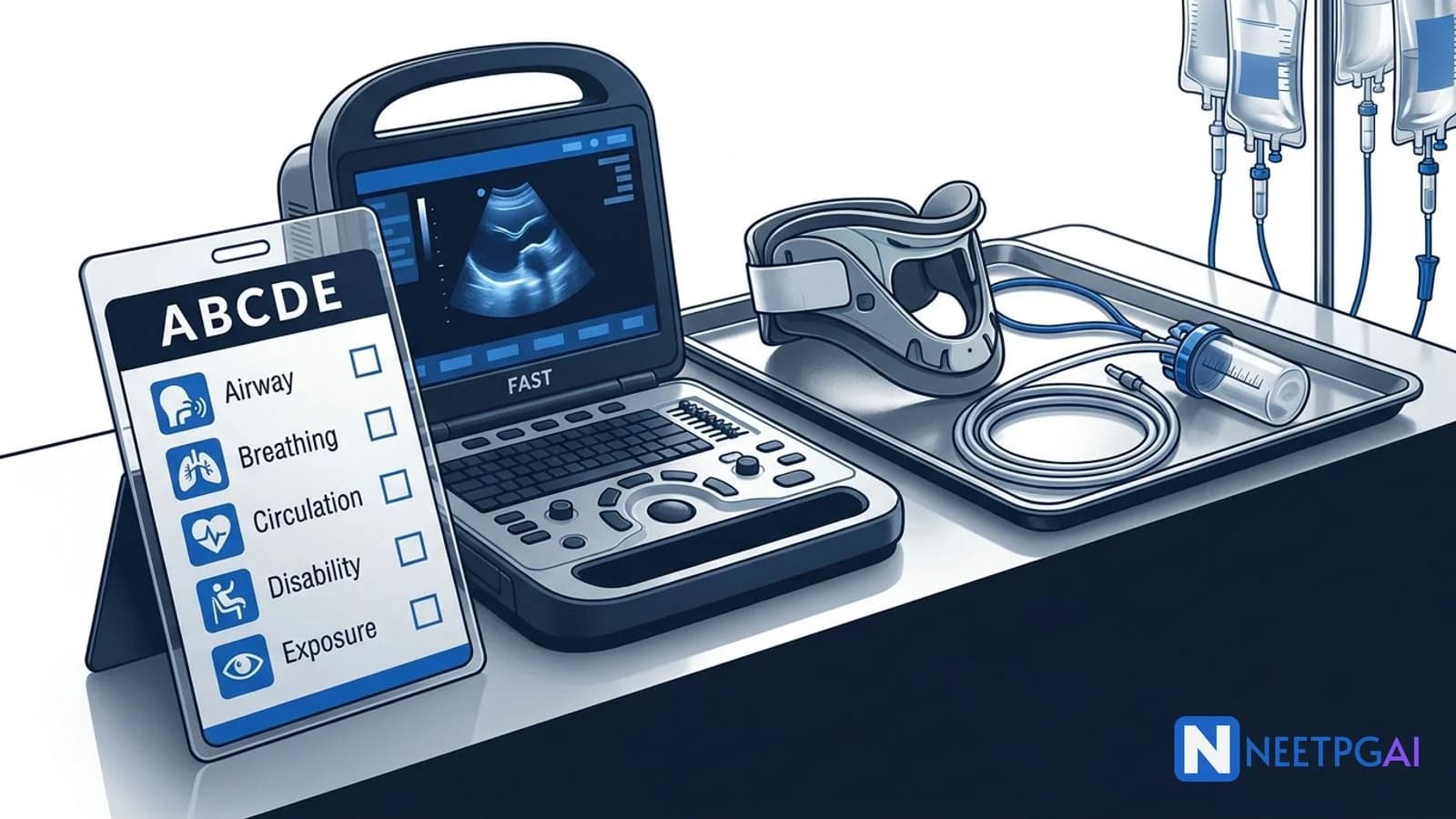
Primary survey (ABCDE) — A (airway + C-spine), B (breathing), C (circulation), D (disability), E (exposure). Strict sequence; reassess after every intervention
Circulation — hemorrhage classes — I <15% (<750 mL), II 15–30% (tachycardia), III 30–40% (hypotension), IV >40% (anuria). Hypotension = Class III minimum
Permissive hypotension — SBP 80–90 (or radial pulse) in penetrating truncal trauma pre-surgical control; NOT in TBI (need MAP >80)
Burns (Parkland) — 4 mL × kg × %TBSA RL in 24 h; half in first 8 h from time of burn; urine output 0.5 mL/kg/h adult, 1 mL/kg/h child
Wallace rule of 9 (adult) — head 9, each arm 9, anterior trunk 18, posterior trunk 18, each leg 18, perineum 1. Children: head 18 (infant), each leg 13–14
Trauma is the leading cause of death in Indians aged 1–44 years, and the ATLS protocol is the globally standardised approach for the first hour of trauma care — making it a NEET PG goldmine across surgery, critical care, and emergency medicine. The student who memorises the ABCDE sequence, lethal six, hemorrhage classes, and Parkland formula covers 3–4 marks across papers. Pair this guide with daily MCQ practice on the surgery subject hub, cross-reference the surgery high-yield topics overview, and revise the shock and sepsis management guide for critical care integration.
ATLS primary survey — the ABCDE sequence
The ATLS primary survey is a structured 5-minute assessment that identifies and treats immediately life-threatening injuries in the order of their lethality — airway first, then breathing, circulation, disability, and exposure.
Key principles:
Treat as you go — identify and treat life threats before moving on
Reassess after every intervention (ABCDE-A-B-C…)
Team approach — horizontal (simultaneous) rather than purely sequential in mature trauma centres
Adjuncts — monitoring, labs, imaging added in parallel once stable
Type and cross-match, CBC, coagulation, biochemistry, pregnancy test (all fertile-age females), drug / alcohol screen, troponin, urinalysis
Trauma series X-rays: chest, pelvis; C-spine lateral (if CT not immediately available)
FAST / eFAST bedside ultrasound
After primary survey → secondary survey (head-to-toe, AMPLE history, reassess).
Airway with C-spine control
Airway is the first priority in the primary survey — ensuring patency and protection while maintaining cervical spine immobilisation in every blunt trauma patient until spine is cleared.
Airway assessment:
Talk to the patient — an ability to speak normally confirms patent airway and adequate cerebral perfusion
Induction agents: ketamine (preferred in shock — maintains BP), etomidate (haemodynamically neutral; single dose in septic is controversial), propofol (avoid in hypotension)
Paralysis: rocuronium (1 mg/kg) or succinylcholine (1–1.5 mg/kg — avoid in crush injury, burns >24 h, paraplegia, hyperkalaemia)
In-line manual stabilisation of cervical spine (collar opened anteriorly, neck held manually)
Cricoid pressure (controversial — may obscure view; applied per unit protocol)
Surgical cricothyroidotomy — definitive surgical airway; incision over cricothyroid membrane; 6.0–7.0 ETT; contraindicated in children <12 years (use needle cricothyroidotomy with jet ventilation)
C-spine protection:
Manual in-line stabilisation (do NOT pull)
Hard collar (Philadelphia, Aspen); head blocks; long spine board for transport (remove within 2 h)
NEXUS criteria or Canadian C-Spine Rule to clear low-risk patients without imaging
Breathing and ventilation — the lethal six
Breathing assessment identifies and treats immediate threats to ventilation and oxygenation — the "lethal six" chest injuries — in the B step.
Assessment:
Expose chest
Inspect — asymmetry, paradoxical movement, wounds, seatbelt sign, use of accessory muscles
Pericardiocentesis (subxiphoid, Larrey's point); emergency thoracotomy if penetrating injury with arrest
Seventh "lethal":Tracheobronchial disruption — persistent large air leak despite adequate chest tube, continued pneumothorax; bronchoscopy; surgical repair.
"Deadly dozen" of chest trauma (primary + secondary survey):
Deadly six (primary survey)
Hidden six (secondary survey)
Airway obstruction
Aortic disruption
Tension pneumothorax
Tracheobronchial injury
Open pneumothorax
Myocardial contusion
Massive hemothorax
Diaphragmatic rupture
Flail chest
Oesophageal injury
Cardiac tamponade
Pulmonary contusion
Circulation and hemorrhage control
Circulation assessment identifies haemorrhagic shock, controls external bleeding, establishes IV access, and initiates fluid resuscitation — with permissive hypotension now favoured in select patients.
Assessment:
Level of consciousness (poor perfusion → confusion, agitation, lethargy)
Skin colour, temperature, capillary refill (>2 s abnormal)
Pulse — rate, quality, regularity
BP (late sign of shock — reflects compensated vs decompensated state)
External bleeding — direct pressure, pressure dressing, tourniquet for limb haemorrhage (>2 h ideally, mark time)
Classes of haemorrhagic shock (ATLS):
Parameter
Class I
Class II
Class III
Class IV
Blood loss (mL)
<750
750–1500
1500–2000
>2000
Blood loss (%)
<15%
15–30%
30–40%
>40%
Heart rate
<100
>100
>120
>140
Blood pressure
Normal
Normal
Decreased
Decreased
Pulse pressure
Normal / increased
Decreased
Decreased
Decreased
Respiratory rate
14–20
20–30
30–40
>35
Urine output (mL/h)
>30
20–30
5–15
Negligible
Mental status
Slightly anxious
Mildly anxious
Anxious, confused
Confused, lethargic
Fluid
Crystalloid
Crystalloid
Crystalloid + blood
Crystalloid + blood (massive transfusion)
Key NEET PG takeaway: Hypotension appears only in Class III — a normotensive trauma patient can still have lost up to 30% of blood volume. Tachycardia and narrowed pulse pressure are earlier signs.
Sources of major bleeding (think "blood on the floor and 4 more"):
External (floor)
Chest
Abdomen / pelvis
Long bones (femur: 1.5 L; pelvis: up to 3 L)
Retroperitoneum
Vascular access:
Two large-bore (16 G or larger) peripheral IVs in antecubital fossae
Failed peripheral → intraosseous (IO) or central venous catheter (femoral, subclavian, IJV)
Central line via cutdown of saphenous vein is a backup
Fluid resuscitation:
Initial bolus: 1 L crystalloid (Ringer lactate preferred) in adults; 20 mL/kg in children
Reassess
Responders vs transient responders vs non-responders
Early use of blood products for Class III/IV shock
Massive transfusion protocol — 1:1:1 ratio of PRBC : FFP : platelets (CRASH-2, PROPPR trial evidence)
Tranexamic acid 1 g over 10 min then 1 g over 8 h — within 3 h of injury reduces mortality (CRASH-2)
Permissive hypotension:
Target SBP 80–90 mmHg (or palpable radial pulse) until surgical control
Rationale: prevent clot disruption and dilution
Indicated: penetrating truncal trauma without head injury
Permissive hypotension + balanced transfusion + minimize crystalloid + early surgical haemorrhage control + rewarming + correction of acidosis + correction of coagulopathy
"Lethal triad" — hypothermia + acidosis + coagulopathy — each exacerbates the others
Pelvic binder: Apply at greater trochanter level for suspected pelvic fracture with haemodynamic instability — reduces pelvic volume and tamponades venous bleeding; interventional angioembolisation or preperitoneal packing for refractory bleeding.
Disability — GCS, pupils, glucose
Disability assessment is a rapid neurological exam in the primary survey — GCS, pupils, and glucose — looking for traumatic brain injury and other causes of altered mental status.
Hyperventilation to PaCO2 30–35 (temporarily — causes cerebral vasoconstriction)
Osmotic therapy: mannitol 0.25–1 g/kg IV OR 3% hypertonic saline 250 mL bolus
Neurosurgical consult + non-contrast CT head
Check blood glucose in all patients with altered mental status — hypoglycaemia mimics head injury.
Exposure with environmental control
Exposure is the final step of the primary survey — full undressing and log-roll for complete body inspection, while actively preventing hypothermia.
What to do:
Remove all clothing (cut off if needed; preserve for forensics in penetrating / sexual assault cases)
Log-roll (4-person, C-spine-protected) to inspect back, flanks, perineum; palpate spine for step-offs, tenderness
Rectal exam: tone, blood, high-riding prostate
Limbs: swelling, deformity, open fractures, compartments
External signs of pelvic injury: perineal bruising, scrotal haematoma
Prevent hypothermia:
Warm blankets; forced-air warming (Bair Hugger)
Warm crystalloid (40°C) and blood products
Warm room temperature
Cover patient immediately after inspection
Hypothermia in trauma (<35°C) is part of the lethal triad — worsens coagulopathy and acidosis; each 1°C drop increases blood loss.
Secondary survey and ongoing management
The secondary survey is a systematic head-to-toe examination performed after the primary survey is complete and the patient is stabilised — the AMPLE history anchors it.
Continuous reassessment and monitoring for deterioration.
Missed injuries are common — up to 10% in polytrauma; a tertiary survey within 24 hours is recommended.
Burns — assessment and fluid resuscitation
Burns are thermal, chemical, electrical, or radiation injuries — the acute resuscitation phase depends on accurate TBSA estimation and Parkland formula fluid calculation.
Burn depth classification:
Depth
Layers involved
Appearance
Sensation
Healing
Superficial (1st degree)
Epidermis
Red, dry, painful
Normal
3–6 days, no scar
Superficial partial thickness (2nd)
Upper dermis
Red, moist, blisters, blanches
Severe pain, very sensitive
7–21 days, minimal scarring
Deep partial thickness (2nd)
Lower dermis
Pale, dry, may blister, poor blanching
Decreased
>3 weeks, hypertrophic scar; may need graft
Full thickness (3rd)
Entire dermis + subcutaneous
White / leathery / charred; no blanching
Painless (nerves destroyed)
Grafting required
4th degree
Into muscle, bone
Charred
None
Reconstruction / amputation
Wallace rule of 9 (adult):
Region
% TBSA
Head and neck
9
Each upper limb
9
Anterior trunk
18 (chest 9 + abdomen 9)
Posterior trunk
18
Each lower limb
18
Perineum
1
Total
100
Paediatric differences:
Infant head: 18% (higher surface-to-volume ratio)
Each leg in infant: 14%
Use Lund and Browder chart for precise paediatric TBSA (adjusts for age)
Palm method: the palm of the patient's hand (including fingers) ≈ 1% TBSA — useful for scattered small burns.
Note: Count only partial-thickness and full-thickness burns in TBSA for Parkland formula (exclude first-degree erythema).
Parkland formula:
4 mL × body weight (kg) × %TBSA of Ringer lactate in first 24 hours
Half in first 8 hours (from time of burn, NOT arrival)
Half in next 16 hours
Example: 70 kg with 30% TBSA → 4 × 70 × 30 = 8400 mL/24h → 4200 mL in first 8 h
Monitoring:
Urine output: target 0.5 mL/kg/h in adults, 1 mL/kg/h in children <30 kg
FAST scan — if positive → emergency laparotomy; if negative and persistently unstable → DPL or repeat FAST or other cause
Hemodynamically stable
CT abdomen/pelvis with contrast (gold standard); solid-organ grading by AAST
Non-operative management (NOM) for stable solid organ injury:
Haemodynamically stable
No peritonitis
Grade I–III splenic / hepatic injuries
Monitoring in ICU / surgical ward
Angioembolisation for blush on CT
Operative if deterioration
Penetrating trauma:
Stab wounds and gunshot wounds
Gunshot wound to abdomen: traditionally mandatory laparotomy (high incidence of visceral injury); selective non-operative management in select stable patients in mature centres
Stab wound to abdomen:
Hemodynamically unstable → laparotomy
Evisceration of omentum / bowel → laparotomy
Peritoneal signs → laparotomy
Anterior abdominal stab wound, stable, no peritoneal signs → local wound exploration (assess peritoneal breach); if breach or equivocal → CT and serial abdominal exams or DPL
Thoracoabdominal stab wound (between nipple line and costal margin anteriorly; between inferior scapular tip and costal margin posteriorly) — high diaphragm injury risk; liberal laparoscopy or thoracoscopy
Phase 2: ICU resuscitation — rewarm, correct coagulopathy and acidosis
Phase 3: return to OR within 24–48 h for definitive repair
Zone of injury in neck trauma (penetrating):
Zone I (sternal notch to cricoid) — highest mortality (major vessels)
Zone II (cricoid to angle of mandible) — most common; easiest surgical access
Zone III (angle of mandible to skull base) — distal ICA; difficult exposure; angiography preferred
"No-zone" approach — clinical findings (hard signs like expanding haematoma, pulsatile bleeding, bruit, airway compromise) drive surgical vs imaging decisions rather than anatomical zone alone.
Sources and references
American College of Surgeons Committee on Trauma. Advanced Trauma Life Support (ATLS) Student Course Manual, 10th Edition (2018).
Bailey & Love's Short Practice of Surgery, 28th Edition (Williams, Bulstrode, O'Connell, Eds., 2023) — Chapter on Trauma.
Sabiston Textbook of Surgery, 21st Edition (Townsend, Beauchamp, Evers, Mattox, Eds., 2021) — Chapters on Trauma and Burns.
Schwartz's Principles of Surgery, 11th Edition (Brunicardi et al., 2019) — Chapter on Trauma and Burns.
CRASH-2 Collaborators. Effects of tranexamic acid on death, vascular occlusive events, and blood transfusion in trauma patients with significant haemorrhage (CRASH-2): a randomised, placebo-controlled trial. Lancet 2010; 376:23-32.
Holcomb JB et al. Transfusion of plasma, platelets, and red blood cells in a 1:1:1 vs a 1:1:2 ratio and mortality in patients with severe trauma: the PROPPR randomized clinical trial. JAMA 2015; 313(5):471-482.
American Burn Association. Advanced Burn Life Support (ABLS) Provider Manual (2018).
Frequently asked questions
How many trauma and ATLS questions appear in NEET PG?
Trauma and ATLS contribute 3-4 direct questions per NEET PG paper across surgery, critical care, and emergency medicine. ABCDE primary survey order, lethal six of chest trauma, hemorrhage class classification, GCS scoring, FAST scan indications, and Parkland formula for burns are the most tested subtopics based on 2019-2025 pattern analysis.
What is the ATLS primary survey?
ATLS primary survey is the rapid 5-minute ABCDE assessment that identifies and treats immediately life-threatening injuries in the order of their lethality. A — Airway with cervical spine control (intubate if GCS less than 8), B — Breathing and ventilation (treat tension pneumothorax, open pneumothorax, flail chest), C — Circulation with hemorrhage control (two large-bore IVs, crystalloid, permissive hypotension for penetrating injuries), D — Disability (GCS, pupils), E — Exposure with environmental control (full undressing, prevent hypothermia). Done in strict sequence and reassessed after every intervention.
What is the lethal six of chest trauma?
The lethal six are immediately life-threatening chest injuries identified in the primary survey: airway obstruction, tension pneumothorax, open pneumothorax (sucking chest wound), massive hemothorax (greater than 1500 mL or 1500 mL loss with ongoing 200 mL/h), flail chest, and cardiac tamponade. Some texts include tracheobronchial disruption as a seventh. All require immediate clinical diagnosis and treatment in the B or C step of the primary survey, not radiological confirmation first.
What are the four classes of hemorrhagic shock?
ATLS classes of hemorrhagic shock by percentage of blood volume lost in a 70 kg adult (total 5 L). Class I: less than 15 percent (less than 750 mL), normal vitals. Class II: 15-30 percent (750-1500 mL), tachycardia over 100, narrowed pulse pressure, mild anxiety. Class III: 30-40 percent (1500-2000 mL), tachycardia over 120, hypotension, oliguria, confusion. Class IV: greater than 40 percent (greater than 2000 mL), severe hypotension, anuria, lethargy. Hypotension is a late sign — it appears only in class III or worse.
What is permissive hypotension?
Permissive hypotension is a fluid resuscitation strategy that accepts a lower-than-normal systolic BP (target approximately 80-90 mmHg, or palpable radial pulse) in patients with uncontrolled hemorrhage until definitive surgical control. It reduces dilution of clotting factors, avoids clot disruption, and limits re-bleeding. Indicated in penetrating truncal trauma without head injury. CONTRAINDICATED in traumatic brain injury (need MAP greater than 80) and elderly (baseline higher BP).
What is the Glasgow Coma Scale and when is intubation indicated?
GCS assesses consciousness on three parameters. Eye opening (E, 1-4): spontaneous 4, to sound 3, to pressure 2, none 1. Verbal response (V, 1-5): oriented 5, confused 4, words 3, sounds 2, none 1. Motor response (M, 1-6): obeys 6, localizes 5, normal flexion 4, abnormal flexion (decorticate) 3, extension (decerebrate) 2, none 1. Total 3-15. GCS less than or equal to 8 is an indication for definitive airway (intubation) because airway protection cannot be guaranteed.
What is the FAST scan and what does it assess?
FAST (Focused Assessment with Sonography for Trauma) is a bedside ultrasound done in the primary survey to detect intraperitoneal or pericardial fluid (blood) in 4 standard views: right upper quadrant (Morison pouch between liver and right kidney), left upper quadrant (spleno-renal recess), pelvis (pouch of Douglas), and subxiphoid (pericardium). Extended FAST (eFAST) adds two anterior chest views for pneumothorax. Sensitivity 60-90 percent for intraperitoneal hemorrhage; a positive FAST in an unstable patient is an indication for immediate laparotomy.
What is the Parkland formula for burns?
Parkland formula calculates fluid requirements in the first 24 hours after burn injury: 4 mL Ringer lactate per kg body weight per percent total body surface area burned. Half is given in the first 8 hours from the time of burn (not time of arrival), and half over the next 16 hours. Example: 70 kg adult with 30 percent TBSA burn needs 4 x 70 x 30 = 8400 mL over 24 hours, with 4200 mL in first 8 hours. Target urine output: 0.5 mL/kg/hour in adults, 1 mL/kg/hour in children less than 30 kg.
What is the Wallace rule of 9?
Wallace rule of 9 estimates total body surface area burned in adults. Head and neck 9 percent. Each upper limb 9 percent (total 18 percent). Anterior trunk 18 percent (chest 9, abdomen 9). Posterior trunk 18 percent. Each lower limb 18 percent (total 36 percent). Perineum 1 percent. In children, head is larger (up to 18 percent in infants) and lower limbs smaller (13-14 percent each) — use Lund and Browder chart for accurate pediatric estimation. Alternative: palmar surface (including fingers) equals approximately 1 percent TBSA for scattered burns.
How do blunt and penetrating trauma management differ?
Blunt trauma causes multiple organ injuries from deceleration, compression, or shearing — common organs affected are spleen (most common in abdomen), liver, kidneys, and mesentery. Investigation prefers CT scan in stable patients; FAST or DPL in unstable. Non-operative management for stable patients with solid organ injury is often possible. Penetrating trauma (stab or gunshot) is handled with higher suspicion for immediate operative intervention — gunshot wounds to the abdomen are usually explored; stab wounds in the anterior abdomen with peritoneal breach and hemodynamic instability need laparotomy. FAST is useful in both; CT is for stable patients only.
Explore our pricing plans for unlimited practice across all 19 subjects, AI-powered doubt resolution, and personalized study plans.
This content is for educational purposes for NEET PG exam preparation. It is not a substitute for professional medical advice, diagnosis, or treatment. Clinical information has been reviewed by qualified medical professionals.
Written by: NEETPGAI Editorial Team
Reviewed by: Pending SME Review
Last reviewed: April 2026
This article is reviewed by qualified medical professionals for clinical accuracy and exam relevance. For corrections or updates, contact the editorial team.