## Astigmatism: Optical Basis and Retinoscopy Findings Astigmatism is a refractive error where the cornea or lens has different curvatures along two principal meridians (usually vertical and horizontal), resulting in different refractive powers. This causes light rays to focus at two different planes rather than a single point on the retina. ### Retinoscopy in Astigmatism: - **Key Point:** In astigmatism, the red reflex moves in **opposite directions** along the two principal meridians during retinoscopy. - This occurs because each meridian has a different refractive power and thus a different focal length. - The meridian with greater refractive power (steeper curvature) will have its focal point closer to the retina. - When the retinoscope beam is aligned with one meridian, the reflex moves one way; when rotated 90°, it moves the opposite way. ### Clinical Correlation: The cylinder axis (90° in this case) indicates the axis of the less powerful meridian. The +1.50 D cylinder corrects the astigmatism by adding power to the weaker meridian. **High-Yield:** The opposite movement of red reflex in two perpendicular meridians is **pathognomonic for astigmatism** and is the basis for determining cylinder power and axis during retinoscopy. 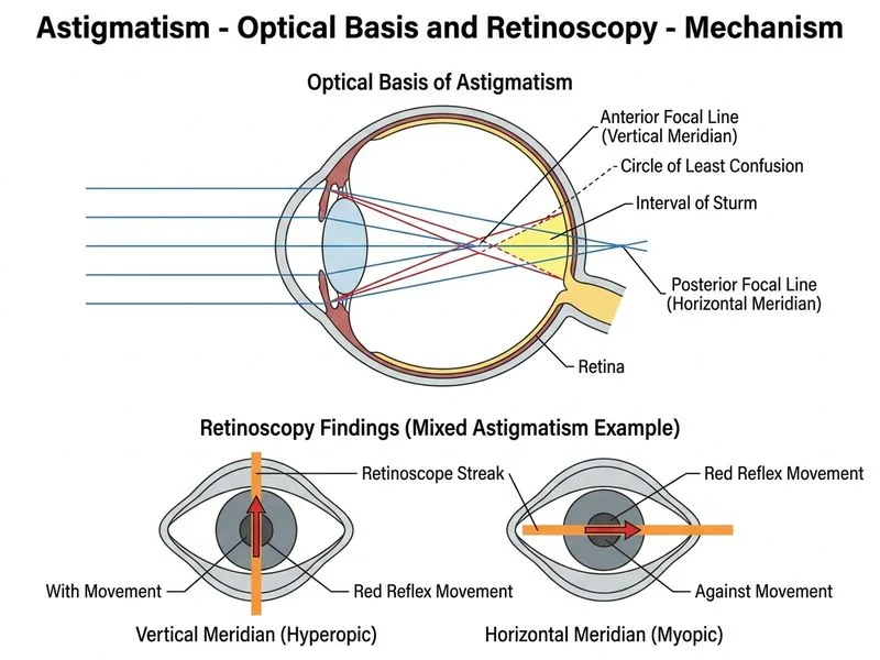
Sign up free to access AI-powered MCQ practice with detailed explanations and adaptive learning.