
## Prognostic Factors in Osteosarcoma ### Overview Osteosarcoma is an aggressive malignancy with high metastatic potential. Understanding prognostic factors is critical for treatment planning and patient counselling. ### Most Important Prognostic Factor: Presence of Metastases **High-Yield:** The single most important prognostic factor in osteosarcoma is the presence or absence of metastatic disease at diagnosis. Patients with metastases (especially pulmonary) at presentation have significantly worse outcomes despite multimodal therapy. **Key Point:** Approximately 20% of osteosarcoma patients have clinically detectable pulmonary metastases at diagnosis, and another 50% have micrometastases. The presence of metastases reduces 5-year survival from ~70% (no metastases) to ~20% (with metastases). ### Prognostic Factors in Osteosarcoma | Factor | Favourable | Unfavourable | |--------|-----------|---------------| | **Metastases** | Absent | Present (especially lung) | | **Tumour size** | < 8 cm | > 8 cm | | **Location** | Distal femur, proximal tibia | Pelvis, spine | | **Chemotherapy response** | > 90% necrosis | < 90% necrosis | | **Age** | Younger | Older (> 25 years) | | **Alkaline phosphatase** | Normal/mildly elevated | Markedly elevated | | **Surgical margins** | Wide/adequate | Marginal/intralesional | **Clinical Pearl:** Necrosis of > 90% of the primary tumour after neoadjuvant chemotherapy is a strong predictor of better overall survival and is used to guide adjuvant therapy intensity. ### Why Metastases Trump Other Factors 1. **Biological aggressiveness:** Presence of metastases indicates a more aggressive tumour phenotype with higher proliferation rate and greater chemoresistance potential 2. **Treatment impact:** Even with aggressive multimodal therapy (neoadjuvant chemotherapy, surgery, adjuvant chemotherapy), metastatic disease carries a fundamentally worse prognosis 3. **Survival data:** The 5-year survival difference is dramatic (70% vs. 20%), far exceeding the impact of other individual factors ### Other Prognostic Factors (Secondary Importance) **Tumour size > 8 cm:** Associated with worse prognosis but less predictive than metastatic status. Large tumours may still be curable with adequate chemotherapy and surgery if no metastases are present. **Alkaline phosphatase elevation:** Reflects osteoblastic activity and tumour burden but is not as powerful a prognostic indicator as metastatic status. Markedly elevated levels (> 500 IU/L) suggest more aggressive disease but are not independently predictive. **Sex:** No significant prognostic difference between males and females in modern series. **Mnemonic: METASTASES = Most important, Eliminates cure potential, Therapy-resistant phenotype, Aggressive biology, Survival dramatically reduced, Thoracic imaging mandatory, Alters treatment strategy, Systemic disease burden, Early detection critical, Staging essential** ### Clinical Management Implications ```mermaid flowchart TD A[Osteosarcoma diagnosed]:::outcome --> B{Metastatic workup}:::decision B -->|Metastases present| C[Intensified chemotherapy]:::action B -->|No metastases| D[Standard neoadjuvant chemotherapy]:::action C --> E[Surgery if feasible]:::action D --> E E --> F[Assess chemotherapy response]:::decision F -->|Good response >90% necrosis| G[Standard adjuvant therapy]:::action F -->|Poor response <90% necrosis| H[Intensified adjuvant therapy]:::action G --> I[5-year survival ~70%]:::outcome H --> J[5-year survival ~40-50%]:::outcome ``` **High-Yield:** Chest CT is mandatory at diagnosis to exclude pulmonary metastases, as they are the most common site of spread and their presence fundamentally changes prognosis and treatment intensity. 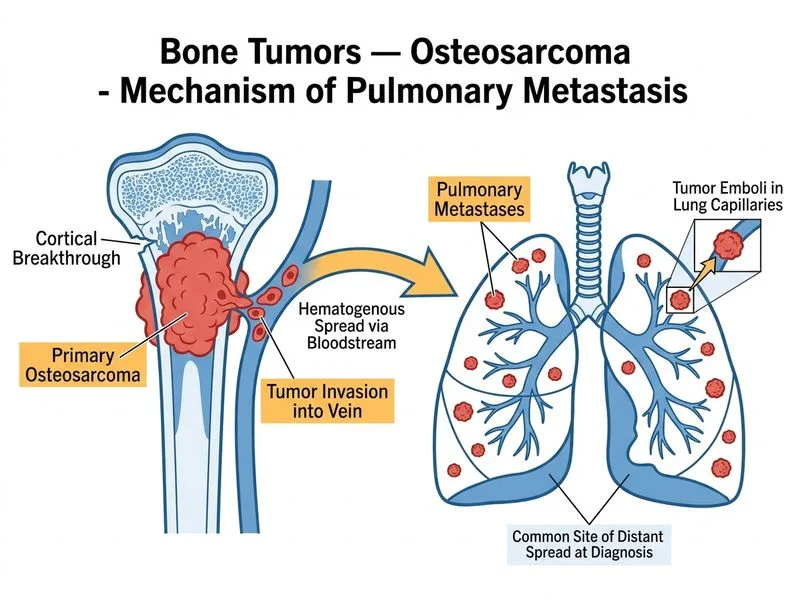
Sign up free to access AI-powered MCQ practice with detailed explanations and adaptive learning.