## Calot's Triangle Anatomy Calot's triangle is a critical landmark in cholecystectomy, bounded by: - **Medially:** Common hepatic artery - **Laterally:** Cystic artery - **Inferiorly:** Cystic duct **Key Point:** The accessory right hepatic artery (also called the right hepatic artery variant) is a common anatomical variant that runs through or near Calot's triangle in approximately 25–30% of the population. This artery arises from the superior mesenteric artery (SMA) or gastroduodenal artery and crosses through the triangle to supply the right lobe of the liver. **Clinical Pearl:** Failure to identify this vessel during cholecystectomy can result in hepatic ischemia and subsequent liver necrosis. It is the most commonly injured aberrant hepatic vessel during biliary surgery. **High-Yield:** The standard right hepatic artery (option 0) runs posterior to the cystic artery and is not typically found within Calot's triangle itself. The cystic artery (option 1) forms the lateral boundary. The right gastric artery (option 2) supplies the stomach and does not traverse this region. 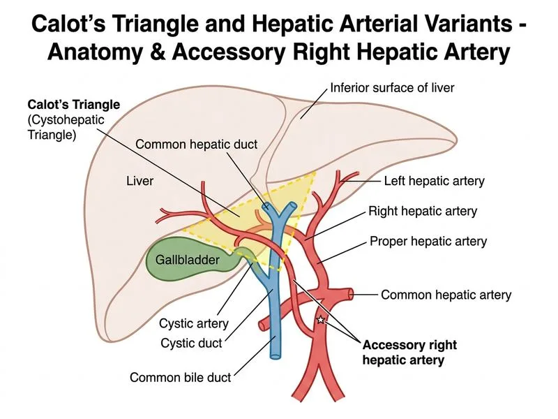
Sign up free to access AI-powered MCQ practice with detailed explanations and adaptive learning.