## Diagnosis: Left Ventricular Hypertrophy (LVH) Secondary to Chronic Hypertension ### Pathological Mechanism **Key Point:** Chronic hypertension causes **hypertrophy** of cardiomyocytes — an increase in the SIZE of individual myocardial cells — NOT an increase in their NUMBER. The left ventricle thickens but the chamber volume decreases (concentric hypertrophy). ### Hypertrophy vs. Hyperplasia in Cardiac Tissue | Feature | Hypertrophy (LVH) | Hyperplasia | |---------|------------------|----------| | **Cell number** | Normal/unchanged | Increased | | **Individual cell size** | **Markedly increased** | Normal | | **Mechanism** | ↑ Protein synthesis; ↑ sarcomeres in parallel | Increased mitotic activity | | **Stimulus** | Pressure/volume overload | Growth factors, hormones | | **Reversibility** | Partially reversible if load removed | Reversible if stimulus removed | | **Cardiomyocyte nuclei** | Enlarged, polyploid | Normal | | **Histology** | Large cells, normal architecture | More cells, normal size | **High-Yield:** Adult cardiomyocytes are **terminally differentiated and non-proliferative**. They cannot undergo hyperplasia in response to chronic load. Instead, they enlarge (hypertrophy) by increasing sarcomeric protein content. ### Clinical Pearl The echocardiographic findings are diagnostic: - **Thickened LV wall** (14 mm) = hypertrophy - **Reduced chamber volume** = concentric pattern - **Preserved EF initially** = compensation is still adequate - **Elevated LVEDP** = diastolic dysfunction (stiff, hypertrophied ventricle) This is **concentric LVH** — the classic response of the heart to sustained systemic hypertension. ### Histological Features of LVH 1. **Enlarged cardiomyocytes** with increased cytoplasmic volume 2. **Enlarged, often polyploid nuclei** (nuclear enlargement parallels cell enlargement) 3. **Increased sarcomeric proteins** (actin, myosin, Z-disc proteins) 4. **Interstitial fibrosis** (secondary; due to impaired perfusion and neurohumoral activation) 5. **Normal myocyte number** (no hyperplasia) **Mnemonic: HyPERtrophy = PERsonal growth (cell gets bigger); HyPERplasia = PERcentage increases (more cells)** ### Why NOT Hyperplasia? Adult cardiomyocytes lack significant regenerative capacity. They respond to chronic overload by: - Increasing protein synthesis (hypertrophy) ✓ - NOT by dividing (hyperplasia) ✗ Hyperplasia of cardiomyocytes occurs only during fetal development and early infancy, not in adult chronic hypertension. 
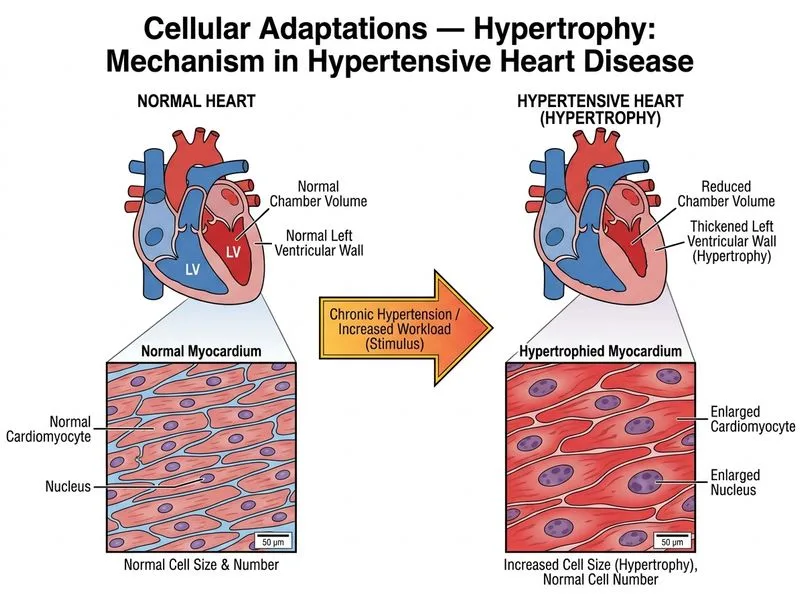
Sign up free to access AI-powered MCQ practice with detailed explanations and adaptive learning.