Fracture Classification — Types and Patterns MCQ — NEET PG Practice Question | NEETPGAI
Fracture Classification — Types and Patterns
medium
bone Orthopedics
A 32-year-old male construction worker sustains a crush injury to his left forearm after a steel beam falls on it. Plain radiographs show a comminuted fracture of the radius and ulna with soft tissue swelling. The attending surgeon suspects an intra-articular extension at the wrist joint. Which investigation is most appropriate to confirm the intra-articular involvement and guide surgical planning?
A. High-resolution CT scan with 3D reconstruction
B. Repeat plain radiographs in different projections
C. Ultrasound of the wrist
D. MRI of the wrist
Explanation
Investigation of Choice for Intra-articular Fracture Assessment
Why CT with 3D Reconstruction is Optimal
Key Point
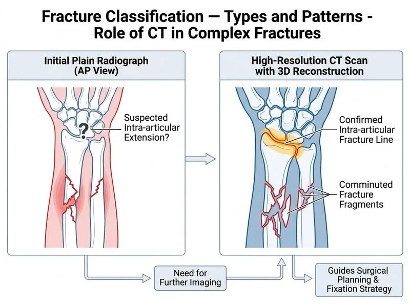
High-resolution CT with 3D reconstruction is the gold standard for evaluating comminuted fractures with suspected intra-articular extension, particularly in the wrist and forearm.
Clinical Pearl
CT provides:
Precise delineation of fracture fragments and their spatial relationships
Clear visualization of intra-articular involvement
Accurate assessment of articular surface displacement
3D reconstruction aids preoperative surgical planning and implant selection
Why CT Excels in This Scenario
Table
Feature
CT with 3D
MRI
Ultrasound
Repeat X-rays
Bone detail
Excellent
Poor
Poor
Limited
Fragment count
Precise
Moderate
Poor
Inadequate
Intra-articular extension
Excellent
Moderate
Poor
Inadequate
Surgical planning
Optimal
Moderate
Poor
Suboptimal
Speed
Fast
Slow
Fast
Fast
Cost-effectiveness
Moderate
High
Low
Low
High-YieldNEET PG
In comminuted fractures with suspected intra-articular involvement, CT is superior because:
1.
It shows the exact number and position of bone fragments
2.
It reveals subtle articular surface steps (even <2 mm)
3.
It guides fixation strategy (ORIF vs. external fixation)
4.
3D reconstruction allows virtual surgical planning
Mnemonic: CT-PLAN — Clear Three-dimensional Picture for Large Articular Navigation
Loading illustration…
Practice similar questions
Sign up free to access AI-powered MCQ practice with detailed explanations and adaptive learning.