A 35-year-old woman with biopsy-proven Hodgkin lymphoma (nodular sclerosis subtype) presents for staging workup. Physical examination reveals no hepatosplenomegaly. Which investigation is most appropriate to assess for occult abdominal and pelvic lymph node involvement and guide treatment planning?
A. Abdominal ultrasound
B. Contrast-enhanced CT chest, abdomen, and pelvis
C. Diagnostic laparotomy with splenectomy
D. Bone marrow biopsy
Explanation
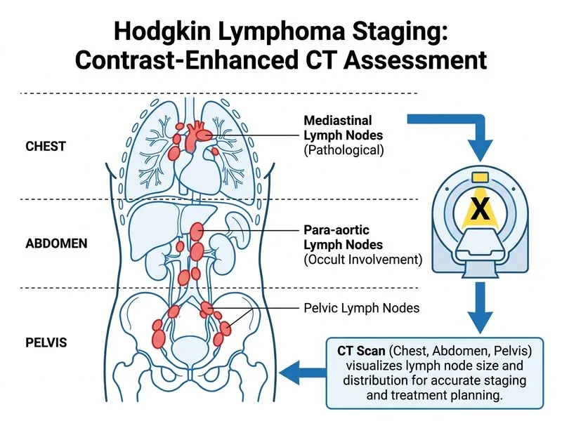
Staging Investigation in Hodgkin Lymphoma
Key Point
Contrast-enhanced CT (CECT) of the chest, abdomen, and pelvis is the standard imaging modality for staging Hodgkin lymphoma and detecting occult nodal and organ involvement.
Why CECT is the Investigation of Choice
1.
Superior nodal assessment:
Detects lymph nodes >1 cm short axis (size criterion for pathological involvement)
Evaluates mediastinal, hilar, abdominal, pelvic, and inguinal nodes
Assesses contiguity of disease (important for radiation planning)
2.
Organ involvement detection:
Hepatic infiltration (nodular, diffuse, or miliary pattern)
Splenic involvement (focal lesions, diffuse infiltration, or splenomegaly)
Defines radiation field boundaries (involved-field radiotherapy)
Identifies sites requiring chemotherapy boost
Baseline for treatment response assessment
High-YieldNEET PG
CECT is mandatory for staging all patients with Hodgkin lymphoma. It is more sensitive than clinical examination (which misses ~20% of abdominal disease) and more practical than PET-CT for initial staging in most centers.
Staging Investigations Hierarchy
Table
Investigation
Indication
Role in HL
CECT chest/abdomen/pelvis
All patients
Mandatory staging
PET-CT
Baseline + end-of-treatment response
Prognostic; increasingly used
Abdominal ultrasound
Limited access to CT
Operator-dependent; lower sensitivity
Bone marrow biopsy
Advanced stage (IIB, III, IV)
Assess marrow involvement
Diagnostic laparotomy
Historical; now obsolete
Replaced by imaging + PET-CT
Clinical Pearl
In the modern era, diagnostic laparotomy with splenectomy is no longer performed for staging Hodgkin lymphoma. Non-invasive imaging (CT + PET-CT) has made it obsolete. Splenectomy carries morbidity (post-splenectomy sepsis risk) and does not change treatment in most cases.