
## Diagnosis: Nodular Sclerosis Hodgkin Lymphoma ### Clinical Presentation **Key Point:** The constellation of B symptoms (fever, night sweats, weight loss), painless progressive lymphadenopathy, and mediastinal involvement in a young adult is classic for Hodgkin lymphoma. **High-Yield:** Nodular sclerosis is the most common subtype of classical Hodgkin lymphoma (~70% of cases), with a predilection for young adults and anterior mediastinal involvement. ### Histopathologic Features The biopsy findings are pathognomonic for Hodgkin lymphoma: | Feature | Significance | |---------|-------------| | Large multinucleated cells ("owl's eye" nuclei) | Reed-Sternberg (RS) cells — hallmark of classical HL | | Prominent eosinophilic nucleoli | "Mirror-image" or "owl's eye" appearance of RS cells | | Background of small lymphocytes, histiocytes, eosinophils | Inflammatory milieu; RS cells are sparse | | Nodular pattern with fibrosis | Nodular sclerosis subtype | **Clinical Pearl:** Reed-Sternberg cells are CD30^+^, CD15^+^, and typically CD45^−^, CD20^−^ (unlike B-cell lymphomas). Hodgkin cells (mononuclear variants) are also present. ### Nodular Sclerosis Subtype **Mnemonic:** NSHD = **N**odular **S**clerosis **H**odgkin **D**isease - Most common classical HL subtype - Mediastinal involvement frequent (60–80%) - Generally favorable prognosis with modern therapy - Age: typically 20–40 years ### Why This Is Hodgkin (Not Non-Hodgkin) **Key Point:** The sparse population of large atypical cells in a rich inflammatory background is characteristic of Hodgkin lymphoma. Non-Hodgkin lymphomas typically show diffuse infiltration of neoplastic cells. [cite:Robbins 10e Ch 13] 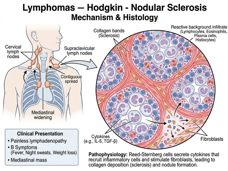
Sign up free to access AI-powered MCQ practice with detailed explanations and adaptive learning.