NEETPGAI
FeaturesBlogComparePricing
Log inStart Free
NEETPGAI

AI-powered NEET PG preparation platform. Master all 19 subjects with adaptive MCQs, AI tutoring, and spaced repetition.

Product

  • Features
  • Subjects
  • Previous Year Questions
  • Compare
  • Pricing
  • Blog

Features

  • Adaptive MCQ Practice
  • AI Tutor
  • Mock Tests
  • Spaced Repetition

Resources

  • Blog
  • Study Guides
  • NEET PG Updates
  • Contact & support

Legal

  • Privacy Policy
  • Terms of Service

Stay updated

© 2026 NEETPGAI. All rights reserved.
    Subjects/Anatomy/Popliteal Fossa
    Popliteal Fossa
    medium
    bone Anatomy

    A 35-year-old male presents to the anatomy lab for cadaveric dissection of the popliteal fossa. The dissector is asked to identify the boundaries and contents of this region. Which of the following structures forms the medial boundary of the popliteal fossa?

    A. Gracilis and sartorius muscles
    B. Vastus medialis and vastus lateralis
    C. Biceps femoris and its tendon
    D. Semimembranosus and semitendinosus

    Explanation

    ## Boundaries of the Popliteal Fossa The popliteal fossa is a diamond-shaped space on the posterior aspect of the knee. Its boundaries are classically defined as: **Medial boundary:** - **Semimembranosus** (superiorly) - **Semitendinosus** (superiorly) - **Gracilis** (superiorly, contributes minimally) - **Medial head of gastrocnemius** (inferiorly) **Lateral boundary:** - **Biceps femoris** and its tendon (superiorly) - **Lateral head of gastrocnemius** (inferiorly) **Floor:** - Popliteal surface of femur - Posterior knee joint capsule - Popliteus muscle **Roof:** - Skin and fascia - Popliteal fascia (continuation of fascia lata) **Key Point:** The semimembranosus and semitendinosus form the superomedial boundary of the popliteal fossa. This is a high-yield anatomical landmark tested frequently in NEET PG. **Contents:** Popliteal artery, popliteal vein, tibial and common peroneal nerves, lymph nodes, and fat. ![Popliteal Fossa diagram](https://mmcphlazjonnzmdysowq.supabase.co/storage/v1/object/public/blog-images/explanation/2059.webp)

    Practice similar questions

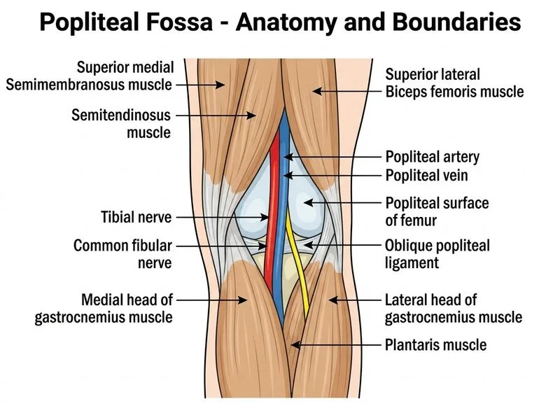
    Sign up free to access AI-powered MCQ practice with detailed explanations and adaptive learning.

    Start Practicing Free More Anatomy Questions