## Esophageal Varices and Portosystemic Anastomoses **Key Point:** The esophageal venous plexus is directly continuous with the **left gastric vein** (also called the coronary vein of the stomach), which drains directly into the **portal vein**. This is the primary site of portosystemic anastomosis in esophageal varices. **Anatomical Pathway:** - **Esophageal venous plexus** → **Left gastric vein** → **Portal vein** - The left gastric vein ascends along the lesser curvature of the stomach and esophagus - It enters the portal vein at the junction of the splenic and superior mesenteric veins **Portosystemic Anastomoses in Portal Hypertension:** | Site | Systemic Vein | Portal Vein Tributary | Clinical Manifestation | |---|---|---|---| | **Esophagus** | Azygos system | Left gastric vein | Esophageal varices | | **Rectum** | Middle/inferior rectal | Superior rectal vein | Rectal varices | | **Umbilicus** | Superficial epigastric | Left/right portal vein | Caput medusae | | **Retroperitoneum** | Lumbar veins | Colic veins | Retroperitoneal varices | **Clinical Pearl:** Esophageal varices are the most dangerous portosystemic anastomoses because they are prone to rupture, causing life-threatening hemorrhage. The left gastric vein is the critical link between the esophageal plexus and the portal system. **High-Yield:** In portal hypertension, blood backs up through the left gastric vein into the esophageal plexus, causing varices. This is why variceal bleeding is a common complication of cirrhosis. 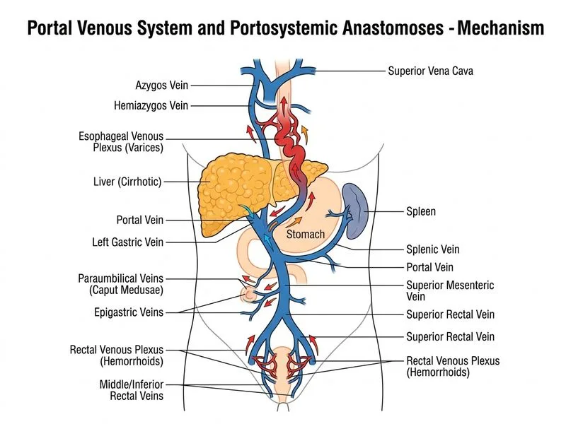
Sign up free to access AI-powered MCQ practice with detailed explanations and adaptive learning.