
## Polymerization Mechanism of Hemoglobin S ### Molecular Basis **Key Point:** Hemoglobin S polymerizes through **hydrophobic interactions** between the valine residue (introduced at position 6 of the β-globin chain) on one HbS molecule and a hydrophobic pocket on an adjacent HbS molecule. ### Step-by-Step Polymerization Process 1. Under normoxic conditions, HbS exists as monomers or dimers and functions relatively normally 2. When oxygen tension decreases (hypoxia), HbS undergoes conformational change and deoxygenation 3. The exposed hydrophobic valine at position 6 interacts with a hydrophobic patch on adjacent deoxygenated HbS molecules 4. These hydrophobic interactions drive the formation of long, rigid polymers 5. Polymer formation distorts the RBC into the characteristic sickle shape ### Why Hydrophobic Interactions? The substitution of glutamic acid (hydrophilic, charged) with valine (hydrophobic, nonpolar) creates an abnormal hydrophobic surface patch that is normally buried in native hemoglobin. This patch becomes the nucleation site for polymerization. **High-Yield:** Polymerization is **oxygen-dependent** and **pH-dependent** — it occurs preferentially in deoxygenated, acidotic environments (tissues with high metabolic demand). **Clinical Pearl:** This is why sickling crises are triggered by hypoxia, infection (acidosis), dehydration, and cold exposure — all conditions that promote HbS polymerization. 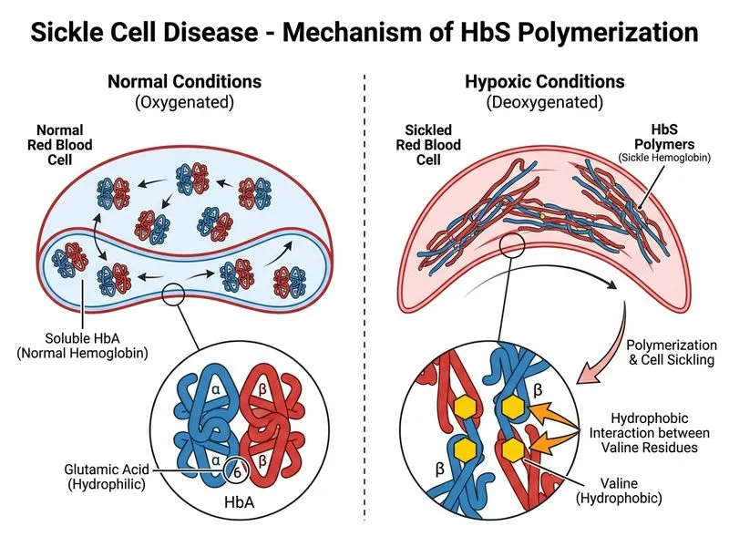
Sign up free to access AI-powered MCQ practice with detailed explanations and adaptive learning.