Version 1.0 — Published April 2026
Quick Answer
Severe aortic stenosis is the commonest valve lesion in elderly Indian patients and one of the most reliably tested NEET PG cardiology vignettes. In a 75-year-old man with exertional syncope, dyspnea, or angina plus a harsh systolic ejection murmur radiating to the carotids, follow this 6-step workflow:
- Recognise the SAD triad — Syncope, Angina, Dyspnea — and remember that symptom onset drops survival to 2-3 years without valve replacement
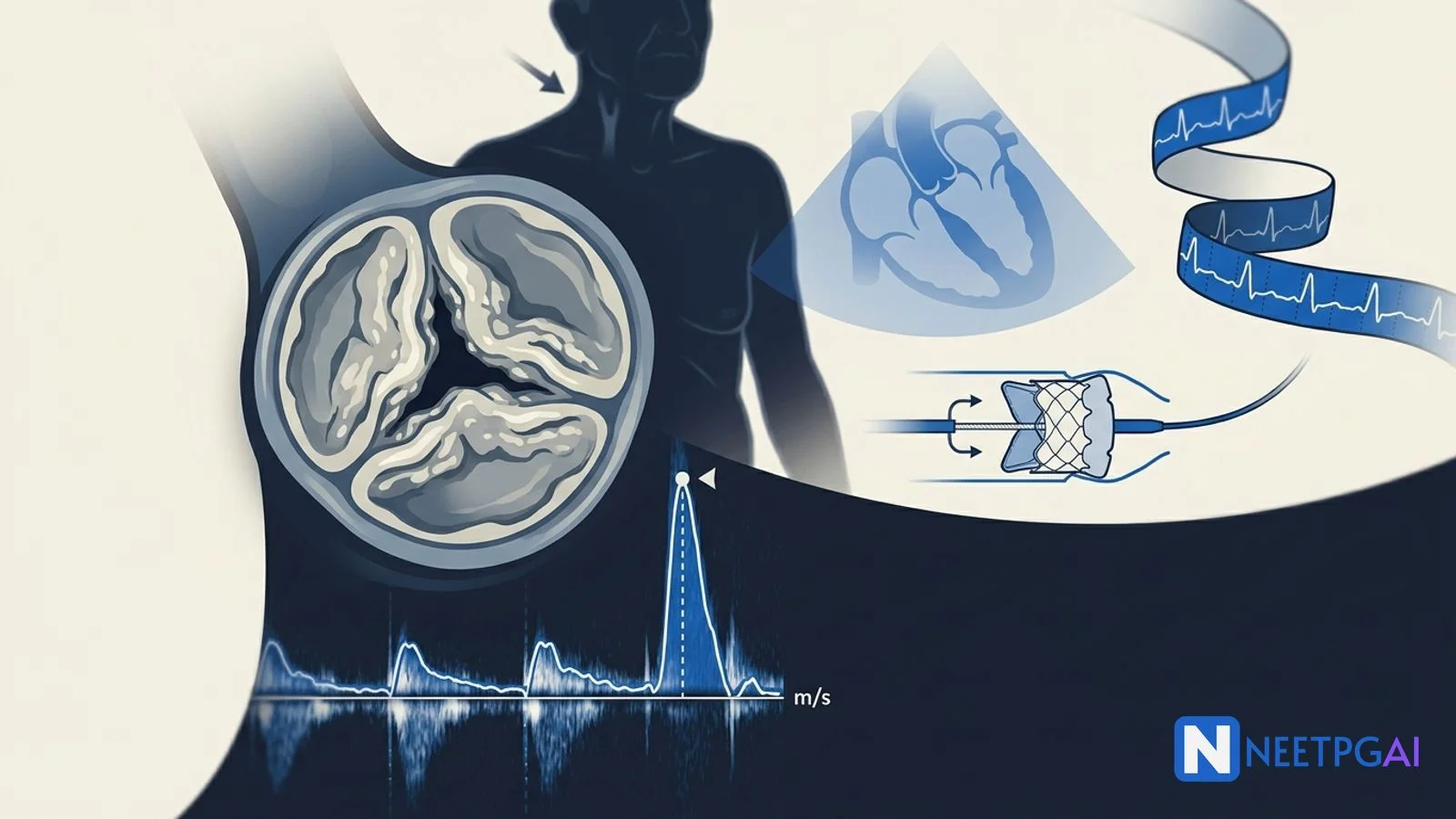
- Confirm with TTE — peak velocity ≥4 m/s, mean gradient ≥40 mmHg, AVA ≤1.0 cm² define severe AS
- Grade further — very severe (peak velocity ≥5 m/s) and low-flow low-gradient subtypes need different workup (dobutamine stress echo)
- Avoid preload and afterload reducers — no nitrates, no dihydropyridines, careful with diuretics
- Plan valve replacement promptly — symptomatic severe AS is a Class I indication for AVR or TAVI regardless of LVEF
- Choose AVR vs TAVI by age, surgical risk, anatomy and patient preference — TAVI dominates above 75 years
The case
A 75-year-old retired bank manager from Pune is brought to the cardiology emergency room by his daughter after a witnessed syncopal episode at the local market that morning. He had walked up a small slope carrying vegetables, felt sudden lightheadedness, broke into a sweat, and lost consciousness for about 30 seconds. He recovered spontaneously without confusion or focal weakness. There was no tongue bite, no urinary incontinence, and no preceding aura. This is his second such episode in the last 8 weeks; the first was during his morning walk and he did not seek care.
Past history: he has had progressively worsening exertional breathlessness for 9 months — initially NYHA II, now NYHA III, walking only one floor before needing to rest. He also describes a tight retrosternal pressure on brisk walking that resolves within 2-3 minutes of rest, present for 6 months, never investigated. He has hypertension on amlodipine 5 mg and ramipril 5 mg, type 2 diabetes on metformin and gliclazide, dyslipidemia on atorvastatin 20 mg. He never smoked, drinks alcohol occasionally, and has no known coronary disease, no prior stroke, no syncope before this year. His daughter mentions that the family GP recently doubled his amlodipine dose because BP was running 150/90 — and that since then he has felt more lightheaded standing up.
On arrival, vitals are: pulse 78/min regular, BP 132/78 mmHg supine, 110/72 mmHg standing (no significant orthostatic drop), respiratory rate 18/min, SpO2 96 percent on room air, temperature 36.8 C, capillary glucose 142 mg/dL. He is oriented, comfortable at rest, no acute distress. JVP not raised. Carotid upstrokes are slow-rising and reduced in amplitude (parvus et tardus) bilaterally. Apex beat is in the 5th intercostal space, mid-clavicular line, sustained heaving in character (pressure overload). On auscultation: a harsh grade 4/6 systolic ejection murmur is heard best at the right second intercostal space, radiating to both carotids, peaking in late systole, with a soft single S2 (the aortic component of S2 is markedly reduced — a hallmark of severe AS). No early diastolic murmur, no S3, no S4 audible at rest. Lung bases are clear; mild pitting edema to mid-shins. Rest of the examination is unremarkable.
Bedside ECG: sinus rhythm at 78/min, left axis, voltage criteria for left ventricular hypertrophy (S in V1 plus R in V5 = 38 mm), strain pattern with ST depression and T-wave inversion in V5-V6 and lead I. No acute ischemic ST elevation, no Q waves.
The on-call resident orders a focused panel and a transthoracic echocardiogram.
ABCD assessment and initial investigations
Severe symptomatic aortic stenosis is a time-sensitive diagnosis — the next syncopal episode could be the last. Resuscitation is gentle (avoid generous fluids that worsen pulmonary congestion in a non-compliant LV) and the diagnostic priority is echocardiography at the bedside.
A — Airway: Patent. GCS 15, no airway compromise. Reassess hourly.
B — Breathing: RR 18, SpO2 96 percent on room air. CXR ordered to look for cardiomegaly, post-stenotic aortic root dilatation, and pulmonary congestion. Continuous SpO2 monitoring; nasal O2 only if SpO2 falls under 92.
C — Circulation: BP 132/78 with parvus et tardus carotids and a pressure-overloaded LV. Two peripheral cannulas. Avoid bolus IV fluids unless overtly volume-deplete. Avoid nitrates for the chest pain — even if angina is the working diagnosis, severe AS is on the table and nitrates can precipitate cardiac arrest. Telemetry for arrhythmia detection.
D — Disability/Dextrose: GCS 15, glucose 142, no focal deficit. The syncope is exertional, brief, and recovers without confusion — much more consistent with cardiac (low-output) syncope than seizure or vasovagal.
Initial investigations (within 60 minutes):
- CBC: Hb 13.4 g/dL, WBC 6,800, platelets 220,000
- BUN: 24 mg/dL; creatinine 1.2 mg/dL; eGFR 58 mL/min (mild CKD)
- Electrolytes: Na 138, K 4.2, Cl 102, HCO3 24
- LFTs: within normal limits
- HbA1c: 7.4 percent
- Lipid profile: LDL 102, HDL 38, TG 168
- TSH: 2.4 mIU/L
- Troponin I high-sensitivity: 18 ng/L (just above the upper reference; consistent with chronic LVH and demand ischemia, not acute MI — serial trend stable at 6 hours)
- NT-proBNP: 2,840 pg/mL (markedly raised — supports cardiac etiology of dyspnea)
- CXR: cardiomegaly with prominent LV contour, mild post-stenotic dilatation of the ascending aorta, no pulmonary edema
- Transthoracic echocardiogram (the diagnostic test):
- Heavily calcified, restricted-motion trileaflet aortic valve
- Peak aortic jet velocity 4.6 m/s
- Mean transvalvular gradient 52 mmHg
- Aortic valve area (continuity equation) 0.78 cm², indexed AVA 0.45 cm²/m²
- LVEF 55 percent (preserved)
- Concentric LVH (septal wall 14 mm, posterior wall 13 mm)
- Mild AR (regurgitation), no MR, no MS
- PASP estimated 38 mmHg (mild pulmonary hypertension)
- LA mildly dilated
- Coronary angiography (planned pre-AVR/TAVI): mid-LAD 60 percent stenosis, LCx and RCA non-obstructive — borderline lesion that may need PCI before or during TAVI
The diagnostic algorithm — why this is severe AS
Aortic stenosis severity grading at NEET PG depth follows the 2020 ACC/AHA and 2021 ESC guidelines. The trio of peak velocity, mean gradient, and aortic valve area must be considered together.
Grading criteria (TTE)
| Severity | Peak velocity (m/s) | Mean gradient (mmHg) | AVA (cm²) |
|---|
| Mild | 2.6 - 2.9 | <20 | >1.5 |
| Moderate | 3.0 - 3.9 | 20 - 39 | 1.0 - 1.5 |
| Severe | ≥4.0 | ≥40 | ≤1.0 |
| Very severe | ≥5.0 | ≥60 | (often ≤0.6) |
Our patient: peak velocity 4.6 m/s, mean gradient 52 mmHg, AVA 0.78 cm² — severe AS, not yet very severe. Indexed AVA ≤0.6 cm²/m² in a small-statured Indian patient (BSA 1.6 m²) reinforces severity classification when raw AVA is borderline.
The four severe AS subtypes (NEET PG favourite)
| Subtype | Velocity / Gradient / AVA | LVEF | Stroke volume index |
|---|
| High-gradient severe AS | V ≥4, G ≥40, AVA ≤1.0 | Any | Any |
| Low-flow low-gradient severe AS, low EF | V <4, G <40, AVA ≤1.0 | <50% | <35 mL/m² |
| Paradoxical low-flow low-gradient severe AS | V <4, G <40, AVA ≤1.0 | ≥50% | <35 mL/m² |
| Low-gradient severe AS with normal flow | V <4, G <40, AVA ≤1.0 | ≥50% | ≥35 mL/m² (often "pseudo-severe") |
Low-flow low-gradient subtypes need dobutamine stress echo to differentiate true severe AS (gradient rises with augmented flow, AVA stays small) from pseudo-severe AS (AVA rises with flow, gradient stays low — the valve was fine; the pump was bad).
Diagnosis
Severe symptomatic aortic stenosis (high-gradient subtype) — peak velocity 4.6 m/s, mean gradient 52 mmHg, AVA 0.78 cm² with classic SAD triad (recurrent exertional syncope, exertional angina for 6 months, NYHA III dyspnea for 9 months) on a background of degenerative trileaflet calcific AS, hypertension, T2DM, and concomitant non-obstructive coronary disease — Class I indication for valve replacement.
This phrasing tells the consultant the lesion, the severity subtype, the symptom complex, and the urgency — the structure NEET PG vignettes test.
Differential diagnosis of syncope in elderly — cardiac vs neurogenic
Syncope is the chief presenting symptom; running the differential matters because the management and prognosis differ wildly.
Cardiac syncope (high-mortality bucket)
- Severe AS — exertional, brief, no aura, normal recovery; harsh ESM; SAD triad; ECG with LVH-strain
- HOCM — younger patient, FH of sudden death, dynamic LVOT obstruction; murmur softens with squat, louder with Valsalva
- Severe pulmonary hypertension or pulmonary embolism
- Tachy- or brady-arrhythmia — sinus pauses, complete heart block, VT; Holter and loop recorder
- Acute MI / ischemia — chest pain, ECG changes, troponin rise
- Cardiac tamponade — Beck triad
- Aortic dissection — tearing chest pain, BP differential
- Atrial myxoma — positional syncope, plop sound, anaemia, raised ESR
Reflex / neurogenic / neurally-mediated syncope (low-mortality bucket)
- Vasovagal — emotional / postural trigger, prodrome (nausea, sweating, blurred vision), brief LOC
- Carotid sinus hypersensitivity — older men, head turning / tight collar, brady-hypotension on carotid massage
- Situational syncope — micturition, defecation, cough, swallow
- Orthostatic hypotension — drug-induced (alpha-blockers, diuretics, vasodilators), volume depletion, autonomic failure (Parkinson, diabetes, MSA)
Why our patient's syncope is cardiac
Three features lock it in: (1) exertional trigger (cardiac syncope is provoked by exertion; vasovagal almost never is), (2) brief duration with no prodrome and no post-event confusion (rules out seizure and most vasovagal), and (3) objective AS findings (parvus et tardus carotids, harsh ESM with soft single S2, ECG LVH-strain). Exertional syncope plus a harsh systolic ejection murmur in an elderly Indian male is severe AS until proven otherwise.
Etiology of aortic stenosis by age
Memorise the age-specific etiology table — NEET PG asks this directly.
| Age band | Most common cause |
|---|
| Infants and children | Congenital unicuspid or dysplastic AV |
| 15-65 years | Bicuspid aortic valve (BAV) with premature calcification |
| >65 years | Senile (degenerative) calcific trileaflet AS |
| Any age | Rheumatic AS (almost always with mitral involvement) |
Bicuspid AV is the commonest congenital cardiac anomaly (1-2 percent of population) and the commonest cause of AS in middle-aged adults; it is associated with aortopathy (root dilatation, dissection risk) requiring closer aortic surveillance. Rheumatic AS in India is often missed because it almost never occurs in isolation — look for concomitant mitral stenosis or regurgitation.
Management — the symptomatic patient gets the valve
Initial stabilisation
- Avoid preload reducers — stop nitrates, hold diuretics if not overtly congested, avoid dihydropyridine CCBs (the doubled amlodipine in this case is a likely contributor to recent lightheadedness and may need de-escalation)
- Avoid afterload reducers acutely — ACE inhibitors and ARBs are not contraindicated in stable severe AS but should not be uptitrated in a freshly symptomatic patient until afterload is restored by valve replacement
- Beta-blockers cautiously — reduce LV contractility and can drop output; reserve for AF rate control or ischemia if needed, with low starting doses
- Treat AF aggressively — loss of atrial kick is poorly tolerated by a stiff hypertrophied LV; rhythm control preferred
- Manage concomitant CAD — high-sensitivity troponin and angiography pre-AVR; plan PCI for significant lesions
- Endocarditis prophylaxis — only for patients with prior IE or prosthetic valves; native AS does not need routine prophylaxis
Definitive treatment — surgical AVR vs TAVI
Symptomatic severe AS = Class I indication for valve replacement. The decision between SAVR and TAVI is now driven by surgical risk, age, anatomy, and patient choice — not by ejection fraction or symptom severity (which are already triggers).
Surgical aortic valve replacement (SAVR)
- Preferred for age <65 years, low surgical risk, bicuspid valve with significant aortopathy, severe concomitant coronary disease needing CABG, infective endocarditis, complex congenital anatomy, severe MR or TR needing repair
- Choice of valve — mechanical for younger patients (durability, lifelong warfarin); bioprosthetic for >65 years (no anticoagulation, but structural valve deterioration in 10-15 years)
- Typical mortality — 1-3 percent in low-risk patients; rises with age, frailty, prior surgery, EuroSCORE II / STS PROM
Transcatheter aortic valve implantation (TAVI / TAVR)
- Preferred for age >75 years, high or intermediate surgical risk, prior cardiac surgery, severe COPD, frailty, porcelain aorta, and increasingly low-risk patients >65 with suitable femoral access and trileaflet anatomy
- Major trials — PARTNER 1 (high risk), PARTNER 2 / SURTAVI (intermediate risk), PARTNER 3 / Evolut Low Risk (low risk >65)
- Approaches — transfemoral (default), transapical, trans-subclavian, trans-caval
- Complications — paravalvular leak (less with newer-generation valves), need for pacemaker (around 10-15 percent — higher with self-expanding valves), vascular access injury, stroke (1-3 percent), valve durability data now extending past 8 years
For our 75-year-old patient with mild CKD, T2DM, and a mid-LAD 60 percent lesion: transfemoral TAVI plus staged or concurrent PCI of LAD is the most likely Heart Team decision after CT angiography of the aortic root and ilio-femoral system.
Asymptomatic severe AS — when to break the watchful-waiting rule
Asymptomatic severe AS is generally observed every 6-12 months. Intervene early if any of the following are present:
- LVEF <50 percent even if asymptomatic (Class I)
- Exercise testing unmasks symptoms (angina, syncope, fall in SBP >20 mmHg, ventricular arrhythmias) — Class I
- Very severe AS (peak velocity ≥5.0 m/s) and low surgical risk — Class IIa
- Rapid progression (peak velocity rise >0.3 m/s per year) — Class IIa
- Markedly elevated BNP (>3x age-sex-adjusted normal) — Class IIa
- Pulmonary hypertension (PASP >60 mmHg attributable to AS) — Class IIa
- Patient already going for cardiac surgery for another indication (CABG, mitral) — Class I
Specific scenarios
- Bicuspid AV — surveil aortic root annually if >40 mm; intervene at root >55 mm (or >50 mm with risk factors) regardless of AS severity
- Pregnancy with severe AS — high risk; ideally counsel against pregnancy until AVR; if presenting pregnant, multidisciplinary team, beta-blockade, avoid epidural-induced hypotension, consider balloon valvuloplasty as a bridge
- End-stage AS with cardiogenic shock — balloon aortic valvuloplasty as bridge to TAVI in selected centres; emergency TAVI in high-volume centres
Complications — pre-op, peri-op, post-op
Pre-op (untreated severe AS)
- Sudden cardiac death — annual rate 1 percent in asymptomatic severe AS, jumping to 8-10 percent once symptoms appear
- Heart failure — initially diastolic from LVH, later systolic with LV decompensation
- Atrial fibrillation — left atrial enlargement; loss of atrial kick poorly tolerated
- GI bleeding from acquired von Willebrand syndrome (Heyde syndrome) — high shear across stenotic valve cleaves vWF multimers; bleeding from colonic angiodysplasia
- Infective endocarditis — calcified valves are less prone than rheumatic ones but still at risk
- Embolic events from valve calcification
Post-AVR / TAVI
- Paravalvular leak — more common with TAVI; significant PVL increases mortality
- Permanent pacemaker need — TAVI 10-15 percent (self-expanding higher than balloon-expandable)
- Stroke — 1-3 percent peri-procedurally
- Vascular access complications — TAVI; iliofemoral injury, retroperitoneal haematoma
- Bleeding and tamponade — both procedures
- Prosthetic valve thrombosis and endocarditis — long-term; lifelong vigilance
- Structural valve deterioration — bioprosthetic 10-15 years; mechanical decades but warfarin-tied
How NEET PG tests aortic stenosis
NEET PG tests this topic through six recurring patterns. Recognise the pattern and the question collapses to a 30-second answer.
Pattern 1 — The diagnostic-criteria question: Vignette gives peak velocity 4.4 m/s, mean gradient 48 mmHg, AVA 0.85 cm². Severity? Severe AS. Trap: answers offering "moderate" — any one of the three criteria meeting threshold qualifies as severe.
Pattern 2 — The symptom-onset prognosis question: Asymptomatic severe AS vs symptomatic severe AS — survival difference? Asymptomatic = <1 percent annual mortality; symptomatic syncope = 3-year median survival; symptomatic heart failure = 2-year median survival. Symptom onset is the inflection point.
Pattern 3 — The murmur-character question: Harsh ESM at RUSB radiating to carotids, soft single S2, parvus et tardus carotids. Diagnosis? Severe AS. Distinguish from HOCM (murmur softens with squat, louder with Valsalva) and aortic sclerosis (no radiation, normal carotids, no symptoms).
Pattern 4 — The drug-to-avoid question: Severe symptomatic AS, BP 130/80, presenting with chest pain. Best initial management? NOT nitroglycerin. Trap: answers offering sublingual nitrate — can precipitate cardiac arrest by dropping preload across a fixed-output valve.
Pattern 5 — The AVR vs TAVI question: 78-year-old with severe symptomatic AS, EF 50 percent, mild frailty, suitable femoral access. Best treatment? TAVI. Trap: answers offering "medical management until LVEF drops" — wrong; symptom onset is the trigger.
Pattern 6 — The Heyde-syndrome question: Elderly patient with severe AS and recurrent lower GI bleeds; colonoscopy shows angiodysplasia. Diagnosis? Heyde syndrome — acquired von Willebrand syndrome from high shear across the stenotic valve. Definitive treatment: AVR (corrects vWF cleavage and resolves bleeding).
High-yield one-liners:
- Severe AS = peak velocity ≥4 m/s, mean gradient ≥40 mmHg, AVA ≤1.0 cm² — any one
- SAD triad (Syncope, Angina, Dyspnea) — symptom onset is the inflection point
- Symptomatic severe AS = Class I indication for AVR/TAVI regardless of LVEF
- Bicuspid AV is the commonest cause of AS in 15-65; senile calcific in >65
- Avoid nitrates, dihydropyridines, generous diuresis in severe AS
- Parvus et tardus carotids, soft single S2, harsh late-peaking ESM = severe AS
- Low-flow low-gradient severe AS needs dobutamine stress echo
- TAVI dominates above 75 years; SAVR for low-risk under 65 and bicuspid with aortopathy
- Pacemaker need post-TAVI 10-15 percent
- Heyde syndrome — severe AS plus GI bleeding from angiodysplasia, vWF multimer cleavage
Frequently Asked Questions
What is the SAD triad in aortic stenosis and why is it prognostically critical?
SAD stands for Syncope, Angina, and Dyspnea — the three classic symptoms of severe aortic stenosis. Once any of these appear, mean survival without valve replacement drops to 3 years for angina, 3 years for syncope, and 2 years for heart failure / dyspnea. Asymptomatic severe AS has a mortality of less than 1 percent per year, but the rate of symptom development is around 8 percent per year. Symptom onset is the single most powerful indication for AVR or TAVI in severe AS — irrespective of LVEF — because the survival curve drops vertically once symptoms begin.
What are the echocardiographic criteria for severe aortic stenosis?
Severe AS on transthoracic echo requires at least one of: peak transvalvular velocity at least 4.0 m/s, mean transvalvular gradient at least 40 mmHg, or aortic valve area no greater than 1.0 cm² (indexed AVA no greater than 0.6 cm²/m²). Very severe AS uses peak velocity at least 5.0 m/s or mean gradient at least 60 mmHg. Low-flow low-gradient severe AS (AVA at or below 1.0 cm² but mean gradient under 40 mmHg) needs dobutamine stress echo to differentiate true severe AS from pseudo-severe AS.
When is surgical AVR preferred over TAVI in severe AS?
Surgical AVR is preferred for low-surgical-risk patients under 65 years, bicuspid aortic valves with significant aortopathy, severe concomitant coronary disease needing CABG, infective endocarditis, complex congenital anatomy, severe mitral regurgitation needing repair, and patients in whom bioprosthetic durability beyond 10 to 15 years is required. TAVI is preferred for high-risk and intermediate-risk patients above 75 years, prior cardiac surgery, severe COPD or frailty, porcelain aorta, and increasingly for low-risk patients above 65 to 70 with suitable femoral access and trileaflet anatomy.
Why must vasodilators and nitrates be used cautiously in severe aortic stenosis?
Severe AS produces a fixed cardiac output across the stenosed valve. Systemic vasodilators (nitrates, ACE inhibitors, dihydropyridine CCBs) reduce afterload and venous return without a compensatory rise in cardiac output, leading to abrupt hypotension, syncope, and even cardiac arrest. Nitroglycerin given for presumed angina in undiagnosed severe AS is a classic preventable cause of in-hospital collapse. Beta-blockers are also relatively risky as they reduce contractility. The safe rule is: avoid preload and afterload reduction in symptomatic severe AS until the valve is fixed.
How does asymptomatic severe AS get watched and when do you intervene?
Asymptomatic severe AS gets serial clinical and echo follow-up every 6 to 12 months. Indications to intervene even without classic symptoms include: LVEF below 50 percent, exercise-test-unmasked symptoms or a fall in systolic BP during exercise, very severe AS (peak velocity at least 5.0 m/s) with low surgical risk, rapid AS progression (peak velocity rise above 0.3 m/s per year), markedly elevated BNP, concomitant cardiac surgery for another indication, and pulmonary hypertension above 60 mmHg attributable to AS. Otherwise watchful waiting with patient-driven symptom reporting is reasonable.
This content is for educational purposes for NEET PG exam preparation. It is not a substitute for professional medical advice, diagnosis, or treatment. Clinical information has been reviewed by qualified medical professionals.
Written by: NEETPGAI Editorial Team
Reviewed by: Pending SME Review
Last reviewed: April 2026