clinical case
pediatrics
ophthalmology
retinoblastoma
leukocoria
RB1 gene
NEET PG 2026
NEET PG pediatric leukocoria case: 2-yo with white pupillary reflex, differential, MRI orbit (never CT), Reese-Ellsworth + IIRC staging, focal therapy, chemoreduction, enucleation, RB1 testing.
Version 1.0 — Published May 2026
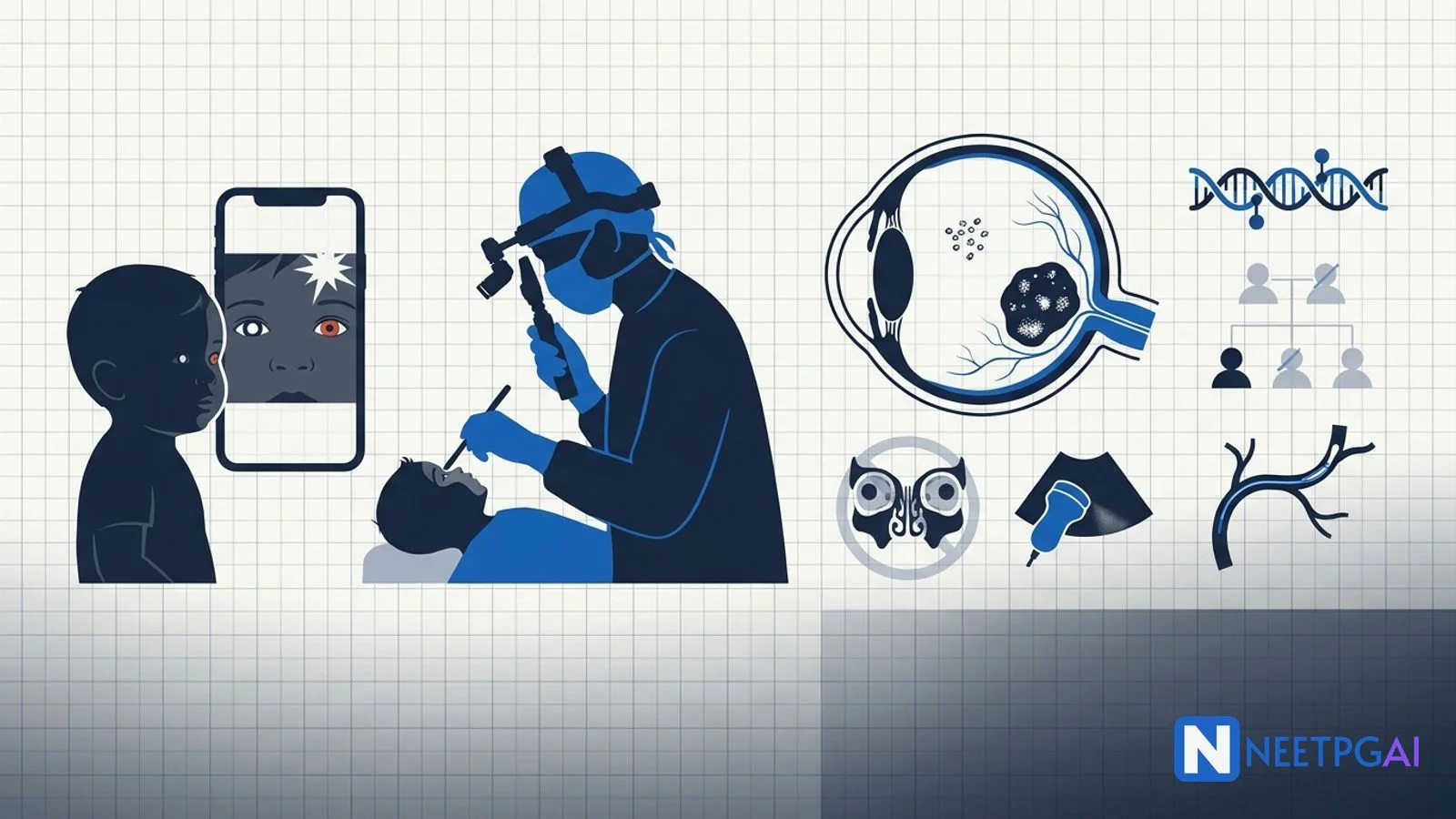
Leukocoria (white pupillary reflex) in a child is retinoblastoma until proven otherwise. The mother's flash-photograph observation is the commonest presenting sign in India and one of the highest-yield NEET PG paediatric ophthalmology stems. In a 2-year-old with leukocoria, follow this 8-step workflow:
A 2-year-old boy is brought to the paediatric outpatient clinic in a tertiary children's hospital in Mumbai by his mother. She reports that for the past 3 weeks she has noticed a "white spot" in her son's right pupil whenever she takes a flash photograph on her smartphone. Initially she thought it was a camera artefact and tried a different phone — the same white reflex appeared on the right eye only. The left eye shows the expected red reflex. The child is otherwise asymptomatic — feeding well, playing normally, no fever, no eye redness, no eye discharge, no head tilt, no apparent visual difficulty, no behavioural change. No history of trauma. No prematurity (term delivery at 38 weeks, birth weight 3.1 kg, uneventful neonatal period). No supplemental oxygen at birth, no NICU stay. Fully immunised per the National Immunisation Schedule. Developmental milestones age-appropriate.
Family history is critical and must be elicited carefully. Mother is 28, father is 32, both healthy, non-consanguineous. No family history of childhood eye cancer, no enucleations in any first- or second-degree relative, no childhood blindness. The father reports that his own paternal uncle "lost an eye as a child" but the diagnosis is unknown — a potentially important clue to germline RB1 carriage that warrants careful three-generation pedigree construction.
This content is for educational purposes for NEET PG exam preparation. It is not a substitute for professional medical advice, diagnosis, or treatment. Clinical information has been reviewed by qualified medical professionals.
Start practicing NEET PG MCQs with AI-powered explanations.
Start Free PracticeMaster GI secretions, digestion, absorption transporters, motility patterns, and gut hormones with high-yield NEET PG 2026 traps and India-context examples.
Master labor stages, Friedman vs Zhang curves, WHO partograph, AMTSL, episiotomy and India JSY/LaQshya policies for NEET PG 2026 OBG MCQs.
5 anterior segment ophthalmology image MCQs for NEET PG: hypopyon and Behcet, Kayser-Fleischer ring in Wilson, Brushfield spots in Down, corneal arcus, and pterygium vs pinguecula.
Daily MCQs, study tips, and topper strategies on Telegram.
Join on Telegram →On examination, the child is alert, interactive, age-appropriate for height and weight. Anterior-segment external examination of both eyes: no proptosis, no orbital asymmetry, no eyelid swelling, no scleral injection, no corneal opacity. Both pupils equal and reactive at rest. Red reflex examination with the direct ophthalmoscope from 30 cm — the LEFT eye shows a clear orange-red reflex; the RIGHT eye shows a WHITE reflex (leukocoria) filling the pupil. No strabismus on cover test today, but the mother reports occasional drifting of the right eye outward over the past month. No nystagmus. Visual acuity in a 2-year-old is best assessed by fix-and-follow — left eye fixes and follows a small toy reliably; right eye fixation is poor and the child resists occlusion of the left eye (the classic objective sign of monocular visual deprivation).
The paediatrician immediately arranges urgent referral to the on-call paediatric ophthalmologist with a working impression of "leukocoria, right eye, retinoblastoma until proven otherwise".
This is not an emergency in the resuscitation sense — the child is well, vitals stable, no airway, breathing, or circulatory compromise. But it IS a time-critical oncology emergency in that any delay in diagnosis and treatment can mean the difference between eye-salvaging therapy and enucleation, and in advanced cases the difference between cure and metastatic death. The 5-year survival in India for intraocular retinoblastoma diagnosed and treated promptly approaches 95 percent; survival in extra-ocular and metastatic disease drops to 30-50 percent.
A — Airway, B — Breathing, C — Circulation: All normal. Proceed to focused ocular oncology workup.
D — Disability: Confirm leukocoria objectively. Assess fixation behaviour. Document any strabismus, head tilt, or signs of orbital extension. Document any neurological signs (cranial nerve palsies, raised ICP signs) — pinealoblastoma in trilateral RB and CNS extension produce headache, vomiting, papilledema, and posturing.
E — Exposure: Full body examination for café-au-lait macules (rule out neurofibromatosis), for haemangiomas (rule out Sturge-Weber), for general health and nutrition status, and for any palpable lymph nodes (preauricular and cervical — metastatic spread route).
A 2-year-old cannot tolerate the prolonged, painful, dilated, indirect ophthalmoscopy with 360-degree scleral depression that is required to fully document intraocular retinoblastoma. EUA is mandatory for any suspected intraocular tumour in a young child. EUA components:
EUA findings in this child: Right eye — anterior segment normal; IOP 14 mmHg; single large intraretinal-exophytic tumour involving 40 percent of the retina inferotemporally, 8 disc-diameters in size, extending from the inferotemporal periphery towards but not involving the optic disc or foveola, with associated focal subretinal fluid and a small cluster of vitreous seeds in the inferior vitreous cavity; no anterior segment involvement, no neovascular glaucoma, no retinal detachment beyond the tumour. Ultrasound confirms a solid mass with high-amplitude internal calcific foci. Left eye on EUA — completely normal, no tumour. MRI orbit confirms the intraocular mass with internal calcification, optic nerve appears normal with no post-laminar invasion, no extra-ocular extension, no pinealoblastoma, no CNS disease.
Staging: Unilateral right-eye retinoblastoma, IIRC Group D (diffuse subretinal seeds or vitreous seeds in the absence of massive tumour filling), IRSS Stage 0 (intraocular, no extra-ocular spread).
NEET PG tests the differential of leukocoria directly. Memorise the 7-cause list and the distinguishing features.
| Diagnosis | Laterality | Key features | Imaging signature |
|---|---|---|---|
| Retinoblastoma | Unilateral 60%, bilateral 40% | White reflex, strabismus, occasionally proptosis or red eye late; family history may be positive | Calcified intraocular mass on ultrasound and MRI |
| Congenital cataract | Often bilateral | Opacified lens, leukocoria visible from birth; rubella, galactosaemia, idiopathic | Lens opacity on slit lamp; no retinal mass |
| Coats disease | Unilateral, boys, age 4-8 | Exudative retinopathy, telangiectatic retinal vessels, yellow subretinal exudate (not white), no family history | No calcification; subretinal exudate on USG; fluorescein angiogram shows telangiectasias |
| Persistent fetal vasculature (PFV) | Unilateral | Microphthalmic eye, retrolental fibrovascular tissue, cataract, glaucoma | Microphthalmia + retrolental tissue, no calcification |
| Retinopathy of prematurity (ROP) Stage 5 | Bilateral usually | Preterm, low birth weight, supplemental-oxygen history; total retinal detachment with retrolental tissue | History critical; no calcification |
| Ocular toxocariasis | Unilateral | Older child (5-10 years), peripheral granuloma, posterior pole granuloma, or endophthalmitis; raised serum Toxocara titres | No calcification; tractional retinal detachment, serology |
| Retinal detachment | Variable | Trauma, FEVR, Norrie disease (X-linked, boys); white reflex from detached retina | Detachment on USG; no mass, no calcification |
Five features lock it in: (1) age 2 (peak incidence 1-3 years), (2) isolated leukocoria with poor fixation on the affected side, (3) white intraocular mass with high-amplitude internal calcification on ultrasound — calcification in an intraocular mass in a young child is virtually pathognomonic for retinoblastoma, (4) MRI confirms calcified intraretinal-exophytic mass with vitreous seeds, and (5) no alternative diagnosis fits (no microphthalmia, no telangiectasias, no prematurity, no trauma, no lens opacity). RB1 mutation analysis is pending.
Unilateral right-eye retinoblastoma, single large intraretinal-exophytic tumour with focal vitreous seeds — IIRC Group D, IRSS Stage 0 — in a 2-year-old boy with no apparent family history but a suggestive paternal-side history requiring three-generation pedigree analysis and RB1 mutation testing on tumour and peripheral blood lymphocytes to determine germline status and guide cascade testing of family members.
The two parallel goals are (1) save life and (2) save eye and vision. Life always trumps eye. Modern multimodal therapy at high-volume centres achieves both in the majority of intraocular Group A-D disease.
| Group | Description | Standard approach |
|---|---|---|
| A | Small intraretinal tumour, away from foveola and disc | Focal therapy (laser photocoagulation, cryotherapy, transpupillary thermotherapy) |
| B | Larger intraretinal tumour, any location | Systemic chemoreduction + focal consolidation |
| C | Focal subretinal fluid or seeds | Systemic or intra-arterial chemoreduction + focal consolidation + intravitreal melphalan for vitreous seeds |
| D | Diffuse subretinal fluid or seeds | Intra-arterial chemotherapy (IAC) + intravitreal melphalan + focal consolidation; selected enucleation |
| E | Massive intraocular tumour, neovascular glaucoma, anterior segment extension | Primary enucleation (eye salvage rarely possible); adjuvant chemotherapy if high-risk histology |
Seven recurring patterns. Recognise the pattern and the question collapses to a 30-second answer.
Pattern 1 — The leukocoria-differential question: Vignette gives a 2-3 year old with white pupillary reflex on flash photography. First differential to exclude? Retinoblastoma. Imaging of choice? MRI orbit with gadolinium (NEVER CT).
Pattern 2 — The Knudson two-hit question: Bilateral retinoblastoma in a 1-year-old. Inheritance pattern? Autosomal dominant (germline RB1 mutation), Knudson two-hit hypothesis. Risk of transmission to each child? 50 percent. Trap: bilateral disease in a child of unaffected parents is still germline — a de novo mutation.
Pattern 3 — The imaging question: Why is CT avoided in suspected retinoblastoma? Radiation increases second-malignancy risk in germline RB1 carriers (osteosarcoma, sarcoma, melanoma). What replaces it? MRI orbit with gadolinium plus ultrasound B-scan.
Pattern 4 — The staging question: Vignette gives a tumour filling 70 percent of the globe with neovascular glaucoma. IIRC group? Group E. Treatment? Primary enucleation. Trap: aggressive eye-salvage attempts at Group E lead to extraocular spread and metastatic death.
Pattern 5 — The chemotherapy question: Standard systemic chemoreduction regimen for retinoblastoma? Vincristine + Etoposide + Carboplatin (VEC), 6 cycles. What revolutionised advanced eye salvage? Intra-arterial chemotherapy with melphalan delivered super-selectively to the ophthalmic artery.
Pattern 6 — The trilateral-RB question: Child with bilateral retinoblastoma develops headache and papilledema at age 3. Diagnosis? Trilateral retinoblastoma — pinealoblastoma. Surveillance? MRI brain every 6 months until age 5 in germline carriers.
Pattern 7 — The second-malignancy question: Adult who survived bilateral retinoblastoma in childhood (treated with EBRT) presents with a thigh mass at age 22. Most likely diagnosis? Osteosarcoma in the radiation field — the commonest second malignancy in germline RB1 carriers.
High-yield one-liners:
CT delivers ionising radiation directly to the orbit, and children with hereditary (germline RB1) retinoblastoma have a markedly elevated lifetime risk of second non-ocular malignancies, particularly osteosarcoma, soft-tissue sarcoma, and melanoma. Radiation exposure further increases this risk. MRI orbit with gadolinium is the imaging investigation of choice — it has no radiation, characterises the tumour, detects optic-nerve invasion, identifies pinealoblastoma (trilateral retinoblastoma), and screens for CNS extension. CT is used only when MRI is unavailable and calcification must be confirmed urgently, and even then it is the exception, not the rule. Ultrasound B-scan is the bedside investigation that demonstrates intraocular mass and calcification with no radiation.
Leukocoria (white pupillary reflex) has seven major causes that NEET PG tests repeatedly. (1) Retinoblastoma — most important, malignant, calcified intraocular mass, can be bilateral, RB1 mutation. (2) Congenital cataract — opacified lens, can be detected early, surgical removal restores vision if treated within the critical period. (3) Coats disease — unilateral, boys, exudative retinopathy with subretinal lipid exudates, no calcification, telangiectatic retinal vessels. (4) Persistent fetal vasculature (PFV, previously PHPV) — unilateral, microphthalmic eye, retrolental fibrovascular tissue. (5) Retinopathy of prematurity (ROP) — preterm, low birth weight, supplemental-oxygen history, vascularised retinal-detachment retrolental tissue. (6) Ocular toxocariasis — Toxocara canis larva migrans, posterior pole granuloma or endophthalmitis, often older child. (7) Retinal detachment from trauma, FEVR, or Norrie disease. Retinoblastoma must be ruled out first because of life-threatening malignancy.
Reese-Ellsworth was the original classification (1963) used to predict eye salvage with external-beam radiotherapy — it has 5 groups (I-V) based on tumour size, location, and vitreous seeding. It is now considered obsolete because modern chemoreduction has replaced external-beam radiotherapy and outcomes do not map cleanly to Reese-Ellsworth groups. The International Intraocular Retinoblastoma Classification (IIRC) — also called the International Classification of Retinoblastoma (ICRB) — was developed in the 2000s to predict eye salvage with chemoreduction plus focal therapy. It has 5 groups (A-E). Group A: small intraretinal tumour away from foveola and disc. Group B: larger intraretinal, any location. Group C: focal subretinal fluid or seeds. Group D: diffuse subretinal fluid or seeds. Group E: massive intraocular tumour filling more than 50 percent of globe, neovascular glaucoma, or extension to anterior segment — usually requires enucleation. For metastatic and extraocular disease, the International Retinoblastoma Staging System (IRSS) Stages 0-IV is used.
Enucleation is indicated for Group E disease (massive intraocular tumour filling more than 50 percent of globe, neovascular glaucoma, severe hyphema with no view, anterior segment extension, optic nerve invasion suggested on imaging, or no useful visual potential), failed conservative therapy, or when conservative therapy is unsafe. Surgical principle: remove the globe with the longest possible optic-nerve stump (15-20 mm), avoid violating the globe, send for histopathology including optic-nerve margin assessment. Intra-arterial chemotherapy (IAC, super-selective ophthalmic artery cannulation with melphalan plus or minus topotecan) has revolutionised eye salvage for advanced disease (Groups C, D, and selected E). IAC delivers high-dose chemotherapy directly to the eye with minimal systemic toxicity, reduces the need for enucleation and external-beam radiation, and is now standard of care for advanced unilateral retinoblastoma at high-volume centres. Intravitreal melphalan or topotecan injection is added for vitreous seeds resistant to IAC. In India, IAC is available at AIIMS Delhi, Sankara Nethralaya, LV Prasad Eye Institute, Aravind Eye Hospital, and a few other tertiary centres.
Approximately 40 percent of retinoblastoma cases are hereditary (germline RB1 mutation), and 60 percent are sporadic (somatic RB1 mutation in retinal cell only). All bilateral cases, all unilateral multifocal cases, all familial cases, and all unilateral cases under 1 year are presumed germline until proven otherwise. Germline-mutation carriers have approximately 50 percent chance of transmitting the mutation to each child, a lifetime risk of trilateral retinoblastoma (pinealoblastoma) of 5-15 percent, and a substantially elevated risk of second non-ocular malignancies (osteosarcoma, soft-tissue sarcoma, melanoma) particularly if treated with external-beam radiation. RB1 testing is performed on tumour tissue (if available from enucleated eye) and on peripheral blood lymphocytes. If a germline mutation is identified, siblings and the parents are tested; future children should undergo cord-blood RB1 testing at birth and repeated dilated fundus examination under anaesthesia from birth. In India, free or subsidised RB1 testing is available through Children's Hospital networks, AIIMS Delhi, NIMHANS, and tertiary eye hospitals — counsel families to access these networks because private RB1 testing can cost Rs 25,000-40,000.
This content is for educational purposes for NEET PG exam preparation. It is not a substitute for professional medical advice, diagnosis, or treatment. Clinical information has been reviewed by qualified medical professionals.
Written by: NEETPGAI Editorial Team Reviewed by: Pending SME Review Last reviewed: May 2026