clinical case
medicine
cardiology
STEMI
acute MI
primary PCI
NEET PG 2026
NEET PG inferior STEMI case: 45-yo male smoker, ECG ST elevation in II/III/aVF, V4R for RV involvement, primary PCI vs fibrinolysis, DAPT, Killip class, MCQ traps.
Version 1.0 — Published April 2026
Acute STEMI is the highest-mortality cardiology diagnosis NEET PG tests, and inferior STEMI in a young Indian smoker is the most-tested vignette across recent papers. In a 45-year-old male smoker with crushing retrosternal chest pain radiating to the left arm, diaphoresis, and 2 hours of symptoms with ST elevation in II, III, and aVF, follow this 7-step workflow:
Door-to-balloon time is the single most important variable: every 30-minute delay increases 1-year mortality by 7.5 percent.
A 45-year-old marketing manager from Mumbai is brought to the emergency department by his wife at 7:45 AM with sudden onset crushing retrosternal chest pain that started about 2 hours ago while he was getting ready for work. He describes it as a heavy "elephant sitting on my chest," radiating to the left arm and jaw, associated with profuse sweating, nausea, and one episode of vomiting. He took a domperidone tablet thinking it was indigestion. The pain has not relented despite rest. He smokes 15 cigarettes per day for 20 years (15 pack-years), is a social drinker, and has a strong family history — his father died of a heart attack at age 52. He has hypertension diagnosed 3 years ago on telmisartan 40 mg, but is non-compliant. No diabetes, no prior cardiac event, no medication allergies.
On arrival, vitals are: pulse 56/min regular, BP 96/62 mmHg, respiratory rate 22/min, SpO2 95 percent on room air, temperature 36.6 C, capillary glucose 138 mg/dL. He is anxious, diaphoretic, in obvious distress, clutching his chest. Pain score 9/10. JVP appears slightly elevated. Carotid pulses normal in volume and contour. Apex beat is in the 5th intercostal space, mid-clavicular line, normal in character. On auscultation: heart sounds S1 and S2 normal, no S3, no S4, no murmurs, no pericardial rub. Lungs are clear bilaterally — no crackles, no wheeze. Abdomen soft, non-tender. No peripheral edema. Distal pulses 2+ bilaterally. Neurologically intact.
This content is for educational purposes for NEET PG exam preparation. It is not a substitute for professional medical advice, diagnosis, or treatment. Clinical information has been reviewed by qualified medical professionals.
Start practicing NEET PG MCQs with AI-powered explanations.
Start Free PracticeMaster GI secretions, digestion, absorption transporters, motility patterns, and gut hormones with high-yield NEET PG 2026 traps and India-context examples.
Master labor stages, Friedman vs Zhang curves, WHO partograph, AMTSL, episiotomy and India JSY/LaQshya policies for NEET PG 2026 OBG MCQs.
5 anterior segment ophthalmology image MCQs for NEET PG: hypopyon and Behcet, Kayser-Fleischer ring in Wilson, Brushfield spots in Down, corneal arcus, and pterygium vs pinguecula.
Daily MCQs, study tips, and topper strategies on Telegram.
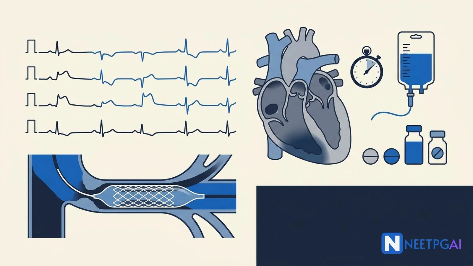
Join on Telegram →The triage nurse hands over the 12-lead ECG done within 7 minutes of arrival.
ECG findings: Sinus bradycardia at 56/min. ST elevation of 3 mm in lead III, 2 mm in lead II, and 2.5 mm in aVF with hyperacute T waves. Reciprocal ST depression of 2 mm in leads I and aVL. No ST elevation in V1-V6. No pathological Q waves yet. PR interval normal. QRS narrow.
The on-call cardiologist is paged immediately, the cath lab is activated, and the right-sided ECG is ordered.
Acute inferior STEMI in a 45-year-old smoker is a time-critical diagnosis where every minute of delayed reperfusion translates into infarcted myocardium. Resuscitation runs parallel to diagnostic workup — they are not sequential.
A — Airway: Patent. GCS 15. No airway compromise. Oxygen by nasal cannula at 2 L/min (SpO2 95 percent — give O2 only if SpO2 below 90; routine high-flow O2 in normoxic patients increases infarct size).
B — Breathing: RR 22, SpO2 95 on 2 L. CXR ordered to rule out aortic dissection (mediastinal widening) and pulmonary edema. Continuous SpO2 monitoring.
C — Circulation: BP 96/62 with bradycardia — concerning for inferior STEMI with right ventricular involvement and Bezold-Jarisch reflex (vagally-mediated bradycardia and hypotension common in RCA territory infarcts). Two large-bore peripheral IV lines (18G). Cautious IV fluid bolus of normal saline 250 mL with re-assessment — RV infarction patients are preload-dependent. Avoid nitrates until RV involvement is excluded. Telemetry continuous monitoring for arrhythmia. Pads on for defibrillation readiness.
D — Disability/Dextrose: GCS 15, glucose 138. Pain score 9/10 — IV morphine 2-4 mg titrated cautiously (aware that it slows P2Y12 absorption and may worsen hypotension).
Initial pharmacotherapy (within 10 minutes):
Right-sided ECG (V4R): ST elevation of 1.5 mm in V4R confirms RV involvement. This changes management — fluid loading becomes the mainstay; nitrates and high-dose diuretics are contraindicated.
Initial labs (within 30 minutes):
NEET PG tests the 4th Universal Definition of MI explicitly. STEMI diagnosis requires:
| Lead group | ST elevation threshold |
|---|---|
| V2-V3 (men ≥40 years) | ≥2 mm at the J-point |
| V2-V3 (men <40 years) | ≥2.5 mm at the J-point |
| V2-V3 (women, all ages) | ≥1.5 mm at the J-point |
| All other leads (men and women) | ≥1 mm in two contiguous leads |
| Posterior leads V7-V9 | ≥0.5 mm |
| Right-sided V3R-V4R | ≥0.5 mm (≥1 mm in men <30) |
Plus ischemic symptoms or new LBBB equivalent (Sgarbossa criteria). Troponin is not required for STEMI diagnosis at presentation — reperfusion should not wait for biomarker confirmation.
| Leads with ST elevation | Likely culprit |
|---|---|
| II, III, aVF | RCA (80%) or LCx (20%) |
| V1-V4 | Proximal LAD |
| V5-V6, I, aVL | LCx or distal LAD/diagonal |
| V1-V6 + I + aVL | Proximal LAD ("widow-maker") |
| ST depression V1-V3 with tall R waves | Posterior MI (LCx or RCA) — confirm with V7-V9 |
| ST elevation aVR > ST elevation V1, with diffuse ST depression | Left main or 3-vessel disease |
For our patient: III > II elevation, reciprocal depression in I/aVL, RV involvement on V4R — RCA proximal occlusion with right ventricular branch involvement.
| Marker | Rise | Peak | Return to baseline |
|---|---|---|---|
| High-sensitivity troponin I/T | 1-3 hr | 12-24 hr | 7-14 days |
| CK-MB | 3-6 hr | 12-24 hr | 48-72 hr |
| Myoglobin (rarely used now) | 1-2 hr | 6-12 hr | 24 hr |
| LDH (historical) | 24-48 hr | 3-6 days | 8-14 days |
CK-MB is the only marker useful for diagnosing re-infarction within the first 14 days because troponin remains elevated. Modern protocols use 0/1-hour or 0/3-hour high-sensitivity troponin algorithms for non-STEMI rule-out.
Acute inferior wall ST-elevation myocardial infarction (STEMI) with right ventricular involvement, presumed RCA proximal occlusion, Killip Class I, presenting 2 hours from symptom onset in a 45-year-old male smoker with hypertension, dyslipidemia, and strong family history — Class I indication for emergent primary percutaneous coronary intervention.
Running the differential matters because misdiagnosed STEMI mimics carry catastrophic management implications.
Five features lock it in: (1) classic ischemic character (crushing, retrosternal, radiating to left arm and jaw, with diaphoresis and nausea), (2) persistent for over 20 minutes (rules out stable angina), (3) diagnostic ECG with ST elevation in contiguous inferior leads with reciprocal changes, (4) risk profile (smoker, hypertensive, dyslipidemic, strong family history), and (5) rising troponin trajectory. The reciprocal ST depression in I and aVL is particularly helpful — it reduces the chance of pericarditis (which has diffuse ST elevation without reciprocal changes) and early repolarization (which has no reciprocal changes).
Primary PCI (Class I, Level of Evidence A) is preferred when:
Fibrinolysis is acceptable when:
| Agent | Class | Dose | Notes |
|---|---|---|---|
| Tenecteplase (TNK-tPA) | Fibrin-specific | Single weight-adjusted IV bolus (30-50 mg) | Easiest to administer; preferred in India |
| Alteplase (rt-PA) | Fibrin-specific | 15 mg bolus, then 0.75 mg/kg over 30 min, then 0.5 mg/kg over 60 min (max 100 mg) | More complex regimen |
| Reteplase | Fibrin-specific | Two 10 U bolus 30 min apart | Less common in India |
| Streptokinase | Non-fibrin-specific | 1.5 million U over 30-60 min | Cheap; antigenic — cannot repeat within 6 months; widely available in India |
Relative contraindications: active peptic ulcer, recent prolonged CPR >10 min, current anticoagulation (INR >1.7), pregnancy, recent major surgery within 3 weeks, chronic poorly-controlled severe hypertension, dementia or known intracranial pathology.
Mumbai with a PCI-capable centre 25 minutes away. The decision is straightforward: transfer to cath lab for primary PCI. While transferring:
Cath findings: Total occlusion of proximal RCA before the RV branch; mid-LAD 40 percent non-obstructive lesion; LCx unobstructed. DES (drug-eluting stent) deployed in RCA with TIMI 3 flow restored. Door-to-balloon time: 78 minutes. Post-procedure echo: LVEF 48 percent, residual inferior wall hypokinesia, mild TR.
Dual antiplatelet therapy (DAPT) for 12 months:
Beta-blocker within 24 hours if no contraindications (HF, hypotension, bradycardia, AV block):
ACE inhibitor / ARB within 24 hours, especially if anterior MI, EF <40, HF, DM:
High-intensity statin (atorvastatin 80 mg or rosuvastatin 20-40 mg) — not LDL-driven; mortality benefit independent of lipid lowering
Mineralocorticoid receptor antagonist (eplerenone 25-50 mg or spironolactone 25 mg) if EF ≤40 with HF or DM (EPHESUS)
Cardiac rehabilitation, smoking cessation (varenicline + counseling), Mediterranean diet, weight loss, BP control to <130/80, LDL target <55 mg/dL post-MI (2019 ESC, 2022 AHA)
Arrhythmias (commonest, contribute most to early mortality)
Pump failure
| Complication | Timing | Murmur / sign | Management |
|---|---|---|---|
| Free-wall rupture | Day 2-7 | Sudden tamponade, PEA arrest | Emergent surgical repair; almost always fatal |
| Ventricular septal rupture | Day 3-7 | New harsh holosystolic murmur at LSB, thrill | Diuretics, IABP, urgent surgical or percutaneous closure |
| Papillary muscle rupture | Day 2-7 | Acute MR — soft systolic murmur, flash pulmonary edema (often no audible murmur from rapid LA pressure equilibration) | IABP, afterload reduction, urgent MV repair/replacement |
| LV pseudoaneurysm | Days to weeks | Persistent ST elevation, contained rupture | Surgical repair |
| True LV aneurysm | Weeks to months | Persistent ST elevation, double impulse, S3, mural thrombus | ACE-I, anticoagulation if thrombus, rarely surgical |
| Dressler syndrome | 2-10 weeks | Pleuropericarditis, fever, pleural effusion (immune-mediated) | NSAIDs, colchicine; avoid corticosteroids early |
NEET PG tests this topic through seven recurring patterns. Recognise the pattern and the question collapses to a 30-second answer.
Pattern 1 — The ECG-localization question: Vignette gives ST elevation in II, III, aVF with ST depression in I and aVL. Culprit artery? RCA in 80 percent (LCx in 20 percent if II > III with ST depression in V1-V3). Trap: answers offering "LAD" — that produces precordial ST elevation, not inferior.
Pattern 2 — The RV-involvement question: Inferior STEMI with hypotension, clear lungs, and raised JVP. Next investigation? Right-sided ECG (V4R). Best initial management of confirmed RV infarction? IV fluid loading; AVOID nitrates and morphine. Trap: answers offering "IV nitroglycerin for chest pain" — can precipitate cardiac arrest in preload-dependent RV infarct.
Pattern 3 — The reperfusion-strategy question: STEMI patient at non-PCI hospital, transfer-to-balloon estimated 90 minutes. Best management? Transfer for primary PCI. Same scenario but transfer-to-balloon 150 minutes? Fibrinolysis on-site, then transfer for routine PCI within 24 hours. Trap: answers offering "wait for cardiologist consultation" — delay kills myocardium.
Pattern 4 — The contraindication question: STEMI presenting 2 hours after onset, recent ischemic stroke 6 weeks ago. Reperfusion? Primary PCI (fibrinolysis contraindicated within 3 months of ischemic stroke). Trap: answers offering "tenecteplase" — absolute contraindication.
Pattern 5 — The mechanical-complication question: Day-5 post-MI patient with new harsh holosystolic murmur at left sternal border and acute pulmonary edema. Diagnosis? Ventricular septal rupture. Acute MR from papillary muscle rupture often has NO audible murmur because of rapid LA pressure equilibration — beware this trap.
Pattern 6 — The Killip-class question: Post-MI patient with bibasal crackles up to mid-zones and S3. Killip class? III (pulmonary edema). Mortality? ~38 percent. Class IV is cardiogenic shock with mortality 60-80 percent.
Pattern 7 — The medication-after-MI question: Best antiplatelet duration after PCI for STEMI? DAPT for 12 months, then aspirin lifelong. Statin choice? High-intensity (atorvastatin 80 mg or rosuvastatin 20-40 mg) regardless of baseline LDL. Beta-blocker in HFrEF post-MI? Carvedilol, metoprolol succinate, or bisoprolol — mortality benefit.
High-yield one-liners:
The 4th Universal Definition of MI requires a rise and/or fall of cardiac troponin with at least one value above the 99th percentile upper reference limit, plus at least one of: ischemic symptoms, new ischemic ECG changes (ST elevation, ST depression, T inversion, new LBBB), pathological Q waves, imaging evidence of new loss of viable myocardium or regional wall-motion abnormality, or identification of an intracoronary thrombus on angiography or autopsy. STEMI is defined separately by ischemic symptoms plus persistent ST elevation greater than 1 mm in two contiguous leads (greater than 2 mm in V2-V3 in men over 40, greater than 1.5 mm in women) or new LBBB — and triggers immediate reperfusion without waiting for troponin.
Inferior STEMI shows ST elevation in leads II, III, and aVF, often with reciprocal ST depression in I and aVL. ST elevation greater in lead III than lead II suggests an RCA culprit (the more common scenario, ~80 percent), while ST elevation greater in II than III with ST depression in I and aVL suggests left circumflex involvement. Right ventricular involvement is suggested by ST elevation in V1 and confirmed by ST elevation greater than 1 mm in V4R on a right-sided ECG. RV infarction occurs in 30-50 percent of inferior STEMIs and changes management — patients become preload-dependent, so nitrates are contraindicated and IV fluid loading is the cornerstone.
Primary PCI is preferred whenever it can be delivered with a first-medical-contact-to-balloon time within 90 minutes (or 120 minutes if transferring from a non-PCI hospital). PCI is also preferred regardless of time in cardiogenic shock, severe heart failure (Killip III-IV), contraindications to fibrinolysis, late presentation (over 3 hours from symptom onset), and diagnostic uncertainty. Fibrinolysis is acceptable when PCI is unavailable within 120 minutes, presentation is within 3 hours of symptom onset, and there are no contraindications. Door-to-needle target is 30 minutes; door-to-balloon target is 90 minutes.
Absolute contraindications: any prior intracranial hemorrhage; known structural cerebrovascular lesion (AVM); known malignant intracranial neoplasm; ischemic stroke within 3 months (except acute ischemic stroke within 4.5 hours); suspected aortic dissection; active bleeding or bleeding diathesis (excluding menses); significant closed-head or facial trauma within 3 months; intracranial or intraspinal surgery within 2 months; severe uncontrolled hypertension over 180/110 unresponsive to therapy; for streptokinase, prior streptokinase use within 6 months. Relative contraindications include active peptic ulcer, recent prolonged CPR, current anticoagulation, pregnancy, recent major surgery within 3 weeks, and chronic severe poorly-controlled hypertension.
The Killip classification grades acute heart failure in STEMI based on bedside examination: Class I — no clinical signs of heart failure (in-hospital mortality ~6 percent); Class II — basal lung crackles, S3 gallop, or elevated JVP indicating mild-to-moderate failure (mortality ~17 percent); Class III — frank pulmonary edema with crackles in over half the lung fields (mortality ~38 percent); Class IV — cardiogenic shock with hypotension under 90 mmHg systolic and peripheral hypoperfusion (mortality 60-80 percent without revascularization). Killip class is a Class I indication for risk stratification and a major driver of decisions about IABP, mechanical support, and ICU admission.
This content is for educational purposes for NEET PG exam preparation. It is not a substitute for professional medical advice, diagnosis, or treatment. Clinical information has been reviewed by qualified medical professionals.
Written by: NEETPGAI Editorial Team Reviewed by: Pending SME Review Last reviewed: April 2026