Version 1.0 — Published May 2026
Quick Answer
Dermatology contributes 6-8 questions in NEET PG, often coupled with medicine, microbiology, and paediatrics. The 14 most expensive mistakes cluster around papulosquamous disorders, bullous diseases, severe cutaneous reactions, infections, and pigmentary changes. To protect your marks:
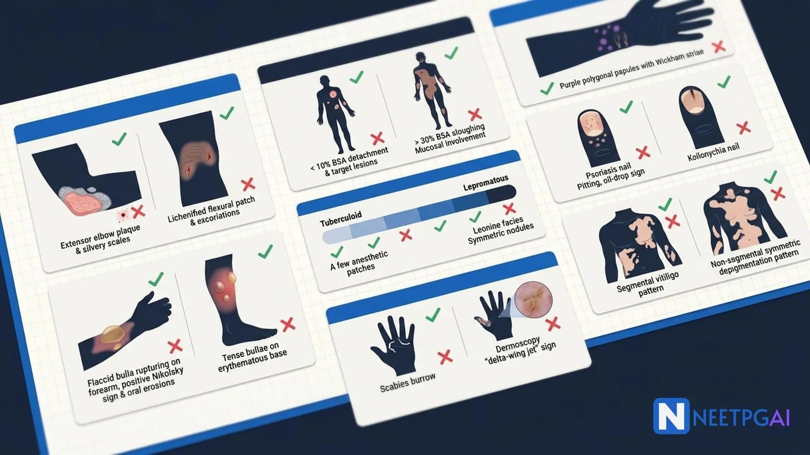
- Do not confuse psoriasis vs eczema — extensor vs flexural; silvery scales vs lichenification; Auspitz vs nothing
- Locate the pityriasis rosea herald patch correctly — single oval scaly plaque on trunk, days to weeks before the Christmas-tree distribution
- Recognise the fixed-drug eruption recurrence pattern — same site every time the offending drug is given
- Remember the 6 P's of lichen planus — Purple, Polygonal, Pruritic, Papular, Planar, Plaques (and Wickham striae)
- Distinguish pemphigus vulgaris from bullous pemphigoid — flaccid vs tense, Nikolsky positive vs negative, intra-epidermal vs sub-epidermal
- Apply the SJS vs TEN BSA cutoffs — under 10 percent vs over 30 percent vs the 10-30 percent overlap zone
- Master the Ridley-Jopling leprosy spectrum — TT to LL, AFB load, Mitsuda reaction, lepra reactions
- Distinguish segmental from non-segmental vitiligo — dermatomal unilateral vs symmetric bilateral
- Differentiate dermatophyte infections from intertrigo — KOH-positive scaling vs satellite lesions/yeast
- Grade acne accurately and respect isotretinoin contraindications — pregnancy is absolute
- Distinguish scabies from prurigo nodularis — web-space burrows vs isolated pruritic nodules
- Apply the ABCDE rule for skin cancer — Asymmetry, Border, Colour, Diameter, Evolving
- Recognise hair loss patterns — androgenetic, telogen effluvium, alopecia areata, scarring
- Read nail signs in systemic disease — clubbing, koilonychia, Beau lines, splinter haemorrhages, Mees lines
Why dermatology mistakes are costly
Dermatology sits at the intersection of medicine, paediatrics, microbiology, and pathology. A single mislabelled bullous disorder or missed SCORTEN risk on an MCQ can cost 1-2 marks; in practice, missing the diagnosis can mean blindness (SJS/TEN), permanent disfigurement (untreated leprosy), or missed systemic disease (porphyria, autoimmune connective tissue disease, internal malignancy). NEET PG and INI-CET examiners use dermatology vignettes to test pattern recognition (skin findings + systemic associations), management decisions (drug withdrawal, immunosuppression, antibiotics), and high-yield trivia (classification spectrums, signs, biopsy findings).
The 14 mistakes below come from analysis of NEET PG 2019-2024 dermatology questions and represent the most frequent error patterns.
Mistake 1: Confusing psoriasis and chronic eczema
What students do: Memorise both as "itchy scaly skin disease" without locking in the distinguishing features.
Why it is wrong: NEET PG specifically tests location, scale type, biopsy findings, and Auspitz sign. Calling a salmon-pink extensor plaque "eczema" is a one-mark loss.
Correct approach:
| Feature | Psoriasis | Chronic eczema |
|---|
| Location | EXTENSOR (elbows, knees, scalp, sacrum, nails) | FLEXURAL (popliteal, antecubital, neck, eyelids) in atopic |
| Lesion | Well-demarcated salmon-pink plaque with silvery scales | Poorly demarcated lichenified skin with excoriations |
| Scale | Silvery, removable | Crust + lichenification (thickened with exaggerated markings) |
| Auspitz sign | POSITIVE (pinpoint bleeding on scale removal) | Negative |
| Koebner phenomenon | POSITIVE | Variable |
| Itching | Variable, often mild-moderate | Severe, often nocturnal |
| Nail involvement | Pitting, oil-drop sign, onycholysis | Usually spared |
| Joint involvement | Psoriatic arthritis in 10-30 percent | None |
| Histology | Parakeratosis, Munro microabscesses, acanthosis, dilated dermal capillaries | Hyperkeratosis with parakeratosis, acanthosis, spongiosis (intercellular oedema) |
| Triggers | Stress, strep throat, drugs (beta-blockers, lithium), trauma | Allergens, irritants, atopy |
How to remember: Psoriasis = pinpoint bleeding (Auspitz), peeled silvery scales, extensor distribution. Eczema = excoriations and lichenification at flexural surfaces.
Mistake 2: Misplacing the pityriasis rosea herald patch
What students do: Either miss the herald patch (and date the rash from the secondary eruption) or place it on the wrong body region.
Why it is wrong: NEET PG tests the herald patch as a discriminator from secondary syphilis and tinea corporis.
Correct approach:
- Herald patch appears 1-2 weeks before the generalised eruption
- Location — TRUNK (chest, abdomen, back) most commonly; occasionally proximal limbs
- Appearance — single, oval, salmon-pink scaly plaque, 2-5 cm in diameter, with central clearing and a fine collarette of scale at the periphery (centripetal scale)
- Secondary eruption — multiple smaller oval plaques on the trunk arranged along skin lines in a "Christmas tree" distribution on the back
- Long axis of secondary plaques follows the lines of Langer/skin cleavage
- Self-limiting — resolves in 6-8 weeks; treatment supportive (emollients, topical steroids, antihistamines for itch)
- Always consider secondary syphilis in the differential — VDRL/RPR if patient is sexually active, rash on palms and soles, lymphadenopathy
- Aetiology — HHV-6 and HHV-7 reactivation in genetically predisposed individuals
How to remember: HERALD = first, big, oval, trunk; then Christmas tree on back.
Mistake 3: Forgetting the fixed-drug eruption recurrence pattern
What students do: Confuse fixed-drug eruption with other drug rashes (urticaria, exanthematous, SJS).
Why it is wrong: Fixed-drug eruption recurs at the SAME site every time the offending drug is given — pathognomonic and frequently tested.
Correct approach:
- Classic presentation — well-demarcated round/oval dusky red to violaceous plaque with central darkening, sometimes with central bullae
- Site — commonly genitals, lips, hands, but anywhere
- Recurrence at SAME site with each re-exposure to the drug; new sites may also appear
- Common culprits — paracetamol, NSAIDs (especially metamizole), sulfonamides, tetracyclines, barbiturates, oral contraceptives
- Histology — basal vacuolar degeneration with melanin incontinence into the upper dermis (gives the post-inflammatory hyperpigmentation), interface dermatitis with apoptotic keratinocytes
- Post-inflammatory hyperpigmentation persists for months at the site after resolution — a key clinical clue when the patient is asymptomatic
- Management — STOP the offending drug; topical or short-course oral steroids; reassurance about pigmentation
- Generalised bullous fixed-drug eruption can mimic SJS/TEN — careful drug history is critical
How to remember: FIXED = same site every time, dusky/violaceous round plaque, post-inflammatory hyperpigmentation.
Mistake 4: Misnaming the lichen planus features
What students do: Remember "P-something" but not all 6 Ps, or confuse Wickham striae with Koplik spots.
Why it is wrong: The 6 Ps mnemonic is a direct exam question. Wickham striae are pathognomonic.
Correct approach — the 6 Ps:
- Purple (violaceous)
- Polygonal
- Pruritic
- Papular
- Planar (flat-topped)
- Plaques
Additional features:
- Wickham striae — fine white lacy lines on the surface of papules (pathognomonic; mucosal LP shows them on buccal mucosa)
- Distribution — flexural surfaces (wrists, forearms, ankles), mucosal (oral, genital), nails (longitudinal ridging, pterygium formation)
- Koebner phenomenon — positive
- Aetiology — idiopathic; associations with hepatitis C, drugs (gold, antimalarials, beta-blockers, thiazides, ACE inhibitors)
- Histology — saw-tooth rete ridges, hyperkeratosis with hypergranulosis (no parakeratosis), basal vacuolar degeneration, band-like (lichenoid) lymphocytic infiltrate at the dermo-epidermal junction, Civatte bodies (apoptotic keratinocytes)
- Oral LP has 6 patterns — reticular (commonest), papular, plaque-like, atrophic, erosive, bullous; erosive oral LP has a 1-3 percent risk of squamous cell carcinoma transformation (frequently tested)
- Treatment — topical/intralesional steroids, oral steroids for severe, topical calcineurin inhibitors, phototherapy for resistant cases
How to remember: 6 Ps + Wickham striae + saw-tooth rete + band infiltrate.
Mistake 5: Confusing pemphigus vulgaris and bullous pemphigoid
What students do: Memorise one as "PEMphigus" with bullae and forget the rest; mix up Nikolsky positivity and DIF patterns.
Why it is wrong: NEET PG asks about clinical signs, histology, DIF, and treatment for both — getting them swapped costs 2-3 marks.
Correct approach:
| Feature | Pemphigus vulgaris | Bullous pemphigoid |
|---|
| Age | Middle age (40-60) | Elderly (over 60) |
| Mucosa | Affected in 50-70 percent (oral first) | Usually spared |
| Bulla type | FLACCID (rupture easily, leave painful erosions) | TENSE (do not rupture easily) |
| Background skin | Normal-appearing | Erythematous, urticarial |
| Nikolsky sign | POSITIVE | NEGATIVE |
| Asboe-Hansen sign | POSITIVE | Negative |
| Histology | INTRA-epidermal suprabasal bulla, ACANTHOLYSIS, Tzanck cells | SUB-epidermal bulla, EOSINOPHIL-rich infiltrate |
| DIF pattern | IgG + C3 in fishnet/chicken-wire pattern AROUND keratinocytes | LINEAR IgG + C3 along basement membrane |
| Antibody target | Desmoglein 1 and 3 (desmosomes) | BP180 and BP230 (hemidesmosomes) |
| Mortality | High if untreated (10-15 percent even with treatment) | Lower; chronic course |
| Treatment | Systemic steroids, rituximab, azathioprine, mycophenolate, IVIG, plasmapheresis | Steroids (oral or topical clobetasol), tetracyclines + nicotinamide, immunosuppressants |
How to remember: PEMphigus = Painful Erosions in Mouth, intra-epidermal, Nikolsky positive. PEMPHIGOID = Pemphigus-like but Outside (sub-epidermal), tense bullae, Nikolsky negative.
Mistake 6: Confusing Stevens-Johnson Syndrome and Toxic Epidermal Necrolysis
What students do: Use SJS and TEN interchangeably or get the BSA cutoffs wrong.
Why it is wrong: The cutoffs determine management intensity, mortality risk, and the SCORTEN score.
Correct approach:
| Feature | SJS | SJS-TEN overlap | TEN |
|---|
| BSA detached/detachable | Under 10 percent | 10-30 percent | Over 30 percent |
| Mortality | 5-10 percent | 25-35 percent | 30-50 percent |
| Triggers | Drugs (90 percent) — allopurinol, sulfonamides, anticonvulsants (carbamazepine, phenytoin, lamotrigine), NSAIDs, abacavir | Same | Same |
| Prodrome | Fever, flu-like illness, sore throat for 1-3 days | Same | Same |
| Skin | Target lesions, painful dusky macules progressing to bullae and detachment; Nikolsky positive | Same | Same |
| Mucosa | Always involved (oral, ocular, genital) | Same | Same |
| Histology | Full-thickness epidermal necrosis, sub-epidermal split | Same | Same |
SCORTEN score — calculated within 24 hours of admission; 7 criteria, each worth 1 point. Age over 40, malignancy, heart rate over 120, BSA over 10 percent on day 1, urea over 28 mg/dL (10 mmol/L), glucose over 252 mg/dL (14 mmol/L), bicarbonate under 20 mmol/L. Higher score = higher mortality.
Management priorities (in order):
- STOP the offending drug IMMEDIATELY — single most impactful intervention
- ICU or burn-unit admission for severe cases
- Fluid and electrolyte management — large insensible losses through detached skin (1-2 mL/kg/percent BSA detached)
- Wound care — non-adherent dressings, no debridement of attached skin, sterile environment
- Ophthalmology consultation early — corneal ulceration causes blindness; daily lubrication, lysis of synechiae
- Nutritional support — NG tube if oral mucosa involved
- Infection surveillance — sepsis is the commonest cause of death
- Immunomodulation — controversial; IVIG, cyclosporine, etanercept in some protocols; high-dose steroids no longer routinely recommended (may worsen sepsis risk)
How to remember: 10-30-30 rule: under 10 percent = SJS, 10-30 percent = overlap, over 30 percent = TEN.
Mistake 7: Mis-classifying leprosy on the Ridley-Jopling spectrum
What students do: Remember tuberculoid and lepromatous but mix up borderline forms; forget the WHO operational classification used in India.
Why it is wrong: NEET PG tests both — Ridley-Jopling for pathology and immunology questions, WHO for management and public health.
Correct approach — Ridley-Jopling spectrum (1966):
| Form | Cell-mediated immunity | Lesions | AFB load | Mitsuda lepromin |
|---|
| TT (tuberculoid) | Strong | 1-3 hypopigmented anesthetic plaques, elevated edges, well-demarcated | Scant or absent | Strongly positive |
| BT (borderline tuberculoid) | Good | A few plaques, may have satellite lesions | Few | Positive |
| BB (mid-borderline) | Unstable | Variable; may have "annular punched-out" lesions | Moderate | Equivocal |
| BL (borderline lepromatous) | Poor | Multiple, less well-defined plaques and nodules | Many | Weakly positive or negative |
| LL (lepromatous) | Poor | Multiple symmetrical macules, papules, nodules; leonine facies; madarosis (loss of eyebrows) | Abundant; foamy histiocytes (Virchow/lepra cells) | Negative |
Indeterminate (I) — single hypopigmented macule, ill-defined; early lesion that may resolve or progress to any pole.
Lepra reactions:
- Type 1 (reversal/upgrading or downgrading reaction) — in borderline forms (BT, BB, BL); altered cell-mediated immunity; existing lesions become erythematous and oedematous; treatment with steroids
- Type 2 (erythema nodosum leprosum, ENL) — in LL and BL; immune complex deposition; painful subcutaneous nodules, fever, neuritis, arthritis, iritis; treatment with steroids, thalidomide (contraindicated in pregnancy), clofazimine
WHO operational classification (1998), used in India under NLEP:
| Class | Criteria | MDT regimen | Duration |
|---|
| Paucibacillary (PB) | 1-5 lesions, smear negative | Rifampicin 600 mg monthly + dapsone 100 mg daily | 6 months |
| Multibacillary (MB) | over 5 lesions OR smear positive | Rifampicin 600 mg monthly + clofazimine 300 mg monthly and 50 mg daily + dapsone 100 mg daily | 12 months |
India-specific: Leprosy remains endemic in India; "elimination" (under 1 per 10,000) was declared in 2005 but pockets of high prevalence persist. NLEP provides free MDT.
Mistake 8: Confusing segmental and non-segmental vitiligo
What students do: Memorise "vitiligo = depigmentation" without specifying the subtype.
Why it is wrong: Subtype determines treatment response and prognosis.
Correct approach:
| Feature | Non-segmental vitiligo (NSV) | Segmental vitiligo (SV) |
|---|
| Distribution | Symmetric, bilateral; often acral, periorificial | Unilateral, along a dermatome |
| Onset | Any age | Childhood/adolescence |
| Course | Slowly progressive; new lesions over years | Rapid onset over 1-2 years then stabilises |
| Autoimmune associations | Common (thyroid, type 1 diabetes, alopecia areata, pernicious anaemia, Addison) | Rare |
| Response to phototherapy/topicals | Good | Less responsive |
| Surgical treatment | Reserved for stable disease | First-line option once stable |
| Koebner phenomenon | Common | Less common |
Treatment options:
- Topical corticosteroids for limited disease
- Topical calcineurin inhibitors (tacrolimus, pimecrolimus) for facial and intertriginous areas
- Narrowband UVB (NB-UVB) phototherapy — first-line for generalised vitiligo
- Excimer laser for limited lesions
- JAK inhibitors (ruxolitinib cream) — newer therapy
- Surgical — punch grafting, suction blister grafting, autologous melanocyte transplant for stable segmental disease
Mistake 9: Confusing tinea cruris with intertrigo
What students do: Diagnose any groin rash as "fungal" without doing KOH or considering bacterial/yeast intertrigo.
Why it is wrong: Treatment differs (antifungal vs antibacterial vs topical steroid + antifungal combination).
Correct approach:
| Feature | Tinea cruris | Candidal intertrigo | Bacterial intertrigo |
|---|
| Border | Active erythematous scaly border with central clearing | Beefy red, satellite pustules, NO central clearing | Erythematous, macerated, no specific pattern |
| Sites | Groin, inner thighs; spares scrotum | Skin folds (intertriginous areas including under breasts, axillae, groin, abdominal folds in obese) | Same as candida |
| Risk factors | Sweating, tight clothing, sharing towels | Diabetes, obesity, immunosuppression, antibiotics | Obesity, maceration |
| KOH | Septate branching hyphae | Pseudohyphae with budding yeasts | Negative |
| Treatment | Topical azole (clotrimazole, ketoconazole) or terbinafine for 2-4 weeks; oral terbinafine/itraconazole for extensive | Topical clotrimazole or nystatin; treat underlying diabetes | Topical antibacterial; address maceration; weight reduction |
Tinea incognito — tinea modified by inappropriate topical steroids; loses classical scaling and central clearing, becomes less itchy but more widespread. Common error in primary care.
Mistake 10: Mis-grading acne and forgetting isotretinoin contraindications
What students do: Lump all acne as "treat with topical retinoid" without grading; forget pregnancy contraindication.
Why it is wrong: Grading determines treatment intensity; pregnancy testing before and during isotretinoin is mandatory.
Correct approach — acne grading:
| Grade | Features | Treatment |
|---|
| Mild (Grade 1-2) | Comedones, few papules (under 30 total lesions) | Topical retinoid (adapalene, tretinoin) + benzoyl peroxide |
| Moderate (Grade 2-3) | Papules, pustules (30-100 lesions); some nodules | Add topical antibiotic (clindamycin) + oral antibiotic (doxycycline) if needed |
| Severe (Grade 3-4) | Multiple nodules, cysts (over 100 lesions); scarring | Oral isotretinoin 0.5-1 mg/kg/day for 16-20 weeks (cumulative 120-150 mg/kg) |
| Nodulocystic | Painful nodules and cysts, sinus tracts | Same as severe; consider intralesional steroids for individual nodules |
Isotretinoin essentials (NEET PG favourite):
- Absolute contraindication — PREGNANCY (teratogenic — craniofacial, cardiac, CNS, thymic anomalies)
- Pregnancy testing — negative test before starting, monthly during, and 1 month after stopping (US iPLEDGE programme; Indian protocols similar)
- Two forms of contraception required during and for 1 month after treatment
- Avoid blood donation during and 1 month after (teratogenicity in transfusion recipients)
- Lipid panel and LFTs at baseline, 1 month, 3 months
- Side effects — cheilitis (universal), dry skin, photosensitivity, raised triglycerides, transaminitis, mood changes (small risk of depression and suicidal ideation), pseudotumor cerebri (avoid combining with tetracyclines)
- Tetracyclines + isotretinoin = increased risk of pseudotumor cerebri; do not combine
Mistake 11: Mistaking scabies for prurigo nodularis or eczema
What students do: Diagnose itching with excoriations as "eczema" without examining for burrows or web-space lesions.
Why it is wrong: Untreated scabies is highly contagious; missing it spreads infection through families and hostels.
Correct approach:
- Itching — severe, NOCTURNAL (key feature)
- Distribution — web spaces of fingers, wrists (flexor surfaces), umbilicus, axillae, groin, genitals, breasts; face spared in adults; in INFANTS the face, palms, and soles are involved
- Burrows — 2-15 mm linear or S-shaped grey-white tracks in the skin; pathognomonic but often hard to find
- Other lesions — papules, vesicles, excoriations, nodules (post-scabietic), secondary impetiginisation
- Multiple household members itchy — major diagnostic clue
- Diagnosis — clinical; skin scraping from a burrow may show mite, eggs, faecal pellets (scybala) under microscopy; dermoscopy shows the "delta-wing jet" sign at the head of a burrow
- Norwegian (crusted) scabies — in immunocompromised (HIV, immunosuppressed, elderly debilitated); thick hyperkeratotic crusts with thousands of mites; HIGHLY contagious
- Treatment — topical permethrin 5 percent (apply head-to-toe, leave 8-12 hours, repeat in 7 days); alternatives — topical benzyl benzoate, oral ivermectin 200 mcg/kg single dose repeated in 7-14 days (mass treatment); treat ALL household and intimate contacts simultaneously; wash all clothes and bedding in hot water on the same day
- Post-scabietic itch can persist for 2-4 weeks despite cure; reassure and treat with topical steroids and antihistamines
Mistake 12: Forgetting the ABCDE rule for skin cancer
What students do: Recognise melanoma as "dark mole" without applying the systematic rule.
Why it is wrong: Early melanoma is curable; late melanoma has under 20 percent 5-year survival in stage IV.
Correct approach — ABCDE:
- A — Asymmetry — one half doesn't match the other
- B — Border — irregular, scalloped, poorly defined
- C — Colour — variation within the lesion (browns, blacks, reds, whites, blues)
- D — Diameter — over 6 mm (about the size of a pencil eraser); smaller melanomas exist but most are over 6 mm
- E — Evolving — changing in size, shape, colour; new symptoms (itching, bleeding)
Other skin cancers:
| Cancer | Features | Treatment |
|---|
| Basal cell carcinoma (BCC) | Pearly papule with telangiectasias; rolled border; rarely metastasises; sun-exposed areas (face) | Excision with 3-5 mm margin; Mohs micrographic surgery for face |
| Squamous cell carcinoma (SCC) | Hyperkeratotic erythematous nodule or ulcer; sun-exposed areas; immunosuppressed; can metastasise to LN | Excision with 4-6 mm margin; Mohs for high-risk |
| Melanoma | ABCDE features; Breslow thickness is key prognostic factor; lymph node involvement, distant metastasis | Excision with margins per Breslow; sentinel lymph node biopsy; immunotherapy (anti-PD-1) for advanced |
Breslow thickness margins:
- In situ — 5 mm
- Under 1 mm — 1 cm
- 1-2 mm — 1-2 cm
- Over 2 mm — 2 cm
Indian context: Melanoma is rare in dark skin (Indian skin types IV-V) but ACRAL LENTIGINOUS melanoma is the commonest subtype — appears on palms, soles, nails (subungual). Pigmented lesions on these sites should be examined carefully.
Mistake 13: Mis-identifying hair loss patterns
What students do: Diagnose all hair loss as "androgenetic" without examining for inflammation, scarring, or systemic illness.
Why it is wrong: Treatment, prognosis, and systemic associations differ dramatically.
Correct approach:
| Pattern | Distribution | Mechanism | Diagnostic clue | Treatment |
|---|
| Androgenetic alopecia (male/female) | Vertex + bitemporal recession in men (Hamilton-Norwood scale); diffuse central thinning in women (Ludwig scale) | Genetic + androgens; miniaturisation of follicles | Gradual onset, family history | Topical minoxidil 5 percent (M) / 2 percent (F); oral finasteride 1 mg (M only — not in pregnancy); hair transplant |
| Telogen effluvium | Diffuse thinning all over scalp | Stress (postpartum, surgery, illness, weight loss, drugs) shifts follicles into telogen | Diffuse + history of stressor 2-3 months earlier; positive hair pull test (over 6 hairs) | Treat underlying cause; reassurance (resolves in 6-12 months) |
| Alopecia areata | Discrete, well-defined round patches; "exclamation mark" hairs at edges | Autoimmune | Sudden onset; nail pitting; alopecia totalis (entire scalp), alopecia universalis (entire body) | Topical/intralesional steroids; topical immunotherapy; JAK inhibitors |
| Scarring (cicatricial) alopecia | Patchy with loss of follicular ostia; permanent | Discoid lupus, lichen planopilaris, frontal fibrosing alopecia, dissecting cellulitis | Loss of follicular openings on dermoscopy; biopsy needed | Treat underlying inflammation; transplant after disease quiescent |
| Anagen effluvium | Sudden diffuse loss including eyebrows | Chemotherapy, radiation | Within days-weeks of insult | Recovers after stopping cause |
| Trichotillomania | Bizarre patches, different hair lengths | Self-induced hair pulling | Hairs of different lengths, broken hairs | Behavioural therapy, SSRIs |
| Tinea capitis | Patchy with scaling, broken hairs ("black dot"), kerion (boggy mass) | Dermatophyte (Trichophyton, Microsporum) | KOH positive; Wood lamp fluorescence for Microsporum | Oral terbinafine/griseofulvin |
Mistake 14: Missing nail signs of systemic disease
What students do: Examine the nails superficially or not at all.
Why it is wrong: Nail changes are pathognomonic for many systemic conditions — clubbing, koilonychia, Beau lines, splinter haemorrhages, leukonychia.
Correct approach — key nail signs:
| Sign | Description | Causes |
|---|
| Clubbing | Loss of Lovibond angle (over 180 degrees); Schamroth window obliterated | Lung cancer, bronchiectasis, cystic fibrosis, IBD, cyanotic heart disease, cirrhosis, infective endocarditis |
| Koilonychia (spoon nails) | Concave nail plate | Iron deficiency anaemia (commonest), Plummer-Vinson, haemochromatosis, hypothyroidism |
| Beau lines | Transverse depressions across nail | Severe illness 2-3 months earlier (fever, surgery, chemotherapy, stress) |
| Onycholysis | Distal separation of nail from bed | Psoriasis, fungal, thyroid disease (hyperthyroidism — Plummer nails), trauma, drugs |
| Splinter haemorrhages | Linear dark lines under nail | Infective endocarditis, trauma, vasculitis |
| Mees lines | TRANSVERSE WHITE lines | Arsenic poisoning, thallium poisoning, severe illness |
| Muehrcke lines | TWO transverse WHITE bands (paired) | Hypoalbuminaemia (nephrotic, cirrhosis) |
| Terry nails | White nails with normal pink distal band | Cirrhosis, CHF, diabetes, ageing |
| Half-and-half nails (Lindsay) | Proximal white, distal pink/red | Chronic kidney disease |
| Yellow nails | Thick, yellow, slow-growing | Yellow nail syndrome (lymphedema + pleural effusion + chronic lung disease) |
| Periungual telangiectasias | Dilated capillaries at nail fold | Dermatomyositis, scleroderma, lupus |
| Subungual hyperkeratosis + pitting + oil-drop sign | Combo | Psoriasis |
| Beau line + onycholysis in fingernails + face changes | Combo | Erythroderma, severe systemic illness |
Habit-tic deformity, hangnail, paronychia are local conditions but worth noting.
Final summary
Dermatology rewards systematic visual pattern recognition more than any other NEET PG subject. The 14 mistakes above are the highest-yield errors to fix. The discipline is:
- Always classify the morphology first — papule, plaque, vesicle, bulla, pustule, macule, nodule, ulcer, scale
- Then note distribution — extensor vs flexural, sun-exposed vs covered, acral vs central, dermatomal vs symmetric
- Apply named signs and classifications — Auspitz, Koebner, Nikolsky, Ridley-Jopling, ABCDE
- Integrate with systemic clues — fever, drug history, family history, immunosuppression, mucosal involvement
- Order targeted investigations — KOH, Tzanck, biopsy with H&E + DIF, patch testing, serology
- Pair with a 100-image dermatology atlas — drill 5-10 images daily for 2 weeks before mock tests
Build a personal photo album of dermatology lesions you have seen during clinical rotations or read about — the visual memory is far more durable than text alone. Pair this guide with the psoriasis vs eczema clinical case and the bullous disorders deep-dive when those land in your prep queue.
Frequently Asked Questions
How many dermatology questions appear in NEET PG?
Dermatology contributes 6-8 questions per NEET PG paper (2021-2024 paper analysis), often overlapping with internal medicine, paediatrics, and microbiology. Question themes cluster around papulosquamous disorders (psoriasis vs eczema vs pityriasis rosea vs lichen planus), bullous disorders (pemphigus vulgaris vs bullous pemphigoid), severe cutaneous adverse reactions (SJS vs TEN vs DRESS), infections (leprosy Ridley-Jopling spectrum, scabies, dermatophytes), pigmentary disorders (vitiligo types), acne and its management, hair loss patterns (androgenetic, telogen effluvium, alopecia areata), and nail signs in systemic disease (clubbing, koilonychia, Beau lines, splinter hemorrhages). The 14 mistakes in this guide cover roughly 60-70 percent of typical dermatology question failures.
What is the difference between psoriasis and chronic eczema on examination?
Psoriasis lesions are well-demarcated, salmon-pink, raised plaques with silvery scales, typically located on extensor surfaces (elbows, knees, scalp, sacrum, nails). Auspitz sign is positive (pinpoint bleeding on scale removal due to suprapapillary thinning). Koebner phenomenon occurs (new lesions in areas of trauma). Itching is variable and often mild. Nail pitting, oil-drop sign, and onycholysis are common. Histology shows parakeratosis, Munro microabscesses, acanthosis with elongation of rete ridges, and dilated dermal capillaries. Chronic eczema (lichen simplex chronicus or chronic dermatitis) is poorly demarcated, with lichenification (thickened skin with exaggerated skin markings from chronic scratching), excoriations, fissuring, and crusts. It typically affects flexural areas (popliteal and antecubital fossa, neck) in atopic eczema. Itching is severe and often nocturnal. Histology shows hyperkeratosis with parakeratosis, acanthosis, spongiosis (intercellular oedema), and a perivascular lymphocytic infiltrate. The distinguishing exam question is location (extensor vs flexural), scale type (silvery vs lichenified), Auspitz sign, and itching severity.
How do you differentiate pemphigus vulgaris from bullous pemphigoid clinically and on biopsy?
Pemphigus vulgaris (PV) and bullous pemphigoid (BP) are both autoimmune blistering disorders but with key differences. Pemphigus vulgaris: middle-aged adults (40-60), affects mucosa first in 50-70 percent (oral erosions for months before skin lesions), flaccid bullae that rupture easily leaving painful erosions, Nikolsky sign POSITIVE (light shear pressure on normal-appearing skin produces blister), Asboe-Hansen sign positive (existing bullae extend laterally with pressure). Histology shows intra-epidermal suprabasal bullae with acantholysis (loss of cell-cell adhesion, free-floating keratinocytes called Tzanck cells). Direct immunofluorescence (DIF) shows IgG and C3 in a fishnet/chicken-wire pattern around keratinocytes (anti-desmoglein 1 and 3 antibodies). Mortality is significant if untreated (10-15 percent even with treatment). Bullous pemphigoid: elderly patients (over 60), mucosa spared in most cases, TENSE bullae on erythematous urticarial base that do NOT rupture easily, Nikolsky sign NEGATIVE. Histology shows subepidermal bullae with eosinophil-rich infiltrate. DIF shows linear IgG and C3 along the basement membrane (anti-BP180 and BP230 antibodies). Better prognosis than PV. Treatment of both involves immunosuppression (steroids, rituximab, azathioprine) but doses and approach differ.
What is the difference between Stevens-Johnson syndrome and toxic epidermal necrolysis, and what is the management priority?
SJS and TEN are severe cutaneous adverse reactions on a spectrum, both characterised by skin and mucosal sloughing. The distinction is BODY SURFACE AREA (BSA) involvement of detached or detachable skin. SJS: under 10 percent BSA. SJS-TEN overlap: 10-30 percent BSA. TEN: over 30 percent BSA. Both share triggers (drugs are commonest: allopurinol, sulfonamides, anticonvulsants like carbamazepine/phenytoin/lamotrigine, NSAIDs, abacavir), Nikolsky sign POSITIVE, mucosal involvement (oral, ocular, genital), and high mortality (SJS 5-10 percent, TEN 30-50 percent). Histology shows full-thickness epidermal necrosis. Management priorities, in order: (1) STOP the offending drug immediately (this is the single most impactful intervention; delay raises mortality); (2) ICU or burn-unit admission for severe cases; (3) fluid and electrolyte management (large insensible losses through detached skin); (4) wound care (non-adherent dressings, no debridement of attached skin, sterile environment); (5) ophthalmology consultation early (corneal ulceration causes blindness); (6) nutritional support (NG tube if oral mucosa involved); (7) infection surveillance (sepsis is the commonest cause of death); (8) immunomodulation is controversial (IVIG, cyclosporine, etanercept in some protocols; high-dose steroids no longer routinely recommended). SCORTEN score predicts mortality.
How is the Ridley-Jopling classification of leprosy structured, and what is the WHO operational classification used in India?
The Ridley-Jopling classification (1966) categorises leprosy along an immunological spectrum based on clinical, bacteriological, histological, and immunological features. Five groups: Tuberculoid (TT) — strong cell-mediated immunity, 1-3 hypopigmented anesthetic plaques with elevated edges, well-demarcated, AFB scant or absent, granulomas in dermis without bacilli, Mitsuda lepromin reaction strongly positive. Borderline tuberculoid (BT). Mid-borderline (BB). Borderline lepromatous (BL). Lepromatous (LL) — poor cell-mediated immunity, multiple symmetrical lesions, leonine facies, loss of eyebrows (madarosis), nodules, AFB abundant in lesions, foamy histiocytes (Virchow cells, lepra cells), Mitsuda reaction negative. Indeterminate is a polar early lesion that may progress to any group. Reactions: Type 1 (lepra reaction) — upgrading or downgrading reactions in borderline forms; Type 2 (erythema nodosum leprosum, ENL) — in LL/BL forms, painful nodules with systemic features. The WHO operational classification (1998, used in India under the NLEP) is simpler: Paucibacillary (PB) — 1-5 lesions, skin smear negative, treated with MDT-PB regimen (rifampicin monthly + dapsone daily) for 6 months. Multibacillary (MB) — over 5 lesions OR skin smear positive, treated with MDT-MB regimen (rifampicin monthly + clofazimine monthly and daily + dapsone daily) for 12 months. NEET PG tests both — Ridley-Jopling for histopathology and immunology questions, WHO for management and public health questions.
This content is for educational purposes for NEET PG exam preparation. It is not a substitute for professional medical advice, diagnosis, or treatment. Clinical information has been reviewed by qualified medical professionals.
Written by: NEETPGAI Editorial Team
Reviewed by: Pending SME Review
Last reviewed: May 2026