mistake guide
ophthalmology
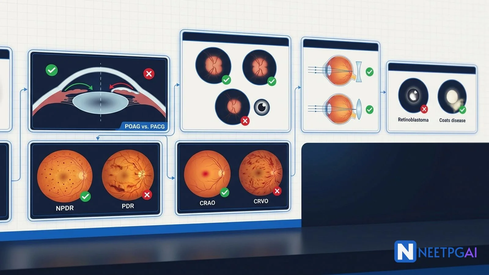
glaucoma
diabetic retinopathy
papilledema
CRAO CRVO
NEET PG 2026
Avoid the costliest ophthalmology mistakes in NEET PG 2026: POAG vs PACG, diabetic retinopathy stages, papilledema vs papillitis, CRAO vs CRVO, refractive errors, leukocoria, anisocoria.
Version 1.0 — Published May 2026
Ophthalmology contributes 10-15 questions in NEET PG, with predictable high-yield clusters in glaucoma, diabetic retinopathy, neuro-ophthalmology, and vascular occlusions. The 14 most expensive mistakes cluster around classification, side-of-pathology, and treatment specifics. To protect your marks:
Ophthalmology questions in NEET PG are often layered — a single fundoscopy image can carry diagnosis, severity grading, and management as three separate testable points. Errors cascade: misclassifying NPDR as PDR drives the wrong management (PRP vs observation); calling papillitis "papilledema" leads to chasing raised ICP instead of starting IV steroids for optic neuritis.
The 14 mistakes below come from analysis of NEET PG 2019-2024 ophthalmology questions and represent the most frequently incorrect answer patterns. For depth on case-based ophthalmology reasoning, pair this with the high-yield diagrams of the anterior chamber angle, the OCT cross-sections of diabetic macular edema, and the fundus photo atlas of vascular occlusions.
What students do: Treat all glaucoma as the same disease and start prostaglandin drops as first-line for every case.
Why it is wrong: POAG and PACG have fundamentally different anatomy, presentation, and management. PACG with a narrow angle worsens on certain drugs (e.g., topical mydriatics, anticholinergics, sympathomimetics) and the first definitive treatment is laser peripheral iridotomy, NOT drops alone.
Start practicing NEET PG MCQs with AI-powered explanations.
Start Free PracticeMaster GI secretions, digestion, absorption transporters, motility patterns, and gut hormones with high-yield NEET PG 2026 traps and India-context examples.
Master labor stages, Friedman vs Zhang curves, WHO partograph, AMTSL, episiotomy and India JSY/LaQshya policies for NEET PG 2026 OBG MCQs.
5 anterior segment ophthalmology image MCQs for NEET PG: hypopyon and Behcet, Kayser-Fleischer ring in Wilson, Brushfield spots in Down, corneal arcus, and pterygium vs pinguecula.
Daily MCQs, study tips, and topper strategies on Telegram.
Join on Telegram →Correct approach — POAG vs PACG comparison:
| Feature | POAG | PACG |
|---|---|---|
| Angle | Open (gonioscopy: trabecular meshwork visible) | Closed or narrow (iris in apposition with TM) |
| Anterior chamber depth | Normal | Shallow |
| Onset | Chronic, asymptomatic | Acute (severe pain, halos) or chronic |
| Symptoms in acute | Usually none | Severe eye pain, headache, nausea, vomiting, halos, blurred vision |
| Pupil | Normal | Mid-dilated, non-reactive (acute) |
| IOP | Mildly to moderately raised (22-35) | Markedly raised in acute (often more than 40) |
| Cornea | Clear | Cloudy/edematous (acute) |
| Fellow eye | Similar | Shallow AC, predisposed |
| First-line treatment | Prostaglandin analogue eye drops | Acute: IV mannitol + acetazolamide + pilocarpine 2 percent + timolol + corticosteroid drops; definitive: laser peripheral iridotomy in both eyes |
| Surgical option | Trabeculectomy if drops fail | Iridotomy/iridectomy; trabeculectomy if combined |
How to remember it correctly: "OPEN → drops" and "CLOSED → laser iridotomy." For any glaucoma question, the first move is to identify whether the angle is open or closed; the entire management ladder flows from that.
What students do: Use vague terms like "mild diabetic retinopathy" without applying the 4-2-1 rule, or treat NPDR as PDR.
Why it is wrong: Diabetic retinopathy classification drives treatment decisions. Severe NPDR (4-2-1 positive) often needs early PRP; mild NPDR is observed. CSME is a separate axis that triggers anti-VEGF treatment regardless of NPDR/PDR stage.
Correct approach — Diabetic retinopathy classification (ETDRS / International Clinical):
| Stage | Findings | Management |
|---|---|---|
| No DR | None | Annual fundus screening |
| Mild NPDR | Microaneurysms only | Annual screening |
| Moderate NPDR | More than mild but less than severe | 6-month screening; tighten glycemic control |
| Severe NPDR (4-2-1 rule) | Any one: diffuse intraretinal hemorrhages in 4 quadrants OR venous beading in 2 quadrants OR IRMA in 1 quadrant | Consider early PRP especially in type 2 DM, single eye, or compliance issues |
| Very severe NPDR | Any 2 of 3 of the 4-2-1 features | Strong indication for PRP |
| Early PDR | NVD/NVE but not high-risk | PRP indicated |
| High-risk PDR | NVD > 1/3 disc area OR any NVD + vitreous hemorrhage OR NVE > 1/2 disc area + vitreous hemorrhage | PRP urgently |
| Advanced PDR | Vitreous hemorrhage, tractional retinal detachment, neovascular glaucoma | Vitrectomy if hemorrhage doesn't clear or detachment threatening macula |
Diabetic macular edema (CSME — ETDRS definition):
Treatment of CSME:
How to remember it correctly: 4-2-1 rule for severe NPDR. High-risk PDR triggers urgent PRP. CSME triggers anti-VEGF — separately from NPDR/PDR staging.
What students do: Call all optic disc swelling "papilledema" and refer for MRI brain regardless.
Why it is wrong: Papilledema is bilateral disc swelling from raised ICP; papillitis (anterior optic neuritis) is inflammation of the optic nerve head. They have different investigations and treatments — and visual acuity is the discriminator.
Correct approach — Papilledema vs papillitis:
| Feature | Papilledema | Papillitis |
|---|---|---|
| Cause | Raised intracranial pressure | Optic nerve inflammation (demyelination, infection, ischemia) |
| Laterality | Bilateral (usually symmetric) | Unilateral or bilateral |
| Visual acuity | Preserved early (may be transient obscurations) | Markedly reduced |
| RAPD | Absent (bilateral disease) | Present in unilateral cases |
| Colour vision | Normal | Reduced (red desaturation) |
| Eye pain on movement | Absent | Present (typical optic neuritis) |
| Visual field | Enlarged blind spot, peripheral constriction | Central scotoma, altitudinal defect |
| Fundus | Bilateral disc swelling, Paton's lines, peripapillary hemorrhages | Unilateral disc swelling with hyperemia, blurred margins |
| Headache | Worse on waking, with vomiting | Absent unless from inflammation |
| Investigations | MRI brain (+/− MR venography), LP for opening pressure | MRI brain + orbits with contrast |
| Treatment | Lower ICP (treat cause: tumor, IIH, venous sinus thrombosis) | IV methylprednisolone 1 g/day × 3 days then oral taper (ONTT for typical optic neuritis) |
Practical clue: Ask "Can the patient see?" — if vision is preserved with disc swelling, think papilledema. If vision is markedly reduced with disc swelling, think papillitis.
How to remember it correctly: "Papilledema = pressure, vision preserved. Papillitis = pathology of the nerve, vision poor." RAPD + reduced VA + pain on eye movement = optic neuritis until proven otherwise.
What students do: Call any CDR > 0.3 "glaucomatous" without considering symmetry, family history, IOP, and visual field.
Why it is wrong: Physiological cupping can reach CDR 0.5-0.6 in some healthy eyes. Glaucomatous cupping has specific features beyond absolute CDR.
Correct approach — Glaucomatous vs physiological cupping:
| Feature | Physiological | Glaucomatous |
|---|---|---|
| CDR | Usually 0.3-0.5; up to 0.6 in some healthy | Often greater than 0.5; progressively increasing |
| Symmetry between eyes | Symmetric (within 0.2) | Asymmetric (more than 0.2 difference) |
| Neuroretinal rim | Follows ISNT rule (Inferior > Superior > Nasal > Temporal) | Loss of ISNT pattern; thinning in inferior/superior pole first |
| Disc hemorrhage | Absent | Present (Drance hemorrhage — small flame hemorrhage at disc margin) |
| Visual field | Normal | Glaucomatous defects (nasal step, arcuate scotoma, paracentral scotoma) |
| OCT RNFL | Normal for age | Thinning in superior/inferior bundle |
| Progression | Stable over years | Progressive over months-years |
Red flags for glaucoma: asymmetric CDR (greater than 0.2 difference between eyes), CDR greater than 0.7, loss of ISNT rule, disc hemorrhage, visual field defect, IOP greater than 21.
How to remember it correctly: Cupping alone is not glaucoma; it is CDR + symmetry + ISNT rule + IOP + visual field + OCT RNFL that together diagnose glaucoma.
What students do: Confuse myopia and hyperopia signs, or get the cylindrical axis wrong.
Why it is wrong: The conventions are arbitrary but examined precisely. Getting the sign wrong is a marks-loss without nuance.
Correct approach — Refractive error conventions:
| Error | Optical defect | Lens correction | Sign |
|---|---|---|---|
| Myopia | Eye too long or refractive power too high; light focuses in front of the retina | Concave (diverging) lens | Minus (−) |
| Hyperopia | Eye too short or refractive power too low; light focuses behind the retina | Convex (converging) lens | Plus (+) |
| Astigmatism | Unequal refractive power in different meridians | Cylindrical lens at the appropriate axis (1-180 degrees) | Plus or minus cylinder + axis |
| Presbyopia | Loss of accommodation with age (over 40) | Reading add (plus) over distance correction | Plus |
| Aphakia | Absence of lens (post-cataract, no IOL) | Strong plus lens (around +10 D) | Plus |
| Pseudophakia | IOL in place | Variable; typically near-emmetropic | Variable |
Axis convention: Cylindrical axis is recorded in degrees from 1 to 180 measured anticlockwise from the patient's right side (TABO convention).
How to remember it correctly: "Minus for Myopia, Plus for Presbyopia and hyperopia." Convex lens converges; concave diverges.
What students do: Confuse esotropia (eye in) with exotropia (eye out), or fail to distinguish concomitant vs incomitant squint.
Why it is wrong: Strabismus terminology is testable verbatim. Wrong direction means wrong answer.
Correct approach — Strabismus types:
| Term | Direction | Example |
|---|---|---|
| Esotropia | Eye deviates inward (nasally) | Convergent squint |
| Exotropia | Eye deviates outward (temporally) | Divergent squint |
| Hypertropia | Eye deviates upward | Right eye higher than left |
| Hypotropia | Eye deviates downward | Right eye lower than left |
| Heterophoria | Latent squint manifest only on cover test | Esophoria, exophoria |
| Heterotropia | Manifest squint visible without cover test | Esotropia, exotropia |
| Concomitant | Same angle of deviation in all directions of gaze | Childhood squint |
| Incomitant (paralytic) | Angle varies with direction of gaze; worse in field of action of paralysed muscle | Cranial nerve palsy (III, IV, VI) |
Common pediatric squints:
How to remember it correctly: "Eso = In (nasally), Exo = Out (temporally)." Hyper = up, Hypo = down — by the eye that is higher/lower. Test for concomitance with the cover-uncover test in 9 cardinal positions.
What students do: Group all "sudden monocular vision loss" together and miss the cherry-red spot, blood-and-thunder fundus, or pale disc of GCA.
Why it is wrong: Treatments diverge sharply. Missing GCA risks blindness in the fellow eye within hours; CRAO has a narrow 90-minute window for ocular massage and IOP-lowering manoeuvres; CRVO needs anti-VEGF for macular edema and PRP for neovascularisation.
Correct approach — Sudden monocular vision loss differential:
| Feature | CRAO | CRVO | GCA (AAION) |
|---|---|---|---|
| Pain | Painless | Painless | Painful — temporal headache, scalp tenderness, jaw claudication |
| Age | Older adult (vascular) | Older adult (50+) | Over 50 (almost never under 50) |
| Onset | Sudden, often seconds-minutes | Sudden | Variable; may have prodromal amaurosis fugax |
| Visual acuity | Counting fingers or worse | Variable (better in non-ischemic, worse in ischemic) | Markedly reduced; may be no perception of light |
| RAPD | Present | Present in ischemic; minimal in non-ischemic | Present |
| Fundus | Cherry-red spot, pale retina, attenuated arterioles, 'boxcar' segmentation | Blood-and-thunder — diffuse intraretinal hemorrhages in all 4 quadrants, dilated tortuous veins, disc swelling, cotton wool spots | Pale swollen optic disc (arteritic AION) |
| Cause | Embolus (carotid, cardiac), thrombosis, GCA (rare), vasculitis | Venous thrombosis (atherosclerosis, hypercoagulable, glaucoma association) | Vasculitis of large/medium arteries (GCA) |
| Acute management | Ocular massage, AC paracentesis, breathe into a bag (CO2 vasodilation), IOP-lowering drops, hyperbaric O2 — narrow 90-min window | Anti-VEGF for macular edema, PRP for neovascular complications | IV methylprednisolone 1 g/day × 3 days then oral 60 mg/day; biopsy temporal artery within 1-2 weeks; never delay steroids waiting for biopsy |
| Investigations | Carotid Doppler, echocardiogram, ESR/CRP (to exclude GCA), lipid profile | OCT (macular edema), FFA, CBC, glucose, lipid, B12, homocysteine, antiphospholipid in young | ESR (usually over 50), CRP, temporal artery biopsy, FDG-PET, MRI/MRA of large vessels |
| Prognosis | Poor visual prognosis if not within 90 min | Variable; non-ischemic better than ischemic | Steroids prevent contralateral blindness but recovered vision rare |
Cherry-red spot differential (NEET PG favourite):
How to remember it correctly: Painless + cherry-red = CRAO. Painless + blood-and-thunder = CRVO. Painful + pale swollen disc + age over 50 + headache + raised ESR = GCA — start steroids first, biopsy later.
What students do: Quote "uncontrolled diabetes" or "active uveitis" as absolute contraindications.
Why it is wrong: Cataract surgery has almost NO absolute contraindications — most are timing issues (controlling co-morbid disease first), not lifetime exclusions.
Correct approach — Cataract surgery considerations:
Relative contraindications / cautions:
Indications timing changed in 2026:
Surgical techniques:
India-specific context: NPCB (National Programme for Control of Blindness, now NPCBVI) targets cataract surgery as the major blindness reduction strategy in India. Approximately 6-7 million cataract surgeries are performed annually in India under NPCBVI. The cataract surgical rate (CSR) is approximately 6,000-7,000 per million per year.
How to remember it correctly: Cataract surgery is rarely "contraindicated for life" — it is mostly "wait until co-morbidity is controlled." Timing, not exclusion.
What students do: Call every white pupil reflex (leukocoria) in a child "retinoblastoma" without considering the full differential.
Why it is wrong: Retinoblastoma is the dominant concern (because life-threatening) but Coats disease, persistent hyperplastic primary vitreous (PHPV), retinopathy of prematurity (ROP), congenital cataract, and toxocariasis can all present with leukocoria. Treatment differs.
Correct approach — Leukocoria differential:
| Condition | Age | Laterality | Features | Treatment |
|---|---|---|---|---|
| Retinoblastoma | Under 5 years (most under 3) | 60 percent unilateral, 40 percent bilateral | Calcifications on USG/CT/MRI; familial RB1 mutation in bilateral cases; metastatic risk | Chemoreduction + focal therapy (cryo, laser, brachytherapy); enucleation for advanced; never biopsy |
| Coats disease | Boys 6-8 years (peak); 80-90 percent male | Unilateral in 95 percent | Retinal telangiectasia with massive subretinal exudate (lipid); retinal detachment in advanced | Laser/cryotherapy for telangiectasia; anti-VEGF; surgery for retinal detachment |
| PHPV (Persistent Hyperplastic Primary Vitreous) | Newborn / infant | Usually unilateral | Microphthalmos, elongated ciliary processes, retrolental fibrovascular tissue | Surgical removal of fibrovascular mass; visual prognosis poor |
| ROP (Retinopathy of prematurity) | Preterm under 32 weeks / 1500 g | Bilateral | History of prematurity, supplemental oxygen; staged retinal vascular disease | Anti-VEGF or laser for threshold/Type 1 ROP |
| Congenital cataract | Newborn / infant | Variable | Lens opacity; may be syndromic (e.g., rubella) | Early surgery (within 6 weeks for visual development) + aphakic correction |
| Toxocariasis | Older child | Usually unilateral | Posterior pole granuloma or peripheral granuloma + intermediate uveitis; history of pica/dog contact | Anti-helminthic + steroids; vitrectomy for traction |
Key discriminator — calcifications on imaging: retinoblastoma has them (USG, CT); Coats disease does NOT have calcifications (subretinal lipid exudate, not calcium).
Retinoblastoma essentials:
How to remember it correctly: Leukocoria in a child is retinoblastoma until proven otherwise. But unilateral + boys + 6-8 years + no calcifications + massive subretinal exudate = Coats. Bilateral + preterm = ROP. Microphthalmos + unilateral newborn = PHPV.
What students do: Use these terms interchangeably.
Why it is wrong: They are distinct entities; one is purely conjunctival and benign, the other invades the cornea and may need surgery.
Correct approach:
| Feature | Pinguecula | Pterygium |
|---|---|---|
| Location | Bulbar conjunctiva, nasal or temporal limbus | Triangular fibrovascular growth extending from the conjunctiva onto the cornea |
| Corneal involvement | None | Yes — onto the cornea |
| Symptoms | Cosmetic only; occasional irritation | Foreign body sensation, redness, astigmatism if visual axis approached |
| Pathology | Elastotic degeneration of conjunctival collagen | Same elastotic degeneration + fibrovascular invasion of cornea |
| Risk factors | UV exposure, dust, wind | Same; "surfer's eye"; outdoor work |
| Treatment | Lubricants; rarely excision for cosmesis | Excision with conjunctival autograft (or mitomycin C) if approaching visual axis or causing significant astigmatism; high recurrence rate |
How to remember it correctly: Pterygium has a "wing" (Greek pteron) that flies onto the cornea. Pinguecula stays on the conjunctiva.
What students do: Call any unequal pupils "anisocoria" without working out which is the abnormal pupil and the underlying cause.
Why it is wrong: Three distinct causes (Horner syndrome, Adie tonic pupil, III nerve palsy) need different investigations and have very different prognostic implications.
Correct approach — Anisocoria differential:
Step 1: Identify which pupil is abnormal (compare in bright and dim light).
Step 2: Examine for associated signs.
| Cause | Pupil | Associated signs | Pharmacological test |
|---|---|---|---|
| Horner syndrome | Small (miosis), normal reaction to light and near | Partial ptosis, anhidrosis (face), apparent enophthalmos, heterochromia (congenital) | Apraclonidine 0.5 percent or cocaine — Horner pupil dilates with apraclonidine (paradoxical pupillary dilation), or fails to dilate with cocaine 4 percent |
| Adie tonic pupil | Large, sluggish or absent reaction to light, slow tonic response to near | Often young women; associated diminished deep tendon reflexes (Holmes-Adie syndrome) | Pilocarpine 0.125 percent (diluted) — denervation supersensitivity causes constriction (normal pupil does not constrict at this dilution) |
| III nerve palsy | Large, fixed (non-reactive to light), often associated with ptosis and down-and-out eye position | Diplopia, ptosis, limited adduction/elevation/depression | Imaging for compressive lesion (PCom artery aneurysm) urgently |
| Pharmacological mydriasis | Large, fixed; recent eye drop exposure (atropine, tropicamide) | History of eye drops or accidental exposure | Pilocarpine 1 percent — pharmacologically blocked pupil does NOT constrict; III nerve palsy pupil DOES constrict |
| Traumatic mydriasis | Large; irregular; history of trauma | Sphincter tears visible on slit lamp | History + slit lamp |
| Physiological anisocoria | Difference less than 1 mm; same in light and dim | None | None |
Horner syndrome localisation:
Adie's syndrome: Adie tonic pupil + diminished deep tendon reflexes; usually idiopathic; young women.
How to remember it correctly: Anisocoria worse in dim light = small pupil abnormal (Horner). Anisocoria worse in bright light = large pupil abnormal (Adie, III nerve, pharmacological). Always look for ptosis (ipsilateral = Horner or III nerve), and the III nerve palsy is a neurosurgical emergency (rule out PCom aneurysm).
What students do: Treat both with the same lubricant drops, or call all anterior segment irritation "dry eye."
Why it is wrong: Dry eye and blepharitis have overlapping symptoms but different mechanisms and treatments. Blepharitis untreated causes recurrent stye, chalazion, and corneal complications.
Correct approach:
| Feature | Dry eye disease | Blepharitis |
|---|---|---|
| Pathology | Decreased tear production (aqueous-deficient) or increased tear evaporation (meibomian gland dysfunction) | Inflammation of eyelid margins; anterior (lashes) or posterior (meibomian glands) |
| Symptoms | Burning, foreign body sensation, fluctuating vision, worse late in day | Burning, crusting on lashes on waking, recurrent stye/chalazion |
| Signs | Reduced Schirmer test, tear film break-up time, punctate epithelial erosions, conjunctival staining | Lid margin erythema, telangiectasia, collarettes around lashes, meibomian gland inspissation, frothy tears |
| Associations | Sjogren syndrome, autoimmune disease, hormonal changes, computer screen use | Rosacea, seborrhoeic dermatitis, atopic dermatitis, Demodex mite |
| Treatment | Artificial tears (preservative-free preferred), warm compresses, omega-3, punctal plugs, cyclosporine 0.05 percent drops (Restasis), lifitegrast, autologous serum tears (severe) | Lid hygiene (warm compresses + lid scrubs), antibiotic ointment (erythromycin or chloramphenicol) on lid margins, oral doxycycline for severe meibomian dysfunction, topical steroids for acute inflammation, tea tree oil for Demodex |
Sjogren syndrome: dry eyes + dry mouth + autoantibodies (anti-Ro/SSA, anti-La/SSB); refer to rheumatology.
How to remember it correctly: Dry eye = tear deficiency (treat the tear); blepharitis = lid inflammation (treat the lid). Many patients have both — treat both.
What students do: Quote a single CDR value as "normal" or "glaucoma" without comparing eyes or tracking change.
Why it is wrong: Progression matters more than the absolute number. CDR 0.6 with prior CDR 0.5 is glaucomatous progression even if the absolute value is borderline; CDR 0.6 stable over 5 years may be physiological.
Correct approach:
How to remember it correctly: Glaucoma is a clinical diagnosis combining CDR + symmetry + ISNT + IOP + visual field + OCT. A single number is rarely enough.
What students do: Refer pediatric strabismus too late, missing the visual development window.
Why it is wrong: Amblyopia (lazy eye) can develop in any child with persistent strabismus, anisometropia, or media opacity before age 7-8. Failure to treat in the critical window leaves permanent monocular vision loss.
Correct approach — pediatric strabismus workflow:
Critical age windows:
How to remember it correctly: Any child with squint, white pupil reflex, or asymmetric red reflex needs prompt ophthalmology referral. The amblyopia treatment window closes by school age — don't wait.
| Mistake | Quick fix |
|---|---|
| 1. POAG vs PACG | Gonioscopy + AC depth + open/closed = drops vs laser iridotomy |
| 2. NPDR vs PDR vs CSME | 4-2-1 rule, PRP for high-risk PDR, anti-VEGF for CSME |
| 3. Papilledema vs papillitis | VA preserved (papilledema) vs VA reduced (papillitis); MRI + LP vs IV steroids |
| 4. CDR interpretation | Symmetry, ISNT, progression, hemorrhage — not absolute number |
| 5. Refractive error signs | Myopia minus, hyperopia plus, presbyopia plus add |
| 6. Strabismus terminology | Eso = in, exo = out, hyper = up, hypo = down; concomitant vs incomitant |
| 7. CRAO vs CRVO vs GCA | Painless cherry-red, painless blood-and-thunder, painful pale disc + raised ESR |
| 8. Cataract surgery contraindications | Mostly timing, not absolute; optimise co-morbidities |
| 9. Leukocoria differential | Retinoblastoma until proven otherwise; calcifications differentiate from Coats |
| 10. Pterygium vs pinguecula | Pterygium invades cornea; pinguecula stays on conjunctiva |
| 11. Anisocoria | Dim light → Horner; bright light → Adie/III nerve; III nerve = neurosurgical emergency |
| 12. Dry eye vs blepharitis | Tear deficiency (tears) vs lid inflammation (lid hygiene) |
| 13. CDR progression | Track over time + ISNT + symmetry + visual field + OCT |
| 14. Pediatric strabismus | Amblyopia treatment window closes by age 7-8; refer early |
Seven recurring patterns. Recognise the pattern and the question collapses.
Pattern 1 — The acute red eye question: Severe eye pain + halos + nausea + raised IOP + shallow AC + mid-dilated pupil = acute angle closure glaucoma. First-line: IV mannitol + acetazolamide + pilocarpine 2 percent + timolol + steroid drops; definitive: laser peripheral iridotomy.
Pattern 2 — The diabetic retinopathy stage question: Apply the 4-2-1 rule for severe NPDR. NVD greater than 1/3 disc or NVE with vitreous hemorrhage = high-risk PDR — PRP urgently. CSME on its own = anti-VEGF.
Pattern 3 — The optic disc swelling question: Bilateral, vision preserved, no RAPD = papilledema (raised ICP) — MRI + LP. Unilateral, vision poor, RAPD, painful eye movement = papillitis (optic neuritis) — MRI + IV methylprednisolone (ONTT).
Pattern 4 — The sudden monocular vision loss question: Painless + cherry-red spot = CRAO (90-min window; ocular massage, AC paracentesis, IOP-lowering, hyperbaric O2). Painless + blood-and-thunder = CRVO (anti-VEGF + PRP). Painful + pale disc + raised ESR + age over 50 = GCA (IV methylprednisolone immediately; biopsy later).
Pattern 5 — The leukocoria question: Calcifications on USG/CT = retinoblastoma (chemoreduction + focal therapy; never biopsy). Unilateral + male + 6-8 years + massive subretinal exudate without calcifications = Coats disease (laser/cryotherapy).
Pattern 6 — The anisocoria question: Worse in dim light + ptosis + anhidrosis = Horner syndrome (work out the level — carotid dissection if Horner alone, Pancoast if anhidrosis present). Worse in bright light + sluggish tonic constriction in young woman = Adie pupil. Down-and-out eye + ptosis + dilated pupil = III nerve palsy with pupillary involvement (compressive — PCom aneurysm until proven otherwise; urgent imaging).
Pattern 7 — The pediatric strabismus / amblyopia question: Refractive correction + patching/atropine penalisation of better eye, before age 7-8. Accommodative esotropia often resolves with hyperopic correction; non-accommodative needs surgery.
High-yield one-liners:
Ophthalmology contributes 10-15 questions in NEET PG (based on 2021-2024 paper analysis), with predictable high-yield areas. The clusters are anterior segment (glaucoma, cataract, anterior uveitis) 3-4 questions; posterior segment (retina including diabetic retinopathy, vascular occlusions, retinal detachment, retinoblastoma) 3-4 questions; refractive errors and orbit 1-2; neuro-ophthalmology (papilledema vs papillitis, optic neuritis, anisocoria) 1-2; pediatric ophthalmology (leukocoria, strabismus, congenital cataract) 1-2; and miscellaneous (drugs, surgery indications, India-specific schemes like NPCB) 1-2. The 14 mistakes in this guide cover roughly 70 percent of typical ophthalmology question failures.
Gonioscopy is the gold standard — direct visualisation of the anterior chamber angle. In primary open angle glaucoma (POAG), the trabecular meshwork is visible and open; in primary angle closure glaucoma (PACG), the angle is narrow or closed with peripheral iridotrabecular contact. A simpler bedside screen is the van Herick test (slit-lamp estimation of peripheral anterior chamber depth as a fraction of corneal thickness) — grades less than or equal to 1/4 of corneal thickness suggest a narrow angle and warrant gonioscopy. Other useful pointers: POAG is asymptomatic and chronic with gradually progressive visual field loss; PACG can present acutely with severe eye pain, headache, nausea, blurred vision, halos around lights, a mid-dilated non-reactive pupil, raised IOP often greater than 40 mmHg, and a shallow anterior chamber on the contralateral eye. Management differs fundamentally: POAG starts with topical drops (prostaglandin analogues), PACG needs emergent IOP lowering plus laser peripheral iridotomy.
Diabetic retinopathy is classified into non-proliferative (NPDR) and proliferative (PDR) stages, with diabetic macular edema (DME) graded separately. NPDR is subdivided into mild (microaneurysms only), moderate (more than microaneurysms but less than severe), severe (4-2-1 rule — diffuse intraretinal hemorrhages in all 4 quadrants OR venous beading in 2 or more quadrants OR intraretinal microvascular abnormalities/IRMA in 1 quadrant), and very severe (any 2 of the 4-2-1 features). PDR shows neovascularisation of the disc (NVD), neovascularisation elsewhere (NVE), vitreous hemorrhage, or pre-retinal hemorrhage. Pan-retinal photocoagulation (PRP) is indicated for high-risk PDR (NVD greater than 1/3 disc area, any NVD with vitreous hemorrhage, or NVE greater than 1/2 disc area with vitreous hemorrhage) and increasingly for severe NPDR. Clinically significant macular edema (CSME) is treated with anti-VEGF intravitreal injections (ranibizumab, bevacizumab, aflibercept) as first-line; focal/grid laser is now adjunctive.
Papilledema is bilateral optic disc swelling secondary to raised intracranial pressure (ICP) — the optic nerve sheath is continuous with the subarachnoid space, so raised ICP transmits to both optic discs. Visual acuity is usually preserved early; visual obscurations and an enlarged blind spot on perimetry are typical; pupils are normal-reactive; and there is no relative afferent pupillary defect (RAPD). Look for systemic features of raised ICP — headache worse on waking, vomiting, papilledema bilateral and symmetric. Papillitis is unilateral or bilateral optic disc swelling due to inflammation of the optic nerve head (anterior optic neuritis) — visual acuity is markedly reduced, there is a relative afferent pupillary defect (RAPD), red colour desaturation, and pain on eye movement (in typical demyelinating optic neuritis). The discriminator is visual acuity (preserved in papilledema, markedly reduced in papillitis), RAPD (absent in papilledema, present in unilateral papillitis), and systemic features (raised ICP in papilledema, demyelination/inflammation in papillitis). Investigation: MRI brain plus orbits with contrast; lumbar puncture for opening pressure if papilledema; treatment with high-dose IV methylprednisolone for typical optic neuritis (ONTT protocol).
All three cause sudden monocular visual loss but have very different mechanisms and treatments. Central retinal artery occlusion (CRAO) is painless and produces a cherry-red spot on fundoscopy (the macula's intact choroidal supply remains red against the surrounding pale ischemic retina), with attenuated arterioles, segmentation of the blood column ('boxcar segmentation'), and a relative afferent pupillary defect (RAPD). Cause is usually embolic (cardiogenic or carotid plaque). Central retinal vein occlusion (CRVO) is painless and produces a 'blood-and-thunder' fundus appearance with diffuse intraretinal hemorrhages in all four quadrants, dilated tortuous veins, optic disc swelling, cotton wool spots, and possible macular edema; non-ischemic CRVO has visual acuity better than 6/60 with little or no RAPD, while ischemic CRVO has visual acuity 6/60 or worse, dense RAPD, and risk of neovascular glaucoma. Giant cell arteritis (GCA, temporal arteritis) is painful — temporal headache, scalp tenderness, jaw claudication, polymyalgia rheumatica, raised ESR/CRP, age over 50 — and can produce arteritic anterior ischemic optic neuropathy with a pale swollen optic disc. The discriminating triad is painless plus cherry-red spot (CRAO), painless plus blood-and-thunder (CRVO), painful plus pale disc plus raised ESR (GCA). All three are emergencies; GCA needs high-dose corticosteroids immediately to protect the fellow eye.
This content is for educational purposes for NEET PG exam preparation. It is not a substitute for professional medical advice, diagnosis, or treatment. Clinical information has been reviewed by qualified medical professionals.
Written by: NEETPGAI Editorial Team Reviewed by: Pending SME Review Last reviewed: May 2026