Quick Answer
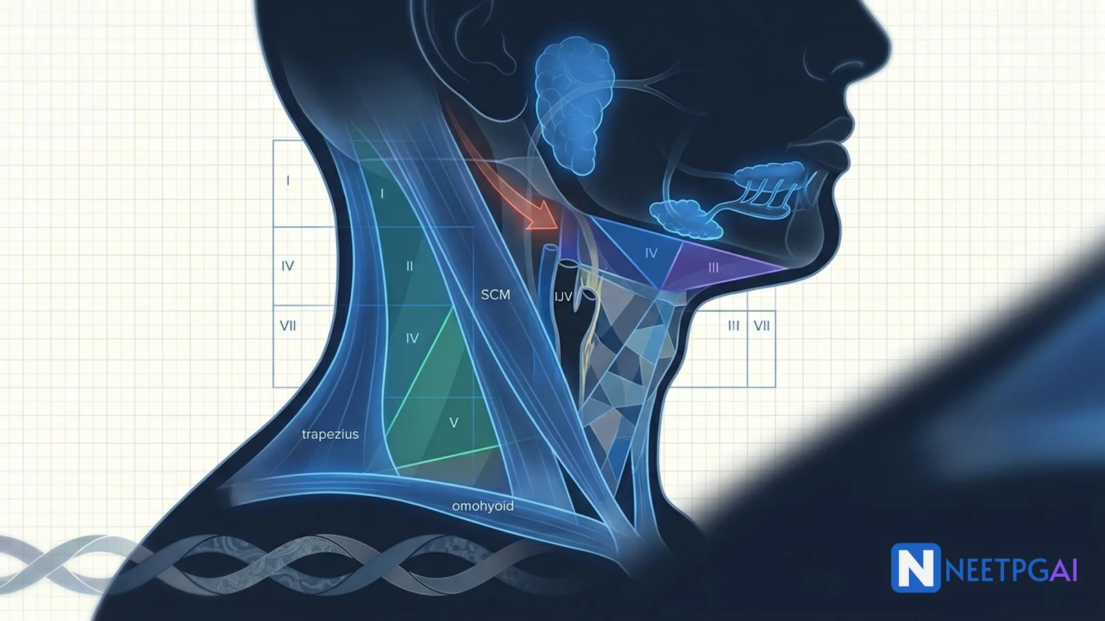
Head and neck anatomy is a 2 to 3 question topic per NEET PG paper and links directly to ENT, general surgery (thyroid), and oncology. Lock these:
- Anterior triangle — bounded by midline, mandible, SCM anterior border; 4 subtriangles (submental, submandibular, muscular, carotid).
- Posterior triangle — bounded by SCM, trapezius, clavicle; contains spinal accessory (crosses roof — vulnerable in lymph node biopsy), brachial plexus roots and trunks, subclavian artery third part.
- Deep cervical fascia — investing, pretracheal, prevertebral layers + carotid sheath (contribution from all three).
- Retropharyngeal danger space — infection spreads to posterior mediastinum → mediastinitis.
- Thyroid arteries — superior thyroid + external laryngeal nerve pair; inferior thyroid + recurrent laryngeal nerve pair.
- Parathyroid glands — superior from pouch IV (stable position), inferior from pouch III (variable, migrates with thymus).
- Salivary glands — parotid (facial nerve inside), submandibular (Wharton duct, stones common), sublingual (multiple ducts).
- Cervical lymph node levels I to VII — level VI first for thyroid; level II first for oral cavity.
Head and neck anatomy is a mainstay of the NEET PG anatomy paper because it links directly to ENT (thyroidectomy, laryngectomy, parotid surgery), general surgery (neck dissections), and oncology (cervical lymph node metastasis mapping). In India, where head and neck cancers are the most common male cancers driven by tobacco and areca nut, this anatomy is not academic — it dictates surgical planning every day.
This NEETPGAI deep dive walks through the neck triangles → deep cervical fascia → retropharyngeal danger space → thyroid and parathyroid → salivary glands → cervical lymph node levels → surgical anatomy correlations. Pair this with the head and neck cancer guide and the lower limb neurovascular guide for the anatomy paper cluster.
Neck triangles — anterior and posterior
The sternocleidomastoid (SCM) divides each side of the neck into an anterior triangle and a posterior triangle.
Anterior triangle
Boundaries
- Anterior — anterior midline of neck
- Posterior — anterior border of SCM
- Superior (base) — inferior border of mandible
- Apex — sternal angle (jugular notch)
- Roof — skin, platysma, superficial cervical fascia
- Floor — pharynx, larynx, thyroid, and the middle constrictor + hyoglossus + mylohyoid muscles
Subtriangles (from superior to inferior)
| Subtriangle | Boundaries | Contents |
|---|
| Submental | Anterior bellies of digastric + hyoid bone | Submental lymph nodes (level Ia), small tributary veins |
| Submandibular (digastric) | Two bellies of digastric + inferior mandible | Submandibular gland, submandibular LN (level Ib), facial artery + vein, marginal mandibular nerve, mylohyoid nerve |
| Muscular (infrahyoid) | Midline + SCM + superior belly of omohyoid | Infrahyoid ("strap") muscles: sternohyoid, sternothyroid, thyrohyoid, omohyoid; overlies thyroid, larynx, trachea |
| Carotid | Posterior belly of digastric + superior belly of omohyoid + anterior SCM | Carotid sheath (common carotid, internal jugular vein, vagus), external carotid + branches, hypoglossal, spinal accessory, deep cervical LN |
Contents of the anterior triangle (overall) — hyoid, thyroid gland, larynx, trachea, cervical part of oesophagus, submandibular gland, tonsils, deep cervical LN, and the carotid sheath structures.
Posterior triangle
Boundaries
- Anterior — posterior border of SCM
- Posterior — anterior border of trapezius
- Inferior (base) — middle third of clavicle
- Apex — meeting of SCM and trapezius on the superior nuchal line
- Roof — skin, platysma, investing layer of deep cervical fascia
- Floor — muscles from superior to inferior — splenius capitis, levator scapulae, scalenus posterior, medius, anterior; all covered by the prevertebral fascia
Subdivision by the inferior belly of omohyoid (running from scapula to hyoid, crossing the triangle):
- Occipital triangle (upper, larger) — above omohyoid
- Supraclavicular (subclavian, omoclavicular) triangle (lower, smaller) — below omohyoid
Contents
- Nerves — spinal accessory (CN XI) crosses the roof superficially in the middle third — vulnerable to injury during posterior triangle lymph node biopsy; brachial plexus roots and trunks (emerging between anterior and middle scalene); phrenic nerve on scalenus anterior; supraclavicular nerves (C3-C4 sensory to skin over clavicle and shoulder)
- Arteries — third part of subclavian artery (behind the middle third of clavicle), transverse cervical, suprascapular, occipital
- Veins — external jugular vein (descends across SCM to enter subclavian vein in supraclavicular triangle)
- Lymph nodes — occipital, transverse cervical, supraclavicular (level V)
Clinical
- Spinal accessory nerve injury in posterior triangle LN biopsy — paralysis of trapezius, dropped shoulder, winging on abduction, difficulty raising the arm above shoulder height
- Brachial plexus injury in penetrating supraclavicular wounds
- Virchow's node (Troisier sign) — left supraclavicular level V node enlargement in gastric cancer (thoracic duct drains here)
Carotid sheath and its contents
The carotid sheath is a tubular fascial condensation that receives contributions from all three layers of the deep cervical fascia (investing, pretracheal, and prevertebral). It extends from the base of the skull to the arch of the aorta.
Contents
- Common carotid artery medial (bifurcates at the upper border of thyroid cartilage — C3/C4 — into internal and external carotid)
- Internal jugular vein lateral
- Vagus nerve posterior between the two
- Deep cervical lymph nodes along the IJV
- Ansa cervicalis on the anterior wall (superior root C1 travels with hypoglossal, inferior root C2-C3)
- Sympathetic chain lies POSTERIOR to the sheath (not inside)
Clinical
- Carotid endarterectomy — access through the anterior triangle
- Central venous catheterisation of the internal jugular vein — target the apex of the triangle formed by the two heads of SCM (Sedillot triangle); ultrasound-guided is now standard
- Horner syndrome from injury to the sympathetic chain — ptosis, miosis, anhidrosis, and enophthalmos; part of the Pancoast tumour syndrome
Deep cervical fascia — three layers plus the carotid sheath
The neck has THREE named layers of deep cervical fascia (all derived from the deep cervical fascia proper). Learn them by what they wrap:
1) Investing (superficial) layer
- Wraps — SCM, trapezius, parotid, submandibular gland
- Attachments — superior nuchal line, mastoid process, mandible, hyoid, clavicle, manubrium, spine of scapula, ligamentum nuchae, spinous processes
- Splits to enclose the parotid (parotid capsule — parotidomasseteric fascia) and the submandibular gland
2) Pretracheal (middle, visceral) layer
- Wraps — thyroid, trachea, larynx, oesophagus (the visceral column)
- Extends from the hyoid down into the superior mediastinum, blending with the fibrous pericardium
- Buccopharyngeal fascia is the posterior continuation covering the pharynx and oesophagus
3) Prevertebral layer
- Wraps — the vertebral column and prevertebral muscles (longus colli, longus capitis)
- Continues laterally to cover the scalene muscles and forms the axillary sheath around the brachial plexus and subclavian artery
- Alar fascia is a subdivision — a thin fascia between the pretracheal and prevertebral layers, forming the posterior wall of the retropharyngeal space
Carotid sheath
- Formed by contributions from all three layers
- Contains carotid artery + IJV + vagus (see above)
Fascial spaces of the neck
Understanding the deep cervical fascia layers immediately explains the potential spaces between them — and how neck infections spread.
Retropharyngeal space
- Between the buccopharyngeal fascia (pretracheal continuation) and the alar fascia (prevertebral subdivision)
- Extent — skull base to approximately T4 (tracheal bifurcation)
- Contents — retropharyngeal lymph nodes (regress after age 4-5)
- Infection — retropharyngeal abscess in children from tonsillitis or upper respiratory infection; in adults from foreign-body impaction (fishbones — common in coastal India — needle-pointed wooden foreign bodies), dental abscess, cervical vertebral osteomyelitis (Pott disease). Presents with stiff neck, dysphagia, drooling, torticollis, muffled voice, and fever. Lateral X-ray neck shows widening of the prevertebral soft tissue. Emergency drainage via a transoral approach.
Danger space
- Between the alar fascia and the true prevertebral fascia
- Extent — skull base to the diaphragm (through the posterior mediastinum)
- Infection from the retropharyngeal space can penetrate the alar fascia and rapidly descend into the posterior mediastinum → acute descending necrotising mediastinitis with high mortality
- Contrast-enhanced CT is diagnostic; treatment is surgical drainage plus broad-spectrum antibiotics
Parapharyngeal (pharyngeal maxillary) space
- Lateral to the pharynx, between the pterygoid muscles and the visceral fascia
- Contains the internal carotid artery + IJV + CN IX, X, XI, XII + cervical sympathetic chain
- Deep neck infection from tonsils, teeth, parotid
- Lemierre syndrome — septic thrombophlebitis of the IJV from Fusobacterium necrophorum, classically following pharyngitis — septic pulmonary emboli, high mortality; treat with broad-spectrum antibiotics + anticoagulation
Submandibular space (Ludwig angina)
- Ludwig angina — a rapidly spreading cellulitis of the submandibular, sublingual, and submental spaces from a dental (mandibular molar) source; bilateral, brawny neck swelling, elevated tongue, drooling, airway obstruction; emergency airway management + IV antibiotics + surgical drainage.
Thyroid gland — surgical anatomy
Gross anatomy
- Paired lateral lobes united by an isthmus at the level of the 2nd-4th tracheal rings
- Pyramidal lobe (present in about 50 percent) extends upwards from the isthmus, a remnant of the thyroglossal duct
- Enveloped by the pretracheal fascia — moves with swallowing (this is why the gland moves with deglutition on examination)
Development
- Endodermal outgrowth from the floor of pharynx at the foramen caecum (junction of anterior 2/3 and posterior 1/3 of tongue)
- Descends anterior to the hyoid and larynx to reach its adult position
- Thyroglossal duct cyst — remnants of the descent tract; presents in the midline (at or below the hyoid), moves with tongue protrusion; treatment is Sistrunk operation (cyst + central hyoid + tract up to the foramen caecum)
Arterial supply
- Superior thyroid artery — first branch of the external carotid artery; descends to the UPPER pole; runs closely with the EXTERNAL BRANCH OF THE SUPERIOR LARYNGEAL NERVE (cricothyroid muscle innervation) — ligate CLOSE to the capsule to protect the nerve
- Inferior thyroid artery — from the thyrocervical trunk (branch of subclavian); enters from below and behind; intimately related to the RECURRENT LARYNGEAL NERVE — ligate CLOSE to the capsule to protect the RLN and the blood supply to inferior parathyroid glands
- Thyroidea ima artery — rare (2-10 percent), arises from the brachiocephalic trunk or aortic arch, enters the isthmus — a risk in emergency tracheostomy
Venous drainage
- Superior + middle thyroid veins → internal jugular vein
- Inferior thyroid veins → brachiocephalic vein
Nerves at risk in thyroidectomy
- Recurrent laryngeal nerve — runs in the tracheoesophageal groove, enters the larynx behind the cricothyroid joint. Unilateral injury → hoarseness with paramedian cord (compensated over months). Bilateral injury → stridor and airway obstruction (emergency tracheostomy). Sensory to the mucosa below the cords; motor to all intrinsic laryngeal muscles except cricothyroid
- Non-recurrent right laryngeal nerve (0.5-1 percent) — associated with aberrant right subclavian artery (arteria lusoria) arising from the aortic arch as the last branch; the nerve enters the larynx directly without looping
- External branch of the superior laryngeal nerve — supplies cricothyroid; injury produces a subtle voice change (loss of high-pitched phonation)
Parathyroid glands
- Usually 4, occasionally 3 to 6
- Superior parathyroid glands — derived from the FOURTH pharyngeal pouch; lie behind the middle-to-upper part of the thyroid; relatively CONSTANT position; supplied mainly by the inferior thyroid artery
- Inferior parathyroid glands — derived from the THIRD pharyngeal pouch (along with the thymus); migrate with the thymus, so the position is more VARIABLE — anywhere from the carotid bifurcation to the mediastinum; supplied by the inferior thyroid artery
- Blood supply — mainly inferior thyroid artery; ligation of the inferior thyroid artery too far from the capsule can devascularise the parathyroids and produce transient (or permanent) hypocalcaemia — the classic post-thyroidectomy complication
Salivary glands
Parotid gland
- Largest salivary gland
- Serous secretion — enzymatically rich saliva
- Duct — Stensen (parotid) duct — traverses the buccinator to open opposite the 2nd upper molar
- Facial nerve — passes THROUGH the substance of the gland, dividing it into an artificial superficial and deep lobe; divides into 5 branches — Temporal, Zygomatic, Buccal, Marginal mandibular, Cervical (mnemonic: "To Zanzibar By Motor Car")
- Landmarks for facial nerve identification during parotidectomy — tragal pointer (nerve 1 cm deep and inferior), tympanomastoid suture (nerve immediately deep), posterior belly of digastric attachment
- Frey syndrome — gustatory sweating after parotidectomy; misdirected regeneration of parasympathetic secretomotor fibres of the auriculotemporal nerve into denervated sweat glands
Submandibular gland
- Mixed serous-mucous secretion, predominantly serous
- Duct — Wharton duct — opens on the floor of the mouth beside the frenulum of the tongue
- Sialolithiasis most common here (80 percent of salivary stones) — because Wharton duct is long, angled upwards, and drains a mucus-rich secretion against gravity
- Removal involves care of the marginal mandibular branch of the facial nerve, the lingual nerve (loops around the duct), and the hypoglossal nerve
Sublingual gland
- Mostly mucous secretion
- Multiple small ducts (of Rivinus) opening on the floor of the mouth along the sublingual fold
- Ranula — mucous retention cyst of the sublingual gland presenting as a bluish cystic swelling under the tongue
Salivary gland tumours (anatomy correlate)
- Parotid — 20 percent malignant; smallest is the sublingual/minor glands where 70 percent are malignant (the smaller the gland, the higher the malignancy risk)
- Facial nerve palsy + parotid mass — malignant until proven otherwise
- Pleomorphic adenoma — most common benign; parotid; treated with superficial parotidectomy
- Mucoepidermoid carcinoma — most common malignant overall
- Adenoid cystic carcinoma — perineural invasion hallmark; classic in submandibular and minor salivary glands
Cervical lymph node levels I to VII
The Robbins-AAO-HNS level system standardises cervical LN description for oncology.
| Level | Location | Primary drainage sources |
|---|
| Ia — Submental | Between anterior bellies of digastric | Lip, floor of mouth, anterior tongue |
| Ib — Submandibular | Within submandibular triangle | Oral cavity, submandibular gland |
| IIa — Upper jugular, anterior to spinal accessory | Skull base to hyoid, IJV | Oral cavity, oropharynx, larynx, parotid |
| IIb — Upper jugular, posterior to spinal accessory | Skull base to hyoid, IJV | Nasopharynx, oropharynx |
| III — Middle jugular | Hyoid to cricoid | Oral cavity, larynx, pharynx |
| IV — Lower jugular | Cricoid to clavicle | Hypopharynx, thyroid, cervical oesophagus |
| Va — Upper posterior triangle | Above cricoid | Nasopharynx, oropharynx, scalp |
| Vb — Lower posterior triangle (supraclavicular) | Below cricoid | Thyroid, thoracic/abdominal primaries (Virchow) |
| VI — Central compartment | Hyoid to suprasternal notch (prelaryngeal Delphian, pretracheal, paratracheal) | Thyroid, glottis, subglottis, hypopharynx, cervical oesophagus |
| VII — Superior mediastinum | Below suprasternal notch | Thyroid, cervical oesophagus |
First-echelon patterns
- Oral cavity cancer → levels I-III first
- Oropharynx / nasopharynx → level II first
- Larynx → levels II-IV
- Thyroid cancer → level VI first (central), then II-V
- Left supraclavicular (Virchow, level Vb) → gastric, breast, ovarian, testicular primary
Neck dissection — surgical anatomy
Radical neck dissection (Crile)
Removes levels I-V + all three critical structures: SCM, IJV, spinal accessory nerve. Substantial morbidity — dropped shoulder, cosmetic deformity, venous congestion.
Modified radical neck dissection (I-III)
Removes levels I-V but preserves one or more of the three critical structures:
- Type I — preserves spinal accessory
- Type II — preserves spinal accessory + IJV
- Type III (functional) — preserves all three
Selective neck dissection
Removes only involved-risk levels:
- Supraomohyoid (levels I-III) — for oral cavity primaries
- Lateral (levels II-IV) — for laryngeal, hypopharyngeal primaries
- Central compartment (level VI) — for thyroid cancer
- Posterolateral (II-V + occipital nodes) — for scalp posterior neck primaries
India-relevant clinical correlations
- Cervical tuberculous lymphadenitis (scrofula) — the most common cause of cervical lymphadenopathy in India, especially in the posterior triangle (level V); FNAC shows caseating granulomas; ATT for 6 months
- Head and neck cancer — most common cancer in Indian males; oral cavity tongue and buccal mucosa most affected; requires understanding of level I-VI drainage patterns for surgical planning
- Fishbone impaction in coastal India — retropharyngeal abscess and danger-space extension
- Ludwig angina — dental infections in the setting of poor oral hygiene remain common in India
- Thyroidectomy safety — RLN identification and parathyroid preservation are the two surgical priorities; intraoperative nerve monitoring is standard in high-volume centres
- Sistrunk operation for thyroglossal duct cyst — an exam favourite in paediatric surgery scenarios
NEET PG MCQ traps
- Anterior triangle subtriangles — submental, submandibular, muscular, carotid.
- Posterior triangle — spinal accessory nerve crosses the ROOF superficially (dropped shoulder after lymph node biopsy).
- Erb point — 2 finger-breadths above the clavicle at the posterior border of SCM — where several cervical plexus branches emerge.
- Deep cervical fascia — 3 layers: investing, pretracheal, prevertebral; PLUS carotid sheath contribution.
- Retropharyngeal danger space — between alar and true prevertebral fascia; extends to diaphragm.
- Descending necrotising mediastinitis — surgical emergency from retropharyngeal abscess.
- Ludwig angina — bilateral submandibular cellulitis from dental infection.
- Lemierre syndrome — Fusobacterium septic IJV thrombophlebitis with septic pulmonary emboli.
- Virchow node — left supraclavicular (Vb) from gastric cancer.
- Superior thyroid artery — external laryngeal nerve at risk; ligate close to capsule; injury = loss of high-pitched voice.
- Inferior thyroid artery — recurrent laryngeal nerve + parathyroid blood supply at risk.
- Non-recurrent right laryngeal nerve — 0.5-1 percent; associated with aberrant right subclavian (arteria lusoria).
- Bilateral RLN injury — stridor + emergency tracheostomy.
- Superior parathyroid — 4th pouch; stable position behind upper thyroid.
- Inferior parathyroid — 3rd pouch; migrates with thymus; variable position.
- Thyroglossal duct cyst — midline, moves with tongue protrusion; Sistrunk operation.
- Parotid facial nerve landmarks — tragal pointer, tympanomastoid suture, posterior belly of digastric.
- Facial nerve branches — Temporal, Zygomatic, Buccal, Marginal mandibular, Cervical (To Zanzibar By Motor Car).
- Frey syndrome — gustatory sweating after parotidectomy (auriculotemporal nerve misdirected regeneration).
- Sialolithiasis — most common in submandibular gland (Wharton duct, angled upward, mucus-rich).
- Ranula — mucous retention cyst of sublingual gland.
- Cervical LN levels I-VII — thyroid drains to level VI (central) first.
- Oral cavity cancer drains to level I-III first.
- Radical neck dissection removes SCM + IJV + spinal accessory + levels I-V.
- Supraomohyoid selective neck dissection (levels I-III) — for oral cavity primaries.
- Cervical TB lymphadenitis — posterior triangle in India; caseating granulomas on FNAC.
Recent updates and Indian context
- Intraoperative RLN monitoring — standard in high-volume Indian thyroid surgery centres (Tata Memorial, AIIMS, PGI, CMC Vellore)
- Sentinel LN biopsy — evolving role in early oral cavity cancer (level I-II mapping) to avoid elective neck dissection
- Robotic neck dissection — increasingly used for select cases (thyroid, oropharynx) in tertiary Indian centres
- Level VII inclusion in thyroid cancer surgery — routine central compartment dissection for medullary and high-risk papillary thyroid cancers
- Ultrasound-guided FNAC — first-line for thyroid and neck lumps in India; Bethesda classification (I-VI) reported
- PET-CT — increasingly used for HNSCC staging and follow-up
Frequently asked questions
What is the retropharyngeal space and why is it called the danger space?
The retropharyngeal space lies between the buccopharyngeal fascia (posterior wall of pharynx and oesophagus, part of the pretracheal layer) and the alar fascia (a subdivision of the prevertebral layer). It extends from the skull base to the tracheal bifurcation at T4 approximately. Immediately posterior to it, between the alar fascia and the true prevertebral fascia, lies the DANGER SPACE, which extends from the skull base down through the posterior mediastinum to the diaphragm. Infection in the retropharyngeal space can spread through the alar fascia into the danger space and then rapidly descend into the posterior mediastinum, causing mediastinitis — a surgical emergency with high mortality. Common Indian sources include dental infections, tonsillitis, and pharyngeal injury from swallowed foreign bodies (fishbones common in coastal India). The clinical clue is a stiff neck with fever, dysphagia, drooling, and torticollis; contrast CT neck shows retropharyngeal fluid or gas. NEET PG loves the danger-space anatomy and the mediastinitis complication.
Why is the recurrent laryngeal nerve at particular risk during thyroidectomy?
The recurrent laryngeal nerve (RLN) runs in the tracheoesophageal groove behind the thyroid gland, entering the larynx at the level of the cricothyroid joint. On the LEFT, the RLN loops around the arch of the aorta (below the ligamentum arteriosum) and ascends. On the RIGHT, it loops around the subclavian artery. It is intimately related to the inferior thyroid artery — the nerve may pass anterior, posterior, or between its branches. During thyroidectomy the artery is ligated on the capsule of the gland rather than in the main trunk to protect both the RLN AND the inferior parathyroid blood supply. Unilateral RLN injury produces hoarseness with a paramedian vocal cord (compensated by contralateral cord adduction over months). BILATERAL RLN injury is a surgical emergency — both cords assume paramedian position, producing stridor and airway obstruction requiring urgent tracheostomy. A rare but exam-favourite variant is the non-recurrent right laryngeal nerve (0.5-1 percent) — associated with an aberrant right subclavian artery (arteria lusoria) that arises directly from the arch as the last branch; the nerve enters the larynx directly from the vagus without looping. NEET PG tests the RLN course, the tracheoesophageal-groove position, and the non-recurrent variant with aberrant subclavian association.
How is cervical lymph node metastasis classified into levels I to VII?
The Robbins level system (updated by AAO-HNS) divides cervical lymph nodes into 7 levels. Level I — submental (Ia, between the anterior bellies of digastric) and submandibular (Ib, within the submandibular triangle) — drains lip, anterior tongue, floor of mouth, submandibular gland. Level II — upper jugular, along the internal jugular vein from the skull base to the level of the hyoid; subdivided by the spinal accessory nerve into IIa (anterior) and IIb (posterior) — drains oral cavity, pharynx, larynx, parotid. Level III — middle jugular, from the hyoid to the cricoid — drains oral cavity, larynx, pharynx. Level IV — lower jugular, from cricoid to clavicle — drains hypopharynx, thyroid, cervical oesophagus. Level V — posterior triangle nodes, subdivided by the horizontal plane of the cricoid into Va (upper, includes spinal accessory chain) and Vb (lower, includes supraclavicular) — drains nasopharynx, oropharynx, thyroid, scalp. Level VI — central compartment, from the hyoid to the suprasternal notch — includes prelaryngeal (Delphian), pretracheal, paratracheal — drains thyroid, glottis, subglottis, hypopharynx, cervical oesophagus. Level VII — superior mediastinal (below the suprasternal notch) — drains thyroid, cervical oesophagus. NEET PG tests the level VI position for thyroid cancer (first echelon) and the level II position for oral cancer (first echelon).
What is the arterial supply of the thyroid gland and which artery is most at risk of injuring the external branch of the superior laryngeal nerve?
The thyroid gland has a rich dual arterial supply. The SUPERIOR thyroid artery is the first branch of the external carotid artery; it descends to the upper pole of the thyroid alongside the EXTERNAL BRANCH OF THE SUPERIOR LARYNGEAL NERVE (which supplies cricothyroid). During superior pole ligation, the artery must be tied CLOSE TO THE THYROID CAPSULE to avoid injuring this nerve; a lateral ligation risks paralysis of cricothyroid, producing a subtle voice change — loss of high-pitched phonation, described as a monotone or 'opera-singer' voice change (the 'Amelita Galli-Curci' syndrome). The INFERIOR thyroid artery arises from the thyrocervical trunk (a branch of the subclavian artery) and enters the gland from below and behind; it is intimately related to the RECURRENT LARYNGEAL NERVE (which passes anterior, posterior, or between its branches). During inferior pole ligation, the artery must be tied CLOSE TO the thyroid capsule to avoid injuring the RLN AND to preserve the blood supply to the inferior parathyroid glands. A rare THYROIDEA IMA artery (2-10 percent) arises from the brachiocephalic trunk or arch of aorta and enters the isthmus — a risk in emergency tracheostomy. Venous drainage is via superior + middle + inferior thyroid veins; the inferior thyroid vein drains into the brachiocephalic vein. NEET PG loves the superior-thyroid-artery + external-laryngeal-nerve pair and the inferior-thyroid-artery + recurrent-laryngeal-nerve pair.
Why is the facial nerve at risk during parotidectomy and how is it identified surgically?
The facial nerve emerges from the stylomastoid foramen, immediately enters the substance of the parotid gland, and divides into 5 major branches within the gland — temporal, zygomatic, buccal, marginal mandibular, and cervical (the 'To Zanzibar By Motor Car' mnemonic). The nerve DIVIDES the gland into an artificial superficial and deep lobe. Superficial parotidectomy for a benign superficial tumour (typically pleomorphic adenoma) preserves the nerve; total parotidectomy for malignant or deep-lobe tumour requires nerve dissection with a real risk of paresis. The facial nerve is identified surgically by 3 classic landmarks — (1) the TRAGAL POINTER (a pointed cartilage extension of the tragus) — nerve lies 1 cm deep and inferior to it, (2) the tympanomastoid suture line — the nerve lies immediately deep, (3) the POSTERIOR BELLY OF DIGASTRIC — the nerve emerges just above its attachment to the mastoid. FROM WITHIN the gland (retrograde approach), the marginal mandibular branch is identified along the mandibular border and traced backwards. Frey syndrome (gustatory sweating) after parotidectomy — misdirected regeneration of parasympathetic secretomotor fibres of the auriculotemporal nerve into denervated sweat glands; managed with topical glycopyrrolate or botulinum injection. NEET PG loves the tragal pointer, the tympanomastoid suture, and the digastric landmark; also the Frey-syndrome pathophysiology.
This content is for educational purposes for NEET PG exam preparation. It is not a substitute for professional medical advice, diagnosis, or treatment. Clinical information has been reviewed by qualified medical professionals.
Written by: NEETPGAI Editorial Team
Reviewed by: Pending SME Review
Last reviewed: July 2026