Version 1.0 — Published September 2026
Quick Answer
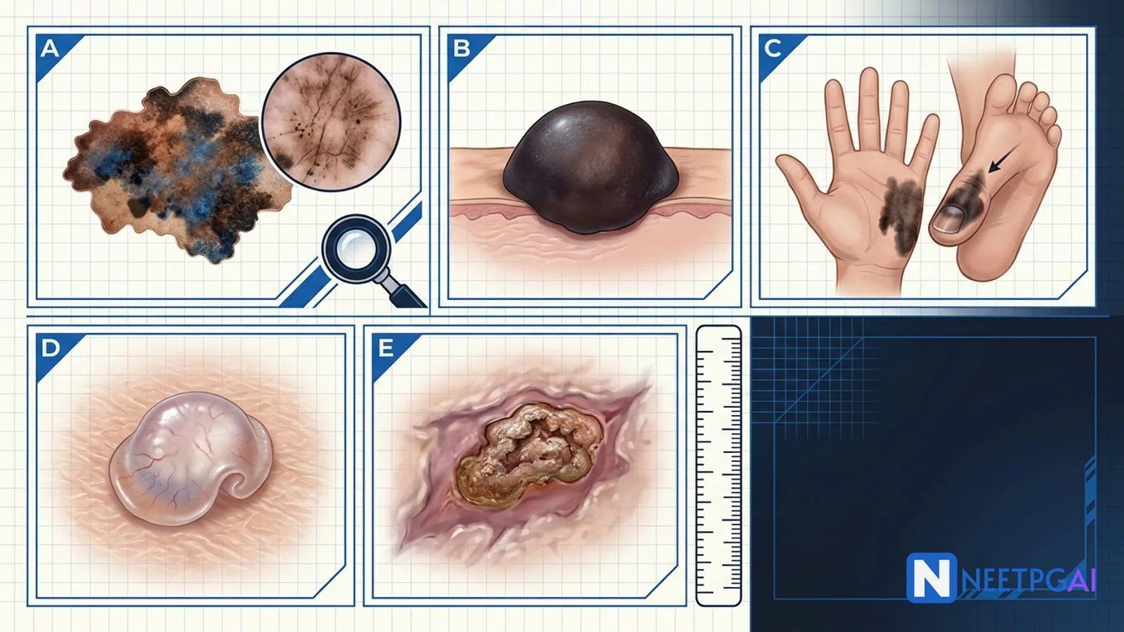
Dermatology image MCQs on skin cancer contribute 2-4 questions per NEET PG paper across dermatology, surgery, and pathology. Five high-yield patterns recur reliably every cycle:
- Superficial spreading melanoma — asymmetric variegated patch, ABCDE-classical, atypical pigment network on dermoscopy; wide excision by Breslow depth plus SLN for depth greater than 0.8 mm
- Nodular melanoma — firm dark or amelanotic pink papule/nodule; skips radial growth phase, poorer prognosis for equal Breslow
- Acral lentiginous melanoma — palm, sole, or subungual; longitudinal melanonychia with Hutchinson sign; commonest melanoma subtype in Indian patients (over 50 percent of Indian series)
- Nodular basal cell carcinoma — pearly telangiectatic papule with rolled border and central rodent ulcer; sun-exposed head/neck; 4 mm excision or Mohs for high-risk sites
- Squamous cell carcinoma — hyperkeratotic ulcerated nodule on sun-exposed skin, immunosuppressed patient, or old burn scar (Marjolin ulcer); 4-6 mm excision or Mohs; cemiplimab for advanced
Locking these 5 patterns plus a handful of benign vs malignant discriminators (Ugly Duckling sign, dermoscopy patterns, biopsy technique) moves dermatology image-MCQ accuracy from 45 to 85 percent.
Why dermatology skin cancer image MCQs are high-yield for NEET PG
Skin cancer is a growing burden globally and, though incidence rates in Indian Fitzpatrick IV-V skin are lower than in Western populations, the presentations are often late, atypical, and prognostically worse. NEET PG tests skin cancer heavily because the visual patterns are stereotyped and diagnostic, and the management pathway (biopsy, staging, definitive surgery, adjuvant therapy) is high-yield across dermatology, surgery, oncology, and PSM. Acral lentiginous melanoma is particularly Indian-relevant and is under-taught in most textbook chapters oriented to Western practice.
Foundational approach — the systematic skin lesion evaluation
The ABCDE plus F and G
| Criterion | Concerning finding |
|---|
| A — Asymmetry | Lesion is not mirror-image across an axis |
| B — Border | Irregular, scalloped, notched, or ill-defined |
| C — Colour | Two or more shades (brown, black, red, white, grey, blue) |
| D — Diameter | Greater than 6 mm (pencil eraser); smaller lesions can still be melanoma |
| E — Evolution | Change in size, shape, colour, elevation, itch, or bleeding over weeks-months — MOST IMPORTANT single criterion |
| F — Funny-looking (Ugly Duckling) | Lesion stands out from the patient's other nevi |
| G — Growing | Actively enlarging lesion |
Dermoscopy-suggestive-of-melanoma features
- Atypical pigment network — thickened, irregular grid pattern
- Blue-white veil — blue-grey structureless area under a white haze
- Regression — white scar-like areas or grey peppering
- Atypical dots and globules — irregular in size, shape, distribution
- Irregular streaks/pseudopods — at the periphery of the lesion
- Multiple colours — three or more shades on dermoscopy
- 7-point checklist (Argenziano) and ABCD dermoscopy score (Stolz) — structured scoring systems
MCQ 1: 34-year-old man with a slowly enlarging back mole showing asymmetry, irregular border, and multiple colours
Image description: [Clinical photograph of the back of a 34-year-old male showing a solitary pigmented lesion measuring approximately 12 mm x 9 mm, located just below the left scapula. The lesion is markedly asymmetric — the upper half is significantly larger and more irregular than the lower half. Border is scalloped and notched with peninsular projections extending into surrounding normal skin. Colour is variegated with tan-brown centrally, dark black in the upper-medial quadrant, a reddish inflammatory rim inferolaterally, and a small white regression area at the 7 o'clock position. Diameter clearly exceeds 10 mm. Paired dermoscopy image shows: atypical pigment network with thickened irregular meshwork, blue-white veil in the upper-medial area, grey peppering (regression) at the 7 o'clock position, and irregular streaks/pseudopods at the 3 and 9 o'clock margins. Surrounding skin shows several small (2-4 mm) regular symmetric benign nevi — the lesion in question is clearly the Ugly Duckling.]
Clinical vignette: A 34-year-old male IT professional with Fitzpatrick type III skin presents with a mole on his back that his wife noticed has changed over the last 8 months — becoming larger, darker in areas, and developing a red rim. He denies bleeding, itching, or pain. He has 30-40 other typical-appearing nevi across his trunk and limbs. Occasional beach holidays; no severe childhood sunburns; no family history of melanoma; no personal history of skin cancer.
Options:
- (a) Benign compound melanocytic nevus
- (b) Seborrhoeic keratosis
- (c) Superficial spreading melanoma
- (d) Blue nevus
Correct answer: (c) Superficial spreading melanoma
Reasoning: The lesion satisfies all five ABCDE criteria — asymmetry, irregular scalloped border, colour variegation with multiple shades including regression, diameter greater than 6 mm, and clear evolution over months. It is the Ugly Duckling in a patient with many benign symmetric nevi. Dermoscopy adds an atypical pigment network, blue-white veil, regression, and irregular streaks — all melanoma-suggestive features. This constellation is the classical superficial spreading melanoma — the commonest melanoma subtype in fair-skinned populations globally (70 percent), typically on the trunk in men and legs in women.
A benign compound nevus is usually symmetric, regularly bordered, single-coloured, and stable. Seborrhoeic keratosis is a stuck-on warty tan-brown plaque with milia-like cysts on dermoscopy, benign. Blue nevus is a uniformly blue-black dome-shaped small papule with a homogeneous blue pigmentation on dermoscopy, benign.
Teaching pearl — superficial spreading melanoma workup and management:
| Step | Action |
|---|
| Diagnostic biopsy | Full-thickness excisional biopsy with 1-2 mm margin to preserve Breslow depth |
| Pathology report must include | Breslow depth, ulceration status, mitotic rate, lymphovascular invasion, perineural invasion, regression, margin status |
| Definitive excision margins by Breslow depth | In situ 5 mm; less than 1 mm depth 1 cm margin; 1-2 mm depth 1-2 cm margin; greater than 2 mm depth 2 cm margin — to and including deep fascia |
| Sentinel lymph node biopsy | Offered for melanoma depth greater than 0.8 mm (or 0.75-1 mm with adverse features) |
| Staging | AJCC 8th edition TNM based on Breslow, ulceration, SLN, distant metastasis |
| Adjuvant systemic therapy for high-risk resected disease | Immunotherapy (nivolumab, pembrolizumab) OR targeted therapy for BRAF V600E mutated (dabrafenib plus trametinib) |
| Advanced/metastatic disease | Immunotherapy (nivolumab-ipilimumab combination) as first-line; BRAF inhibitors for BRAF V600E |
- Prognosis is driven by Breslow depth — 5-year survival greater than 95 percent for depth less than 1 mm; drops to 50-60 percent for depth greater than 4 mm
- NEET PG tests the ABCDE criteria, the excisional biopsy rule, Breslow-based excision margins, and the SLN cut-off of 0.8 mm
MCQ 2: 52-year-old man with a rapidly enlarging firm dark nodule on the calf over 3 months
Image description: [Clinical photograph of the posterior calf of a 52-year-old male. The image shows a firm, dome-shaped, deeply pigmented (blue-black) nodule approximately 8 mm in diameter and 4 mm in height, with a surface ulceration at the apex covered by a small serous-hemorrhagic crust. The border is relatively regular and the colour is nearly uniform (dark blue-black) but with a small amelanotic pink area at the base. The surrounding skin shows no radial growth-phase pigmented patch. There are no other atypical lesions. Paired dermoscopy image shows: blue-black homogeneous pigmentation, irregular vascular pattern with atypical vessels (polymorphous, hairpin, and dotted vessels) in the amelanotic area, and ulceration. There is no pigment network (in contrast to superficial spreading melanoma). Excisional biopsy is performed showing Breslow depth 3.8 mm with ulceration and mitotic rate 8/mm2.]
Clinical vignette: A 52-year-old male labourer presents with a nodule on his calf that he noticed 3 months ago and has grown rapidly. It occasionally bleeds when caught on clothing. He denies preceding trauma. No previous melanoma, no family history. Fitzpatrick type IV skin, mostly outdoor occupation. Physical exam otherwise unremarkable; no palpable inguinal lymphadenopathy.
Options:
- (a) Pyogenic granuloma
- (b) Superficial spreading melanoma
- (c) Nodular melanoma
- (d) Blue nevus
Correct answer: (c) Nodular melanoma
Reasoning: Nodular melanoma is a firm, dark (or occasionally amelanotic pink), rapidly growing dome-shaped papule or nodule that skips the radial growth phase and enters vertical growth from the start. The lesion here is a dome-shaped deeply pigmented nodule with rapid 3-month growth, surface ulceration, and Breslow depth 3.8 mm with ulceration and high mitotic rate on excisional biopsy — the pathognomonic nodular melanoma pattern. The absence of a preceding flat pigmented patch (radial growth phase) and the aggressive rapid growth are the discriminators.
Pyogenic granuloma is a benign vascular proliferation, typically bright red or purple, that grows rapidly after minor trauma and bleeds easily, but is not deeply pigmented; dermoscopy shows a homogeneous red-purple lacuna-like pattern without atypical vessels. Superficial spreading melanoma has a preceding radial growth-phase pigmented patch and the ABCDE-classical pattern. Blue nevus is stable, small, symmetric, and non-ulcerated.
Teaching pearl — nodular melanoma:
- Accounts for 15-30 percent of all melanoma but disproportionately more melanoma-related deaths per unit incidence because of thicker Breslow at diagnosis
- Vertical growth from the start — no radial growth phase — hence rapidly growing dome/nodule
- Amelanotic variant — pink or skin-coloured nodule; easily mistaken for benign lesion (dermatofibroma, pyogenic granuloma, adnexal tumour); high index of suspicion for any new firm rapidly growing nodule
- Poorer prognosis for equivalent Breslow depth than SSM — because thicker at diagnosis and higher mitotic rate
- Treatment identical to any melanoma — wide excision by Breslow, SLN for depth greater than 0.8 mm, adjuvant systemic therapy for high-risk (immunotherapy or BRAF-targeted)
- NEET PG tests the vertical-growth-from-start definition and the amelanotic trap
MCQ 3: 58-year-old man with a dark linear band under the right thumb nail and pigment extending onto the nail fold
Image description: [Clinical photograph of the right thumb of a 58-year-old male. The image shows a longitudinal band of dark brown-black pigmentation approximately 4 mm wide running the full length of the nail plate from the proximal to the distal edge. The band is irregular in width along its length (wider proximally, narrower distally), and shows variegated colour with darker central areas and lighter brown edges. Critically, there is pigmentation extending onto the proximal nail fold (Hutchinson sign) appearing as an irregular smudge of brown-black pigment on the skin overlying the nail matrix. The distal aspect shows nail plate splitting and thinning. Paired dermoscopy of the nail fold shows brown-black pigment with an irregular pattern, some grey-black dots, and disruption of the normal parallel lines. Excisional biopsy of the nail matrix confirms melanoma with Breslow depth 2.4 mm.]
Clinical vignette: A 58-year-old male farmer from Punjab presents with darkening of the right thumb nail over the past 12 months. He initially thought it was a bruise from farming injury but it has not resolved. Over the past 2 months he noticed the pigment extending onto the skin around the nail. No pain, no personal or family history of skin cancer. Fitzpatrick type IV skin.
Options:
- (a) Subungual haematoma
- (b) Onychomycosis
- (c) Benign longitudinal melanonychia
- (d) Subungual acral lentiginous melanoma
Correct answer: (d) Subungual acral lentiginous melanoma
Reasoning: Acral lentiginous melanoma (ALM) arises on the palms, soles, or under the nail (subungual). Subungual ALM presents as longitudinal melanonychia — a dark linear band in the nail plate — with the Hutchinson sign (extension of pigment onto the proximal nail fold or hyponychium), which is a key distinguishing feature from benign melanonychia. This patient has all the features — 12-month history (chronic — not haematoma), longitudinal melanonychia with irregular width and variegated colour (not benign), Hutchinson sign (pigment on the proximal nail fold — the pathognomonic melanoma feature), nail plate thinning, and biopsy-confirmed Breslow 2.4 mm melanoma.
Subungual haematoma clears distally with nail growth (moves distally over months) and typically follows trauma. Onychomycosis causes yellow-brown discolouration with subungual hyperkeratosis and nail plate thickening, no pigment. Benign longitudinal melanonychia is a uniform-width single-toned brown band without Hutchinson sign, common in Fitzpatrick IV-VI skin (physiological) and stable over years.
Teaching pearl — acral lentiginous melanoma:
| Feature | Detail |
|---|
| Global share | 5 percent of all melanoma |
| Indian and Asian share | Over 50 percent of melanoma in Indian series — the commonest subtype |
| Sites | Palm, sole, subungual |
| Not related to UV exposure | Sun protection does not prevent it |
| Subungual ALM red flags | Longitudinal melanonychia with band width over 3 mm, irregular width, variegated colour, Hutchinson sign (pigment on nail fold), nail plate destruction |
| Biopsy | Nail matrix biopsy under nail plate reflection; punch through most representative area; requires dermatologist or hand surgeon technique |
| Definitive treatment | Wide excision by Breslow (in situ 5 mm; less than 1 mm 1 cm; etc); often requires digital amputation for advanced subungual disease |
| SLN biopsy | For depth greater than 0.8 mm — same threshold as other melanoma |
| Prognosis | Worse than SSM because typically thicker at diagnosis (delayed presentation) |
- Hutchinson sign — pigmentation of the proximal nail fold or hyponychium overlying subungual melanoma — pathognomonic, differentiates from benign melanonychia
- Pseudo-Hutchinson sign — pigment visible through translucent nail fold (as in Peutz-Jeghers, Laugier-Hunziker) — must be distinguished
- India-specific — high index of suspicion for any new or changing subungual pigmented band in an Indian adult, given ALM predominance
- NEET PG tests the ALM subtype prevalence in Asians, the Hutchinson sign, and the biopsy approach
MCQ 4: 68-year-old with a pearly translucent papule with rolled border and central ulceration on the ala nasi for 2 years
Image description: [Clinical photograph of the right ala nasi of a 68-year-old male. The image shows a 6 mm pearly translucent, dome-shaped papule with fine telangiectasias coursing over the surface, rolled everted borders, and a central shallow ulcer covered by a thin serous crust. The surrounding skin shows moderate solar elastosis and multiple actinic keratoses. There is no lymphadenopathy. Paired dermoscopy image shows: arborising (tree-branching) telangiectasias — the classical BCC vascular pattern, shiny white-red structureless areas, maple-leaf-like areas at the periphery, and spoke-wheel structures. Punch biopsy confirms nodular basal cell carcinoma with no aggressive histologic features.]
Clinical vignette: A 68-year-old male retired postman with 40 years of outdoor sun exposure and Fitzpatrick type II skin presents with a small nodule on the right side of his nose that has been present for approximately 2 years. He initially ignored it. It occasionally bleeds when he shaves. No pain. No personal history of skin cancer but he has multiple pre-existing actinic keratoses treated with cryotherapy over the years. No family history.
Options:
- (a) Nodular basal cell carcinoma
- (b) Squamous cell carcinoma
- (c) Amelanotic nodular melanoma
- (d) Sebaceous hyperplasia
Correct answer: (a) Nodular basal cell carcinoma
Reasoning: Nodular BCC — the classical pearly, translucent papule or nodule with rolled everted border, telangiectasias on the surface, and central ulceration (rodent ulcer); slow-growing on the face; risk factors chronic UV exposure and fair skin. This patient has all the features — pearly translucent papule with rolled border, telangiectasias, central ulcer, 2-year indolent course, sun-exposed nasal site, Fitzpatrick II skin, 40 years of outdoor UV exposure. Dermoscopy adds arborising telangiectasias, shiny white-red structureless areas, and maple-leaf-like areas — the classical BCC dermoscopic triad. Biopsy confirms.
SCC would be more hyperkeratotic and less translucent, with a more indurated ulcer, and typically arises in an actinic keratosis. Amelanotic nodular melanoma would grow more rapidly (weeks-months not 2 years) and would show polymorphous atypical vessels on dermoscopy rather than arborising telangiectasias. Sebaceous hyperplasia is a benign yellow-white umbilicated papule of middle-aged and older adults, not translucent-pearly and not ulcerating.
Teaching pearl — basal cell carcinoma treatment:
| Modality | Indication | Notes |
|---|
| Standard surgical excision | Typical low-risk BCC | 4 mm margin; cure rate 95-98 percent |
| Mohs micrographic surgery (MMS) | High-risk sites (nose, ears, eyelids, lips, digits, genitals — the "H-zone"), aggressive histology (morpheaform, infiltrative, micronodular), recurrent tumours, size over 2 cm, immunosuppressed | Progressive tangential excision with immediate frozen-section of ENTIRE cut margin; cure rate 98-99 percent |
| Electrodesiccation and curettage | Small superficial BCC | Cannot use on hair-bearing areas or terminal-hair sites |
| Cryotherapy | Small superficial BCC | Elderly non-surgical |
| Topical imiquimod (5%) or 5-fluorouracil | Superficial BCC only | 5-6 week course, moderate cure rate |
| Photodynamic therapy | Superficial BCC | Cosmetically favourable, moderate cure rate |
| Radiotherapy | Elderly non-surgical, adjuvant for positive margins or perineural invasion | Higher recurrence than surgery |
| Hedgehog pathway inhibitors (vismodegib, sonidegib) | Locally advanced or metastatic BCC unamenable to surgery or radiation | Muscle cramps, alopecia, dysgeusia, teratogenicity |
- BCC is the commonest human malignancy globally — vastly more common than melanoma but rarely metastasises (under 0.1 percent)
- Local destruction can be extensive on the face — the reason cure rate matters so much
- Nasal ala BCC needs Mohs by default given cosmetic and functional stakes
- NEET PG tests the nodular BCC classical description, the H-zone indication for Mohs, and the rarity of metastasis
MCQ 5: 62-year-old with a hyperkeratotic ulcerated nodule arising in a 40-year-old burn scar on the leg
Image description: [Clinical photograph of the anterior right leg of a 62-year-old female showing a wide area of atrophic, hyperpigmented scar tissue extending from mid-thigh to mid-calf, consistent with a healed extensive burn. Within the scar there is a 2.5 cm indurated, hyperkeratotic, verrucous nodule with an ulcerated centre, everted rolled borders, and a serosanguinous exudate. The surrounding scar shows contracture and pigmentary changes. There is a palpable 1.5 cm mobile right inguinal lymph node. Paired dermoscopy shows: hyperkeratotic scales, white circles (targetoid appearance), keratin pearls, and polymorphous atypical vessels (dotted, hairpin, glomerular). Punch biopsy shows well-differentiated squamous cell carcinoma with keratin pearl formation and perineural invasion.]
Clinical vignette: A 62-year-old female homemaker sustained a scald burn to the right leg 40 years ago from a kitchen accident. The burn healed with extensive scarring. Over the past 4 months she noticed a wart-like nodule developing within the scar that has grown and started bleeding. No prior skin cancer. She reports occasional dragging pain in the right groin. Fitzpatrick type IV skin.
Options:
- (a) Verrucous seborrhoeic keratosis
- (b) Marjolin ulcer (SCC in a chronic scar)
- (c) Keratoacanthoma
- (d) Melanoma in a scar
Correct answer: (b) Marjolin ulcer (squamous cell carcinoma in a chronic scar)
Reasoning: A Marjolin ulcer is a squamous cell carcinoma arising in a chronic wound, burn scar, sinus tract, or long-standing inflammation; presents as a non-healing ulcer with rolled everted borders in an old scar; median latency 30 years; aggressive with higher metastasis (20-30 percent) than typical UV-induced SCC. This patient has all the features — a 40-year-old burn scar, new indurated hyperkeratotic ulcerated nodule within the scar, palpable inguinal lymphadenopathy suggesting nodal metastasis, and biopsy-confirmed SCC with perineural invasion.
Verrucous seborrhoeic keratosis is a benign stuck-on lesion, not ulcerated and not in a scar. Keratoacanthoma is a rapidly growing dome-shaped nodule with a central keratin plug that spontaneously regresses over months, considered a low-grade SCC variant. Melanoma in a scar would be pigmented and would show melanoma dermoscopy patterns.
Teaching pearl — squamous cell carcinoma:
| Feature | Detail |
|---|
| Precursor lesions | Actinic keratosis (1-10 percent progress to SCC over years), Bowen disease (SCC in situ), erythroplasia of Queyrat (Bowen of the glans penis) |
| Risk factors | Cumulative UV exposure, fair skin, immunosuppression (transplant recipients 65-250 x risk), HPV (genital/periungual), chronic wounds/scars, chronic inflammation, radiation, arsenic, tobacco |
| High-risk sites | Lip, ear, temple, periorbital area — metastasis rate over 3-5 percent overall; over 20 percent in immunosuppressed |
| Marjolin ulcer | SCC in a chronic wound/burn scar; median latency 30 years; 20-30 percent metastasis |
| Diagnostic biopsy | Shave or punch biopsy of the raised/ulcerated area |
| Definitive excision margins | 4-6 mm for low-risk, wider (6-10 mm) or Mohs for high-risk |
| Mohs micrographic surgery | Cosmetically sensitive sites, ill-defined tumours, recurrent tumours, immunosuppressed patients, over 2 cm size |
| Sentinel lymph node biopsy | High-risk features (over 2 mm depth, perineural invasion, poor differentiation, high-risk site, immunosuppression) |
| Adjuvant radiotherapy | Perineural invasion, positive margins, unresectable |
| Systemic therapy for locally advanced or metastatic | Cemiplimab (anti-PD-1 immunotherapy) is standard of care |
- India-specific — Marjolin ulcer is disproportionately common in India due to the high burden of untreated childhood burns; time from burn to Marjolin often 20-40 years
- NEET PG tests the Marjolin ulcer definition, the 30-year median latency, and the higher metastasis rate compared to UV-induced SCC
Common pitfalls in dermatology skin cancer image MCQs
Pitfall 1: Missing amelanotic melanoma
Nodular melanoma can be amelanotic (pink or skin-coloured), easily mistaken for a benign lesion (dermatofibroma, pyogenic granuloma, adnexal tumour). Always keep a high index of suspicion for any new firm rapidly growing nodule and biopsy it. Dermoscopy shows polymorphous atypical vessels.
Pitfall 2: Confusing subungual haematoma with subungual melanoma
Subungual haematoma clears distally with nail growth (moves 3-4 mm per month), typically follows trauma, and has a red-brown to purple colour that transitions to yellow-brown. Subungual melanoma has a persistent longitudinal band with irregular width, variegated colour, and often the Hutchinson sign. Any subungual pigment lasting more than 8 weeks in the absence of clear trauma needs dermatology review.
Pitfall 3: Shaving a suspected melanoma
Shave biopsy transects the base of a melanoma and gives falsely shallow Breslow depth, destroying prognostic information. Full-thickness excisional biopsy with 1-2 mm margin is the standard for suspected melanoma. Shave or punch acceptable only when excision is not feasible (large lesion, subungual, cosmetically sensitive).
Pitfall 4: Missing the Ugly Duckling
Melanoma often stands out from the patient's other nevi. Look at all the nevi together — the one that looks different (larger, darker, more irregular, more asymmetric) is the concerning one. Total-body photography for patients with over 50 nevi facilitates change detection.
Pitfall 5: Assuming Indian skin does not get melanoma
Acral lentiginous melanoma is the commonest melanoma subtype in Indian patients (over 50 percent of Indian series). Palms, soles, and subungual sites need active examination in every skin check, especially in Fitzpatrick IV-V patients — where the classical UV-related melanomas are less common but ALM is disproportionately represented.
How to study skin cancer for NEET PG
- Memorise the ABCDE(FG) criteria cold — the bedside pneumonic every clinical vignette anchors on
- Learn the 4 melanoma subtypes — SSM (commonest globally), nodular (worst prognosis for equal Breslow), lentigo maligna (elderly, sun-damaged), acral lentiginous (commonest in Indians)
- Learn the Hutchinson sign for subungual melanoma — pathognomonic
- Learn Breslow-based excision margins — in situ 5 mm; less than 1 mm 1 cm; 1-2 mm 1-2 cm; over 2 mm 2 cm
- Learn SLN threshold — depth greater than 0.8 mm
- Learn the BCC dermoscopy triad — arborising telangiectasias, shiny white-red areas, maple-leaf-like areas
- Learn the Mohs indications — H-zone (nose, ears, eyelids, lips, digits, genitals), aggressive histology, recurrent, over 2 cm, immunosuppressed
- Learn the Marjolin ulcer — SCC in chronic wound/burn scar, 30-year latency, 20-30 percent metastasis
- Learn cemiplimab as first-line systemic therapy for advanced cutaneous SCC
- Practice 15-20 dermatology image MCQs per day for 2-3 weeks
Key takeaways
- Skin cancer image MCQs contribute 2-4 questions per NEET PG paper
- ABCDE criteria — asymmetry, border, colour, diameter, evolution — plus F (Ugly Duckling) and G (Growing)
- Superficial spreading melanoma — commonest globally, ABCDE-classical
- Nodular melanoma — vertical growth from start, worse prognosis for equal Breslow, amelanotic variant trap
- Acral lentiginous melanoma — commonest melanoma subtype in Indian patients (over 50 percent), palm/sole/subungual, Hutchinson sign pathognomonic
- Basal cell carcinoma — pearly translucent papule with rolled border, telangiectasias, central rodent ulcer; commonest human malignancy; rarely metastasises
- Squamous cell carcinoma — hyperkeratotic ulcerated nodule; higher risk in immunosuppressed and chronic wounds; Marjolin ulcer in old burn scars (20-30 percent metastasis)
- Biopsy — excisional full-thickness for suspected melanoma; shave/punch for BCC/SCC
- Definitive treatment — wide excision by Breslow for melanoma; 4 mm for BCC; 4-6 mm for SCC; Mohs for H-zone or high-risk
- SLN biopsy — melanoma depth over 0.8 mm; high-risk SCC
- Systemic — immunotherapy (nivolumab, pembrolizumab, ipilimumab) for melanoma; hedgehog inhibitors (vismodegib) for advanced BCC; cemiplimab for advanced SCC
Frequently Asked Questions
What is the ABCDE criteria for pigmented lesion evaluation and how does dermoscopy add to it?
The ABCDE criteria are the classical bedside pneumonic. A — Asymmetry. B — Border irregularity. C — Colour variegation (two or more shades — combinations of brown, black, red, white, grey, blue). D — Diameter greater than 6 mm. E — Evolution (change in size, shape, colour, elevation, or new symptoms of itch or bleeding over weeks to months; this is the single most important criterion). Some add F for Funny-looking (the Ugly Duckling sign) and G for Growing. Dermoscopy adds structural information invisible to the naked eye. Key melanoma-suggestive dermoscopic features include atypical pigment network, blue-white veil, regression, atypical dots and globules, irregular streaks/pseudopods, and multiple colours. Structured scoring systems include the 7-point checklist (Argenziano) and the ABCD dermoscopy score (Stolz). Sensitivity of dermoscopy in trained hands is 80-90 percent vs 60 percent for naked-eye examination.
What are the four main clinicopathological subtypes of melanoma and how does acral lentiginous melanoma differ in the Indian setting?
Four main subtypes. Superficial spreading melanoma (SSM) — commonest globally (70 percent), typically on trunk in men and legs in women, ABCDE-classical. Nodular melanoma (15-30 percent) — a firm dark or amelanotic pink papule that skips the radial growth phase, poorer prognosis for equal Breslow. Lentigo maligna melanoma — arises in a long-standing lentigo maligna on chronically sun-damaged skin of the elderly (face, neck). Acral lentiginous melanoma (ALM) — the commonest subtype in Asian, African, and Hispanic populations (over 50 percent in Indian series vs 5 percent globally); arises on palms, soles, or subungual. Subungual ALM presents as longitudinal melanonychia with the Hutchinson sign (extension of pigment onto the proximal nail fold) — pathognomonic for subungual melanoma. ALM is not related to UV exposure. Often diagnosed late because missed on skin surfaces not usually checked, mimics subungual haematoma or onychomycosis, and lacks classical ABCDE features.
How is basal cell carcinoma recognised and treated and what is Mohs micrographic surgery?
BCC is the commonest human malignancy globally. Risk factors — chronic UV exposure, fair skin, immunosuppression, radiation, arsenic, hereditary syndromes. Five subtypes — nodular (60 percent, classical pearly telangiectatic papule with rolled border and rodent ulcer), superficial (flat erythematous patch on trunk), morpheaform (ivory-white indurated scar-like plaque, higher recurrence), pigmented, and fibroepithelioma of Pinkus. Treatment — surgical excision with 4 mm margin for typical low-risk. For high-risk BCC — Mohs micrographic surgery — the definitive treatment for cosmetically or functionally sensitive sites (H-zone: nose, ears, eyelids, lips, digits, genitals), aggressive histology (morpheaform, infiltrative, micronodular), recurrent tumours, tumours over 2 cm, and immunosuppressed patients. MMS involves progressive tangential excision of the tumour with immediate frozen-section examination of the ENTIRE cut margin. Cure rate 98-99 percent for primary BCC. Other options — electrodesiccation and curettage, cryotherapy, topical imiquimod or 5-fluorouracil, photodynamic therapy, radiotherapy, and hedgehog pathway inhibitors (vismodegib, sonidegib) for advanced or metastatic BCC. Metastasis is very rare but local destruction can be extensive.
What are the risk factors for squamous cell carcinoma of the skin and what is a Marjolin ulcer?
Risk factors — chronic UV exposure (cumulative), fair skin, immunosuppression (transplant recipients 65-250 x risk), HPV (especially genital and periungual), chronic wounds and scars (Marjolin ulcer), chronic inflammation (discoid lupus, lichen planus, hidradenitis suppurativa), radiation, arsenic, tobacco, and hereditary conditions. Precursor lesions include actinic keratosis, Bowen disease (SCC in situ), and erythroplasia of Queyrat (Bowen of the glans penis). Classical SCC presents as an indurated, hyperkeratotic, scaly, ulcerated nodule on sun-exposed skin. High-risk sites are the lip, ear, temple, and periorbital area — metastasis rate 3-5 percent overall but greater than 20 percent in immunosuppressed patients and high-risk sites. A Marjolin ulcer is an SCC arising in a chronic wound, burn scar, sinus tract, or long-standing inflammation; median latency 30 years; aggressive with higher metastasis (20-30 percent). Treatment — surgical excision with 4-6 mm margin for low-risk, wider for high-risk; Mohs for cosmetically sensitive sites, ill-defined tumours, recurrent tumours, immunosuppressed patients. Sentinel lymph node biopsy for high-risk features. Systemic therapy — cemiplimab (anti-PD-1) is now standard for locally advanced or metastatic cutaneous SCC.
How is the diagnostic biopsy performed for suspected skin cancer and what are the pitfalls?
For a suspected melanoma, the gold standard is a full-thickness excisional biopsy with a 1-2 mm margin — preserves the entire lesion for accurate Breslow depth measurement. Shave biopsy and punch biopsy should be avoided for suspected melanoma because they can transect the base of the lesion and give a falsely shallow Breslow depth; the exception is when excisional biopsy is not feasible (large lesion, cosmetically sensitive site, subungual location) — then a broad shave to depth, or a punch through the most raised or darkest area, is acceptable. For suspected BCC or SCC, a shave biopsy of the raised or ulcerated area is usually sufficient; a punch biopsy is an alternative. All biopsies should be oriented anatomically. Definitive excision after positive biopsy — melanoma margins are Breslow-based (in situ 5 mm; less than 1 mm depth 1 cm margin; 1-2 mm depth 1-2 cm margin; greater than 2 mm depth 2 cm margin). Sentinel lymph node biopsy is offered for melanomas of depth greater than 0.8 mm. For BCC and SCC, definitive margins are 4 mm (low-risk BCC), 4-6 mm (SCC low-risk), and wider or Mohs for high-risk. Common pitfalls — shaving a suspected melanoma, punching without depth in a subungual pigment, biopsying the least representative area.
This content is for educational purposes for NEET PG exam preparation. It is not a substitute for professional medical advice, diagnosis, or treatment. Clinical information has been reviewed by qualified medical professionals.
Written by: NEETPGAI Editorial Team
Reviewed by: Pending SME Review
Last reviewed: September 2026