Image MCQ Walkthrough: Histopathology — Granulomas on Biopsy (NEET PG)
Step-by-step histopathology image interpretation for NEET PG: systematic approach to identifying granulomas on biopsy, caseating vs non-caseating granulomas, Langhans vs foreign body giant cells, and differential diagnosis of granulomatous diseases with practice MCQs.
NEETPGAI EditorialPublished 28 Dec 202512 min read
Share this article
This content is for educational purposes for NEET PG exam preparation. It is not a substitute for professional medical advice, diagnosis, or treatment. Clinical information has been reviewed by qualified medical professionals.
Ready to put this into practice?
Start practicing NEET PG MCQs with AI-powered explanations.
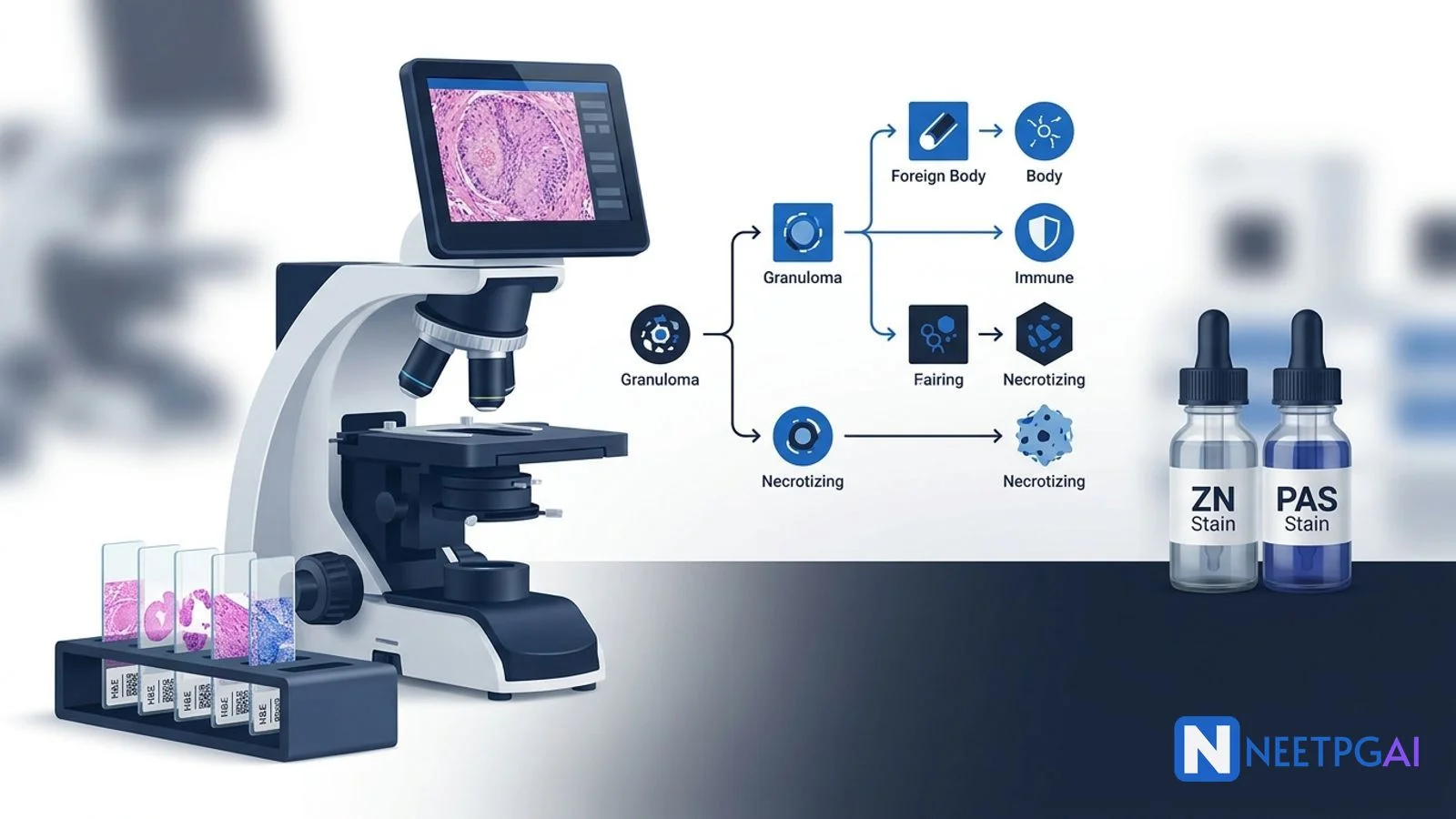
Granulomas are organized collections of activated macrophages (epithelioid histiocytes) that form in response to persistent antigenic stimulation. To correctly identify and classify granulomas in NEET PG image MCQs:
Check for central necrosis — caseating (cheese-like, eosinophilic, acellular) points to TB or fungal infection; non-caseating points to sarcoidosis, Crohn, or foreign body reaction
Identify giant cell type — Langhans giant cells (peripheral horseshoe nuclei) indicate immune granulomas (TB, sarcoidosis); foreign body giant cells (scattered nuclei) indicate inert material reaction
Look for special features — Schaumann/asteroid bodies (sarcoidosis), perineural granulomas (leprosy), foamy macrophages with globi (lepromatous leprosy), birefringent material under polarized light (foreign body)
Clinical image presentation
A 35-year-old male presents with a 3-month history of low-grade fever, night sweats, weight loss (6 kg), and a non-productive cough. Chest X-ray shows bilateral hilar lymphadenopathy. An excisional biopsy of a supraclavicular lymph node is performed.
The histopathology slide (H&E stain, medium power magnification) shows the following findings that a student should identify systematically:
Low-power survey:
The normal lymph node architecture is partially effaced
Multiple discrete, well-formed granulomas are visible
The granulomas are surrounded by a rim of lymphocytes
Medium-power detail:
Each granuloma shows a central zone of amorphous, eosinophilic, acellular material — this is caseous necrosis
The necrotic center is surrounded by epithelioid histiocytes — elongated macrophages with abundant pink cytoplasm and vesicular nuclei that resemble epithelial cells
Langhans giant cells are present — large multinucleated cells with nuclei arranged in a horseshoe or crescentic pattern along the cell periphery
A cuff of lymphocytes and occasional plasma cells surrounds the epithelioid layer
High-power confirmation:
The central necrosis is acellular and granular (no ghost outlines of cells as in coagulative necrosis)
Epithelioid cells have indistinct cell borders and "slipper-shaped" nuclei
The Langhans giant cell nuclei are clustered at one pole of the cell, forming a characteristic arc
MCQ question as it appears in NEET PG
A 35-year-old male with fever, weight loss, and bilateral hilar lymphadenopathy undergoes lymph node biopsy. Histopathology shows well-formed granulomas with central caseous necrosis, epithelioid cells, and Langhans giant cells. Which of the following is the most likely diagnosis?
(a) Sarcoidosis
(b) Tuberculosis
(c) Hodgkin lymphoma
(d) Non-Hodgkin lymphoma
Take a moment to work through this before reading the analysis below.
Step-by-step visual analysis
Systematic histopathology slide reading is critical for NEET PG image MCQs. Jumping to one finding and ignoring the overall pattern is the reason students confuse TB with sarcoidosis or miss a lymphoma. Use this protocol every time:
Step 1: Low-power survey (scan the entire slide)
Before identifying individual structures, assess the overall architecture at low magnification (4x or 10x):
Is the tissue architecture preserved or effaced? In this biopsy, the lymph node architecture is partially effaced by granulomas. Complete effacement would raise concern for lymphoma.
What is the dominant pattern? Granulomatous (discrete collections), diffuse (sheet-like infiltrate), or follicular (nodular). Here: granulomatous pattern.
Are the granulomas discrete or confluent? Discrete, well-formed granulomas suggest an organized immune response. Confluent, poorly formed granulomas may indicate immunosuppression.
Step 2: Evaluate the granuloma center (the defining step)
The center of the granuloma determines the classification — this is the single most important observation:
Caseous necrosis — amorphous, eosinophilic (pink), acellular material with a granular texture. Resembles cottage cheese grossly. Diagnosis: TB until proven otherwise. Also seen in histoplasmosis, coccidioidomycosis, and some other fungal infections.
No necrosis (non-caseating) — the granuloma is composed entirely of epithelioid cells without a necrotic center. Think: sarcoidosis, Crohn disease, berylliosis, foreign body.
Suppurative center — pus (neutrophils) in the center of the granuloma. Think: cat scratch disease (Bartonella), lymphogranuloma venereum, fungal infection (blastomycosis).
In this slide: the center shows classic caseous necrosis — eosinophilic, acellular, granular material.
Step 3: Identify the cellular components
Moving outward from the center:
Epithelioid histiocytes — the defining cell of a granuloma. These are activated macrophages with elongated, pale, vesicular nuclei and abundant eosinophilic cytoplasm. Their cell borders are indistinct, giving a "syncytial" appearance. They are arranged in a palisading pattern around the necrotic center.
Giant cells — identify the nuclear arrangement:
Langhans type: nuclei form a horseshoe/crescent at the periphery. Present here. Characteristic of immune granulomas (TB, sarcoidosis).
Foreign body type: nuclei scattered randomly. NOT present in this slide.
Touton type: nuclei form a wreath/ring with central lipid. Seen in xanthogranulomas, fat necrosis.
Lymphocyte cuff — a rim of T-lymphocytes (CD4+ in TB, CD8+ tend to predominate around sarcoid granulomas in some studies) surrounds the granuloma. A thick lymphocyte cuff is seen in TB; a thin or absent cuff ("naked granuloma") is more typical of sarcoidosis.
Step 4: Look for special features and inclusions
Schaumann bodies — laminated, calcified, conchoidal inclusions within giant cells. Favor sarcoidosis. NOT present in this slide.
Asteroid bodies — star-shaped eosinophilic inclusions within giant cells. Favor sarcoidosis. NOT present here.
Birefringent material under polarized light — indicates foreign body (suture, talc, silicone). Not applicable here.
Perineural involvement — granulomas surrounding nerves indicate leprosy. Not seen in this lymph node biopsy.
Step 5: Correlate with special stains
The histological pattern suggests TB, but special stains confirm:
Ziehl-Neelsen (ZN) stain — look for bright red, beaded, rod-shaped organisms against a blue background. In paucibacillary TB, bacilli may be extremely rare (1 bacillus per 100 oil-immersion fields).
Auramine-rhodamine fluorescence — more sensitive than ZN (sensitivity 60-80% vs 20-60% for ZN on tissue). Bacilli fluoresce bright green/yellow.
GMS and PAS — negative for fungi in this case (rules out histoplasmosis, coccidioidomycosis).
Answer and detailed explanation
Correct answer: (b) Tuberculosis
The combination of caseous necrosis + epithelioid cells + Langhans giant cells + lymphocyte cuff is the classic histological pattern of tuberculosis. The clinical context (young male, fever, weight loss, bilateral hilar lymphadenopathy) is entirely consistent with tubercular lymphadenitis — the most common form of extrapulmonary TB in India.
Why each distractor is wrong:
Option
Why incorrect
(a) Sarcoidosis
Sarcoidosis produces non-caseating granulomas. Central caseous necrosis essentially rules out sarcoidosis. Both conditions cause bilateral hilar lymphadenopathy, but the histology is the differentiator.
(c) Hodgkin lymphoma
Hodgkin lymphoma can cause granulomatous reaction (5-10% of cases show granulomas adjacent to Reed-Sternberg cells), but the granulomas are non-caseating and the diagnostic feature is the RS cell itself (bilobed "owl-eye" nucleus, CD15+, CD30+). No RS cells are described here.
(d) Non-Hodgkin lymphoma
NHL causes diffuse or follicular lymph node effacement by monomorphic lymphoid cells. Granulomas are not a feature. The well-formed granulomatous pattern makes NHL highly unlikely.
NEET PG trap alert: Sarcoidosis vs TB is the most tested differential in granuloma questions. Both cause hilar lymphadenopathy. The distinguishing feature on histology is always the necrosis — caseating = TB, non-caseating = sarcoidosis. If the question stem says "non-caseating granuloma with bilateral hilar lymphadenopathy in a young patient," the answer is sarcoidosis, not TB, regardless of the clinical similarity.
Granulomatous diseases overlap in clinical presentation but diverge on histology. This table covers the five conditions most commonly tested in NEET PG image-based MCQs:
What is the difference between caseating and non-caseating granulomas?
Caseating granulomas have central necrosis that appears cheese-like (caseous) on gross examination and eosinophilic, amorphous, and acellular on histology. They are characteristic of tuberculosis and certain fungal infections. Non-caseating (non-necrotizing) granulomas lack central necrosis and are composed of tightly packed epithelioid histiocytes. They are characteristic of sarcoidosis, Crohn disease, and berylliosis. This distinction is the single most tested granuloma concept in NEET PG.
What is a Langhans giant cell and how does it differ from a foreign body giant cell?
A Langhans giant cell has nuclei arranged in a horseshoe or peripheral crescentic pattern at the cell margin. It is formed by fusion of epithelioid macrophages and is characteristic of granulomatous inflammation, especially tuberculosis. A foreign body giant cell has nuclei scattered haphazardly throughout the cytoplasm without peripheral arrangement. It forms around indigestible material like suture, talc, or silicone. Both are multinucleated giant cells, but the nuclear arrangement is the distinguishing feature.
What are the most common causes of granulomatous inflammation tested in NEET PG?
The top causes tested are: tuberculosis (caseating granuloma, Langhans giant cells, AFB-positive), sarcoidosis (non-caseating granuloma, Schaumann and asteroid bodies), leprosy (tuberculoid: epithelioid granulomas around nerves; lepromatous: foamy macrophages, globi), Crohn disease (non-caseating granulomas in bowel wall, transmural inflammation), and foreign body reaction (foreign body giant cells around suture, talc). Fungal granulomas (histoplasmosis, coccidioidomycosis) and cat scratch disease are also tested occasionally.
What are Schaumann bodies and asteroid bodies?
Schaumann bodies (conchoidal bodies) are laminated calcium and protein inclusions found within the giant cells of sarcoid granulomas. They appear as round, concentrically layered, basophilic structures. Asteroid bodies are star-shaped, eosinophilic inclusions within giant cells, also seen in sarcoidosis. While neither is pathognomonic for sarcoidosis (they can occur in other granulomatous diseases), their presence in a non-caseating granuloma strongly supports the diagnosis.
How do I differentiate TB granuloma from sarcoid granuloma on histology?
TB granuloma shows caseating necrosis centrally, surrounded by epithelioid cells, Langhans giant cells, and a lymphocyte cuff. Staining with Ziehl-Neelsen may reveal acid-fast bacilli. Sarcoid granuloma is non-caseating (no central necrosis), tightly formed ("naked" granuloma with fewer surrounding lymphocytes), and may contain Schaumann or asteroid bodies. The key differentiator: central caseous necrosis = TB until proven otherwise; absence of necrosis with tight granuloma = sarcoidosis.
What stains are used to identify organisms in granulomatous inflammation?
Ziehl-Neelsen (ZN) stain or auramine-rhodamine fluorescence for acid-fast bacilli (Mycobacteria). GMS (Grocott-Gomori methenamine silver) and PAS (periodic acid-Schiff) for fungal organisms. Fite-Faraco stain for lepra bacilli (modified ZN — regular ZN can miss M. leprae because its cell wall is thinner). Warthin-Starry stain for Bartonella (cat scratch disease). Polarized light microscopy for foreign bodies (suture, talc, silicone — these are birefringent).
What is the significance of granulomas in Crohn disease?
Non-caseating granulomas are found in only 30-50% of Crohn disease biopsies, but when present, they strongly support the diagnosis over ulcerative colitis. They are typically small, scattered throughout the bowel wall (transmural), and may be found in mesenteric lymph nodes. In NEET PG, the combination of transmural inflammation + non-caseating granulomas + skip lesions is the classic triad that differentiates Crohn from UC.
What is the histopathology of leprosy and how does it differ between tuberculoid and lepromatous forms?
Tuberculoid leprosy shows well-formed epithelioid granulomas with Langhans giant cells, typically centered around dermal nerves (perineural granulomas). Bacilli are few or absent on Fite stain (paucibacillary). Lepromatous leprosy shows diffuse dermal infiltrates of foamy macrophages (Virchow cells or lepra cells) packed with acid-fast bacilli arranged in clumps called globi. The granuloma spectrum mirrors the immune response: strong cell-mediated immunity in tuberculoid, weak in lepromatous.
This content is for educational purposes for NEET PG exam preparation. It is not a substitute for professional medical advice, diagnosis, or treatment. Clinical information has been reviewed by qualified medical professionals.
Sources and references
Robbins and Cotran Pathologic Basis of Disease, 10th Edition (Kumar, Abbas, Aster, 2021) — Chapter on Inflammation and Repair, granulomatous inflammation classification and morphology.
Rosai and Ackerman's Surgical Pathology, 11th Edition (Goldblum et al., 2018) — Lymph node pathology, differential diagnosis of granulomatous lymphadenitis.
Harrison's Principles of Internal Medicine, 21st Edition (Loscalzo et al., 2022) — Chapters on Tuberculosis and Sarcoidosis, clinical-pathological correlation.
For personalized study guidance on pathology and other high-yield topics, try the AI Tutor — it adapts to your weak areas and explains concepts the way a senior resident would.
Written by: NEETPGAI Editorial Team
Reviewed by: Pending SME Review
Last reviewed: April 2026
This article is reviewed by qualified medical professionals for clinical accuracy and exam relevance. For corrections or updates, contact the editorial team.