Version 1.0 — Published April 2026
Quick Answer
Papilledema and glaucomatous disc are opposite optic disc pathologies tested in NEET PG fundoscopy image questions. To differentiate them:
- Papilledema — disc margins blurred, disc hyperemic and elevated, cup obscured, veins engorged and non-pulsating, flame-shaped hemorrhages, bilateral, vision preserved initially
- Glaucomatous disc — disc margins sharp, cup enlarged (C:D ratio >0.6), neuroretinal rim thinning (ISNT rule violated), disc pallor, lamina cribrosa dots visible, asymmetric between eyes
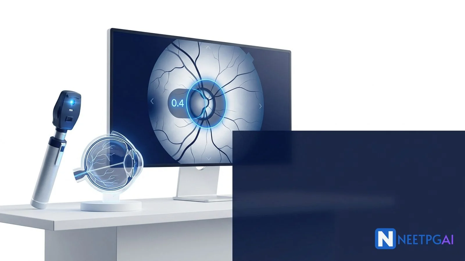
- Cup-to-disc ratio — normal <0.3, glaucoma suspect >0.6, definite concern >0.8, asymmetry >0.2 between eyes
Clinical image presentation
A 45-year-old woman presents to the ophthalmology OPD with headaches for 3 months that are worse in the morning and aggravated by coughing and straining. She has noticed transient visual obscurations (brief episodes of graying out of vision lasting seconds, especially on standing). Her visual acuity is 6/6 in both eyes. Direct ophthalmoscopy reveals bilateral disc changes that require systematic evaluation.
Simultaneously, a 62-year-old man presents for a routine eye check-up. He is asymptomatic but has a family history of glaucoma (father, diagnosed at age 55). His intraocular pressure is 24 mmHg in both eyes (normal <21 mmHg). His visual acuity is 6/6 bilaterally. Fundoscopy reveals optic disc changes that differ significantly from the first patient.
These two presentations illustrate the two most important optic disc pathologies for NEET PG — papilledema and glaucomatous cupping. The systematic fundoscopic approach below will help you differentiate them confidently.
MCQ question as it appears in NEET PG
A 45-year-old obese woman presents with headaches, transient visual obscurations, and diplopia. Fundoscopy shows bilateral disc swelling with blurred margins, engorged veins, and flame-shaped peripapillary hemorrhages. Visual acuity is 6/6. The most likely diagnosis is:
- (a) Bilateral optic neuritis
- (b) Papilledema due to raised intracranial pressure
- (c) Bilateral glaucomatous disc damage
- (d) Foster Kennedy syndrome
Take a moment to work through this before reading the analysis below.
Step-by-step visual analysis
Step 1: Assess the disc margins
The first thing to evaluate on any fundoscopic image is whether the disc margins are sharp or blurred.
Papilledema: The disc margins are blurred, starting with the nasal margin (nasal margin blurring is the earliest sign of papilledema because the nasal retinal nerve fibers are thickest). As papilledema progresses, all margins become obscured. The disc appears elevated above the surrounding retina.
Glaucomatous disc: The disc margins remain sharp and well-defined. The pathology is within the disc, not at its edges. Even in advanced glaucoma, the disc border is crisp. If you see a disc with blurred margins in an image question, glaucoma is effectively ruled out.
Step 2: Evaluate the cup
Papilledema: The optic cup is obscured or absent because disc swelling fills the cup. The normal central depression disappears, and the disc surface appears dome-shaped.
Glaucomatous disc: The cup is enlarged. This is the hallmark of glaucoma — progressive loss of retinal ganglion cell axons leads to enlargement and deepening of the optic cup. Measure the cup-to-disc ratio:
| C:D ratio | Interpretation |
|---|
| 0.0-0.3 | Normal |
| 0.3-0.5 | Normal variant (physiological, especially in large discs) |
| 0.6-0.7 | Suspicious — requires serial monitoring and visual field testing |
| >0.8 | Strongly suggestive of glaucoma |
| Asymmetry >0.2 | Abnormal regardless of absolute value |
Step 3: Examine the neuroretinal rim
The neuroretinal rim is the tissue between the edge of the cup and the disc margin. It contains the retinal ganglion cell axons.
ISNT rule (normal rim thickness order): Inferior > Superior > Nasal > Temporal. The inferior rim is the thickest; the temporal rim is the thinnest. Violation of this rule — especially focal thinning (notching) of the inferior or superior rim — is an early and specific sign of glaucomatous damage.
In glaucoma: Look for focal notching (localized thinning of the rim, especially inferotemporal or superotemporal), generalized rim thinning, and eventual "bean-pot" or "baring" of circumlinear blood vessels (vessels no longer supported by rim tissue and appear to drop off the disc edge).
In papilledema: The rim cannot be properly assessed because disc swelling obscures the cup-rim relationship.
Step 4: Check the vessels
Papilledema:
- Retinal veins are engorged and tortuous (venous congestion from raised ICP impeding venous return)
- Spontaneous venous pulsations are absent (present in ~80% of normal individuals — their absence suggests ICP >190 mmH2O)
- Flame-shaped hemorrhages in the peripapillary retinal nerve fiber layer
- Possible cotton-wool spots (retinal infarcts from capillary compression)
Glaucomatous disc:
- Vessels may show bayoneting — a vessel appears to dip backward as it crosses the rim then reappears at the base of the excavated cup, creating a "bayonet" shape
- Nasal displacement of vessels — as the cup enlarges, the central retinal artery and vein are displaced nasally
- Baring of circumlinear vessels — vessels that normally run along the rim now appear unsupported at the disc edge
Step 5: Look at the surrounding retina
Papilledema: Peripapillary retinal nerve fiber layer (RNFL) swelling causes a striated, glistening appearance around the disc. Flame-shaped hemorrhages radiate from the disc. Paton lines (circumferential retinal folds around the disc) may be visible.
Glaucoma: The peripapillary RNFL shows wedge-shaped defects — dark bands in the RNFL best seen with red-free (green) illumination, corresponding to localized ganglion cell axon loss. These RNFL defects precede visual field loss and are an early diagnostic sign.
Step 6: Assess the lamina cribrosa
Glaucomatous disc: In advanced cupping, the lamina cribrosa dots become visible at the base of the deep cup — these are the fenestrations in the cribriform plate through which retinal ganglion cell axons pass. Their visibility indicates significant excavation and advanced glaucomatous damage (Kanski's Clinical Ophthalmology, 9th Edition).
Papilledema: Lamina cribrosa dots are not visible because the cup is filled by swollen tissue.
Answer and detailed explanation
Answer: (b) Papilledema due to raised intracranial pressure.
Why (b) is correct: The vignette describes a 45-year-old obese woman with headaches (worse in morning, aggravated by Valsalva), transient visual obscurations, and bilateral disc swelling with blurred margins, engorged veins, and flame-shaped hemorrhages. Visual acuity is preserved at 6/6. This is the classic presentation of papilledema from raised ICP — specifically, this matches idiopathic intracranial hypertension (pseudotumor cerebri), which characteristically affects young obese women.
Why (a) is wrong: Bilateral optic neuritis would cause significant visual loss (usually 6/18 or worse), pain on eye movement, and relative afferent pupillary defect. The preserved 6/6 acuity effectively rules out bilateral optic neuritis. Also, bilateral simultaneous optic neuritis is rare — it should raise suspicion for neuromyelitis optica (NMO) rather than typical MS-associated optic neuritis.
Why (c) is wrong: Glaucomatous disc changes show sharp margins with enlarged cupping, not blurred margins with disc swelling. Glaucoma also does not cause hemorrhages in this pattern. The fundoscopic description in the stem is the opposite of glaucoma.
Why (d) is wrong: Foster Kennedy syndrome is disc swelling in one eye (from direct compression of the ipsilateral optic nerve by an olfactory groove or frontal lobe tumor) with optic atrophy in the contralateral eye. This patient has bilateral disc swelling, which rules out the classic Foster Kennedy pattern.
Similar patterns comparison table
| Feature | Papilledema | Glaucomatous disc | Optic atrophy | Papillitis (optic neuritis) |
|---|
| Disc margins | Blurred | Sharp | Sharp | Blurred |
| Disc color | Hyperemic (pink-red) | Pale (especially temporal pallor) | White/chalky pale | Hyperemic |
| Cup | Obscured | Enlarged (C:D >0.6) | Normal or enlarged | May be obscured |
| Laterality | Bilateral | Usually bilateral (may be asymmetric) | Depends on cause | Usually unilateral |
| Visual acuity | Preserved (until late) | Preserved (until late) | Reduced | Significantly reduced |
| Visual field | Enlarged blind spot | Arcuate scotoma, nasal step | Depends on cause | Central scotoma |
| Hemorrhages | Flame-shaped peripapillary | Disc hemorrhages (Drance hemorrhage) | Absent | May be present |
| Venous pulsations | Absent | Present (usually) | Present | Variable |
| IOP | Normal | Elevated (>21 mmHg in POAG) | Normal | Normal |
Frequently asked questions
What is the normal cup-to-disc ratio?
Normal C:D ratio is 0.3 or less. Above 0.6 is suspicious for glaucoma, above 0.8 is strongly suggestive. Asymmetry between eyes of more than 0.2 is a red flag regardless of absolute value. Glaucoma enlarges the cup; papilledema obscures it.
How do I differentiate papilledema from papillitis?
Papilledema: bilateral, vision preserved, raised ICP, enlarged blind spot. Papillitis: usually unilateral, significant visual loss, pain on eye movement, central scotoma, RAPD present. Both show disc swelling on fundoscopy.
What causes papilledema?
Raised ICP from intracranial tumors, idiopathic intracranial hypertension (young obese women), cerebral venous sinus thrombosis, hydrocephalus, or malignant hypertension. First investigation: neuroimaging (CT/MRI) before lumbar puncture.
What is the significance of spontaneous venous pulsations?
SVPs present = ICP likely normal. SVPs absent = nonspecific (20% of normal people lack them). In a patient with disc swelling, absent SVPs support raised ICP but their absence alone does not confirm it.
What visual field defect does glaucoma produce?
Progressive pattern: enlarged blind spot, nasal step, arcuate scotoma (Bjerrum), ring scotoma, tunnel vision. Glaucoma affects peripheral vision first, spares central vision until late. This explains why patients are asymptomatic until advanced disease.
What is the ISNT rule?
Normal neuroretinal rim thickness: Inferior (thickest) > Superior > Nasal > Temporal (thinnest). Violation, especially inferior or superior rim notching, is an early sign of glaucomatous damage. Tested in NEET PG image questions requiring rim assessment (Ophthalmology high-yield topics reference: ophthalmology subject page).
For more ophthalmology image-based practice, see the ophthalmology high-yield topics guide and practice ophthalmology MCQs. Want AI-powered image analysis for fundoscopy images? Explore NEETPGAI Pro.
This content is for educational purposes for NEET PG exam preparation. It is not a substitute for professional medical advice, diagnosis, or treatment. Clinical information has been reviewed by qualified medical professionals.
Written by: NEETPGAI Editorial Team
Reviewed by: Pending SME Review
Last reviewed: April 2026
This article is reviewed by qualified medical professionals for clinical accuracy and exam relevance. For corrections or updates, contact the editorial team.