Version 1.0 - Published October 2026
Quick Answer
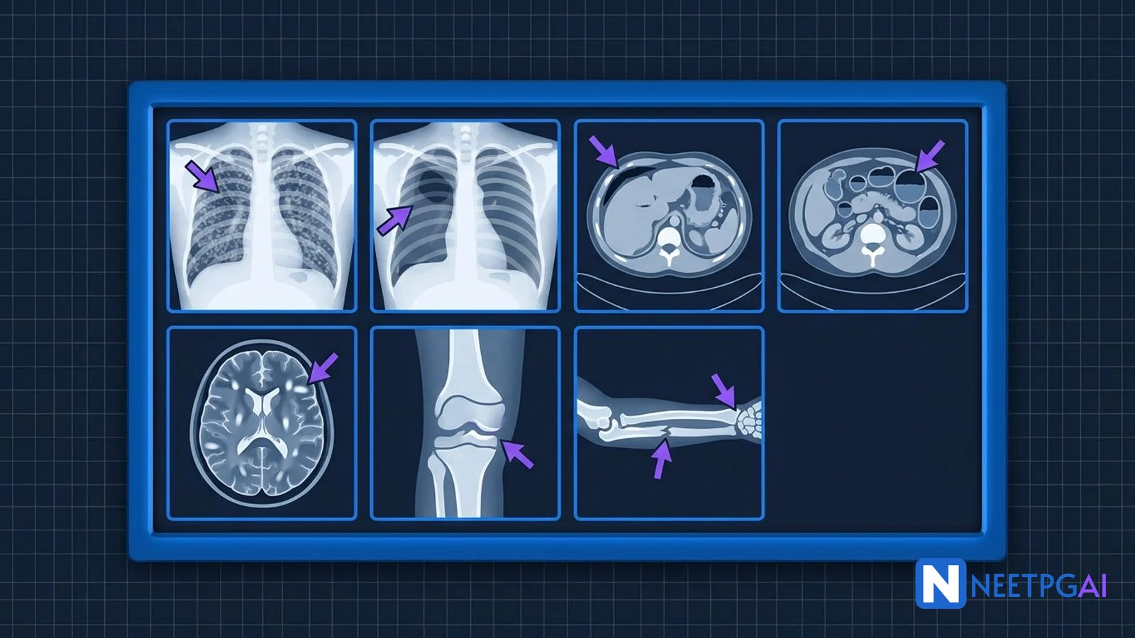
Radiology image MCQs account for 3-5 pure plus 8-10 embedded image stems per INI-CET paper. Seven high-yield patterns dominate - miliary TB, pneumothorax, pleural effusion with meniscus, pneumoperitoneum, SBO coffee bean, acute appendicitis, MS Dawson fingers, acute stroke DWI, HSV encephalitis, SCFE, Monteggia, Galeazzi. Drill the seven MCQs below and you add 5-7 marks on exam day.
Why This Matters for NEET PG & INI-CET
Radiology image MCQs reward one specific skill - pattern recognition on a projection you have seen before. Across INI-CET Nov 2023 to May 2025, radiology contributed 3 pure questions per paper plus 8-10 embedded image stems in Medicine (chest X-ray, ECG), Surgery (CT abdomen, X-ray), Orthopedics (fracture X-rays), Pediatrics (neonatal and infant classics), Neurology (MRI brain), and OBG (ultrasound and MRI pelvis). On NEET PG 2026, image stems are expected to account for roughly 20-25% of the 180 questions.
Repeat density on radiology images is 18% - direct lifts are rare because the archive is large and patient-identifiable, but near-direct lifts (same pathology, different image) are common. The predictability by pathology category is high - six categories account for 70% of all radiology output. Learn each category with its signature imaging modality, projection, and classical imaging finding, and you will recognise most exam-day images on sight.
The seven MCQs below follow the INI-CET pattern - stem with image prompt, four options, one correct answer, detailed explanation, and a one-liner. Each image is written for the NEETPGAI Gemini image pipeline.
MCQ 1 - Chest X-Ray Miliary Pattern
A 24-year-old HIV-positive woman presents with 3 weeks of fever, night sweats, and dry cough. CD4 count is 85 cells/mcL. Chest X-ray is shown above. What is the most likely diagnosis?
A. Pneumocystis jirovecii pneumonia
B. Miliary tuberculosis
C. Metastatic lung disease
D. Pulmonary alveolar proteinosis
Correct answer: B. Miliary tuberculosis
Explanation: The chest X-ray shows diffuse uniform 1-3 mm nodules throughout both lung fields - the hallmark miliary pattern. In an HIV-positive patient with low CD4 count presenting with fever, night sweats, and weight loss, miliary TB is the leading diagnosis. The nodules represent haematogenous spread of Mycobacterium tuberculosis with granulomas forming at random throughout the pulmonary interstitium. PCP produces diffuse bilateral ground-glass opacities on CT, not discrete nodules; chest X-ray in PCP is typically normal or shows subtle perihilar haze. Metastatic lung disease (cannonball metastases from renal, colorectal, or thyroid primaries) usually produces larger (5-20 mm), fewer, and more variable-sized nodules. Pulmonary alveolar proteinosis produces a crazy-paving pattern on CT with ground-glass opacity and septal thickening. Diagnosis is confirmed by acid-fast bacilli in sputum, GeneXpert MTB-RIF, and often bone marrow biopsy showing caseating granulomas with AFB.
High-yield one-liner: Uniform 1-3 mm nodules throughout both lungs in HIV = miliary TB until proven otherwise.
MCQ 2 - Chest X-Ray Pneumothorax
A 23-year-old tall thin man presents with sudden-onset right-sided chest pain and dyspnoea while playing cricket. On examination, he has tachycardia 128/min, BP 86/52 mmHg, trachea deviated to the left, and absent breath sounds on the right. The chest X-ray is shown. What is the next immediate step?
A. Portable CT chest
B. Immediate needle decompression at the second intercostal space mid-clavicular line, followed by intercostal drain
C. Observation and oxygen only
D. CT pulmonary angiogram
Correct answer: B. Immediate needle decompression followed by intercostal drain
Explanation: The chest X-ray shows a right-sided tension pneumothorax with mediastinal shift away from the affected side, flattened right hemidiaphragm, and widened intercostal spaces. In a haemodynamically unstable patient with signs of tension pneumothorax, immediate needle decompression is mandatory before any imaging. The classic site is second intercostal space mid-clavicular line, though the fifth intercostal space anterior axillary line is now preferred by some trauma guidelines (ATLS tenth edition). Needle decompression is followed by definitive intercostal drain (chest tube) insertion in the fifth intercostal space anterior to mid-axillary line (triangle of safety). Spontaneous pneumothorax in a tall thin young man is typically caused by rupture of a subpleural apical bleb; recurrence rate is 30% after first episode and surgical pleurodesis or VATS bullectomy is considered after the second episode.
High-yield one-liner: Tension pneumothorax = immediate needle decompression before imaging. Second ICS MCL or fifth ICS AAL.
MCQ 3 - Pleural Effusion with Meniscus
A 58-year-old man presents with 3 weeks of progressive dyspnoea and dull chest pain. Examination shows decreased breath sounds and stony dull percussion over the right lower chest. Chest X-ray is shown. Thoracentesis reveals pleural fluid LDH 320 U/L (serum LDH 180 U/L), pleural protein 4.2 g/dL (serum protein 6.8 g/dL). What is the fluid type and most likely cause?
A. Transudate; congestive heart failure
B. Exudate; malignant pleural effusion
C. Transudate; nephrotic syndrome
D. Chylous effusion; thoracic duct injury
Correct answer: B. Exudate; malignant pleural effusion
Explanation: The X-ray shows a classic meniscus sign - fluid in the pleural space tracks up laterally against the lateral chest wall because of surface tension and the lower pressure in the apical pleural space. Light criteria classify pleural effusion as exudate if any one of three criteria is met - pleural protein to serum protein ratio >0.5, pleural LDH to serum LDH ratio >0.6, or pleural LDH >two-thirds of the upper limit of normal serum LDH. This patient meets both protein ratio (4.2/6.8 = 0.62) and LDH ratio (320/180 = 1.78) criteria, confirming an exudate. In a 58-year-old smoker with progressive symptoms, malignant pleural effusion (lung cancer with pleural seeding or metastatic from breast, GI, lymphoma) is the leading diagnosis. Pleural cytology and pleural biopsy (closed, VATS, or image-guided) establish the specific primary. Transudate causes are CHF, cirrhosis, nephrotic syndrome, and hypoalbuminaemia. Exudate causes are infection (parapneumonic, empyema, TB), malignancy, pulmonary embolism, and connective tissue disease.
High-yield one-liner: Meniscus sign + Light criteria positive = exudative pleural effusion. Malignancy and infection are the top causes.
MCQ 4 - Pneumoperitoneum CT Abdomen
A 72-year-old man on long-term NSAID therapy for osteoarthritis presents with sudden severe epigastric pain radiating to the shoulder, followed by generalised abdominal rigidity 4 hours later. CT abdomen is shown. What is the next step?
A. Oral PPI and observation
B. Diagnostic laparoscopy and primary closure of perforated duodenal ulcer with Graham patch
C. Upper GI endoscopy
D. CT angiogram
Correct answer: B. Diagnostic laparoscopy and primary closure of perforated duodenal ulcer with Graham patch
Explanation: The CT shows extensive free intraperitoneal air anterior to the liver and under the diaphragm - pneumoperitoneum - which in this clinical scenario of acute severe epigastric pain in an NSAID user indicates perforated peptic ulcer. The classic Graham omental patch repair involves placing an omental pedicle over the perforation and suturing it in place; this is standard for perforated duodenal ulcer. For perforated gastric ulcer, biopsy of the margin is mandatory to exclude malignancy before omental patch. Erect chest X-ray would show free gas under the right hemidiaphragm (classic sign), but CT abdomen is more sensitive and now the preferred investigation in haemodynamically stable patients. Mortality from perforated peptic ulcer rises linearly with the time from perforation to surgical intervention - the 24-hour mortality is 10%, rising to 40% at 48 hours. Oral PPI is not appropriate for a perforated ulcer; endoscopy is contraindicated because insufflation worsens peritoneal contamination.
High-yield one-liner: Free gas under diaphragm or on CT = perforated viscus until proven otherwise. Graham omental patch is standard for perforated duodenal ulcer.
MCQ 5 - MRI Brain Multiple Sclerosis
A 28-year-old woman presents with a 3-day history of painful loss of vision in the right eye with a central scotoma. Fundus examination shows optic disc swelling. Six months earlier she had a brief episode of right lower limb numbness that resolved spontaneously. MRI brain is shown. What is the most likely diagnosis?
A. Neuromyelitis optica spectrum disorder
B. Multiple sclerosis
C. Acute disseminated encephalomyelitis
D. Progressive multifocal leukoencephalopathy
Correct answer: B. Multiple sclerosis
Explanation: The MRI shows multiple ovoid periventricular hyperintense lesions oriented perpendicular to the lateral ventricles - the classic Dawson fingers appearance pathognomonic of multiple sclerosis. The clinical presentation of optic neuritis (painful monocular vision loss with central scotoma) in a young woman with a prior transient neurological episode suggests dissemination in time and in space, meeting the McDonald 2017 criteria for MS. Diagnosis is confirmed by CSF oligoclonal bands (present in 90% of MS, absent in most mimics), MRI dissemination in space (lesions in at least 2 of 4 CNS areas - periventricular, juxtacortical, infratentorial, spinal cord) and in time (new or contrast-enhancing lesion). NMOSD typically presents with longitudinally extensive transverse myelitis spanning 3+ vertebral segments and is positive for aquaporin-4 antibodies. ADEM is monophasic post-infectious inflammatory demyelination, usually in children. PML occurs in immunocompromised patients and shows asymmetric subcortical white matter lesions without mass effect.
High-yield one-liner: Dawson fingers on sagittal FLAIR + optic neuritis + prior neurological episode = multiple sclerosis.
MCQ 6 - Slipped Capital Femoral Epiphysis (SCFE)
A 13-year-old obese boy presents with 6 weeks of insidious-onset left hip and knee pain, walking with an externally rotated foot and antalgic gait. There is no history of trauma. On examination, there is loss of internal rotation of the left hip. X-ray is shown. What is the diagnosis and management?
A. Developmental dysplasia of the hip; closed reduction and spica cast
B. Perthes disease; non-weight bearing and orthosis
C. Slipped capital femoral epiphysis; urgent in situ percutaneous screw fixation
D. Transient synovitis; NSAIDs and bed rest
Correct answer: C. Slipped capital femoral epiphysis; urgent in situ percutaneous screw fixation
Explanation: The frog-leg lateral X-ray shows posteroinferior slippage of the capital femoral epiphysis relative to the femoral neck - the hallmark of SCFE. The Klein line drawn along the superior femoral neck normally intersects the lateral part of the epiphysis; in SCFE it does not, which is a sensitive diagnostic sign. SCFE is a Salter-Harris type I injury occurring at the proximal femoral physis, typically in obese adolescents during the growth spurt (ages 10-14). It presents with hip or knee pain, external rotation deformity, and loss of internal rotation. The classic sign of referred knee pain from a hip problem is pathognomonic for SCFE in this age group - never miss it. Management is urgent surgical stabilisation with percutaneous in situ screw fixation to prevent further slippage and avoid the dreaded complication of avascular necrosis of the femoral head. Delay beyond 48 hours increases AVN risk sharply. The contralateral hip should be prophylactically pinned in high-risk patients (very young onset, endocrine aetiology).
High-yield one-liner: Obese adolescent + hip or knee pain + externally rotated foot = SCFE. Urgent in situ screw fixation; AVN risk rises with delay.
MCQ 7 - Monteggia versus Galeazzi
A 7-year-old child falls on an outstretched hand while playing. He presents with forearm pain and inability to extend his fingers at the MCP joints, though his wrist extension is preserved. X-ray is shown. What is the diagnosis and which nerve is at risk?
A. Galeazzi fracture-dislocation; ulnar nerve
B. Monteggia fracture-dislocation; posterior interosseous nerve (PIN)
C. Colles fracture; median nerve
D. Smith fracture; anterior interosseous nerve
Correct answer: B. Monteggia fracture-dislocation; posterior interosseous nerve (PIN)
Explanation: The X-ray shows fracture of the proximal third of the ulnar shaft with anterior dislocation of the radial head - the Monteggia fracture-dislocation. The mnemonic MUGR helps - Monteggia Ulna, Galeazzi Radius. The radiocapitellar line (drawn along the central axis of the proximal radius) should intersect the capitellum in any projection; failure to do so is diagnostic of radial head dislocation. PIN (posterior interosseous nerve, the deep branch of the radial nerve) is at risk because it passes through the supinator muscle just distal to the elbow joint - radial head dislocation damages the PIN at this site. The clinical pattern of PIN palsy is finger drop (inability to extend MCP joints) with preserved wrist extension (because the extensor carpi radialis longus is innervated proximal to the supinator by the radial nerve proper, not the PIN). In adults, Monteggia requires open reduction and plate fixation of the ulna with closed reduction of the radial head. In children, closed reduction of the radial head often follows acceptable reduction of the ulna. Galeazzi fracture-dislocation is a fracture of the distal third of the radius with dislocation of the distal radioulnar joint (DRUJ) - also requires surgical fixation in adults.
High-yield one-liner: Monteggia = ulnar fracture + radial head dislocation + PIN at risk (finger drop with preserved wrist extension).
High-Yield One-Liners
- Uniform 1-3 mm nodules on chest X-ray = miliary TB, especially in HIV with low CD4
- Tension pneumothorax = clinical diagnosis - needle decompression before imaging
- Needle decompression at second ICS mid-clavicular line or fifth ICS anterior axillary line
- Pleural effusion meniscus sign - fluid rises higher laterally; exudate by Light criteria
- Light criteria exudate - pleural protein/serum protein >0.5 OR pleural LDH/serum LDH >0.6
- Free gas under diaphragm = perforated viscus until proven otherwise
- Graham omental patch is standard for perforated duodenal ulcer
- CT abdomen with IV contrast is first-line for most acute abdomen presentations
- Appendix diameter >7 mm plus periappendiceal fat stranding = acute appendicitis
- Coffee bean sign = sigmoid volvulus
- Dawson fingers on sagittal FLAIR MRI = multiple sclerosis
- McDonald 2017 - MS needs dissemination in space (2+ CNS areas) and in time (new or enhancing lesion)
- Acute stroke on DWI - hyperintense on DWI, hypointense on ADC (restricted diffusion)
- HSV encephalitis - bilateral asymmetric temporal and inferior frontal oedema on MRI
- SCFE in obese adolescent - external rotation deformity; urgent in situ screw fixation
- Klein line fails to intersect epiphysis in SCFE
- Monteggia = ulnar fracture + radial head dislocation + PIN palsy risk
- Galeazzi = radial fracture + DRUJ dislocation + AIN risk
- Radiocapitellar line should intersect capitellum on any X-ray projection
Common Mistakes
- Ordering imaging in tension pneumothorax before needle decompression. Needle decompression is a clinical decision - do not wait for a radiograph in a haemodynamically unstable patient.
- Reading pleural effusion as transudate without Light criteria. Low protein alone does not define transudate - all three Light criteria must be checked.
- Treating referred knee pain in an adolescent as a knee problem. Always examine the hip - SCFE classically presents as knee pain.
- Reversing MUGR mnemonic. Monteggia is Ulna fracture, Galeazzi is Radius fracture.
- Attributing finger drop to ulnar nerve injury. PIN injury (branch of radial nerve) causes finger drop with preserved wrist extension; ulnar nerve causes claw hand with preserved finger extension.
Related Topics on NEETPGAI
Practice This on NEETPGAI
NEETPGAI's open question bank includes 1,500+ radiology image MCQs across chest X-ray, CT abdomen, MRI brain, orthopedic X-ray, obstetric and pediatric imaging - all SME-reviewed with annotated image explanations. Free for every registered aspirant - 28,000+ MCQs, real community-reconstructed PYQs, section-locked INI-CET and NEET PG mocks, and spaced-repetition revision. Pro features (AI Tutor, AI MCQ Generator, Clinical Case Simulator, AI Study Planner) layer on top. Sign up free and start drilling radiology images today.