Version 1.0 — Published April 2026
Quick Answer
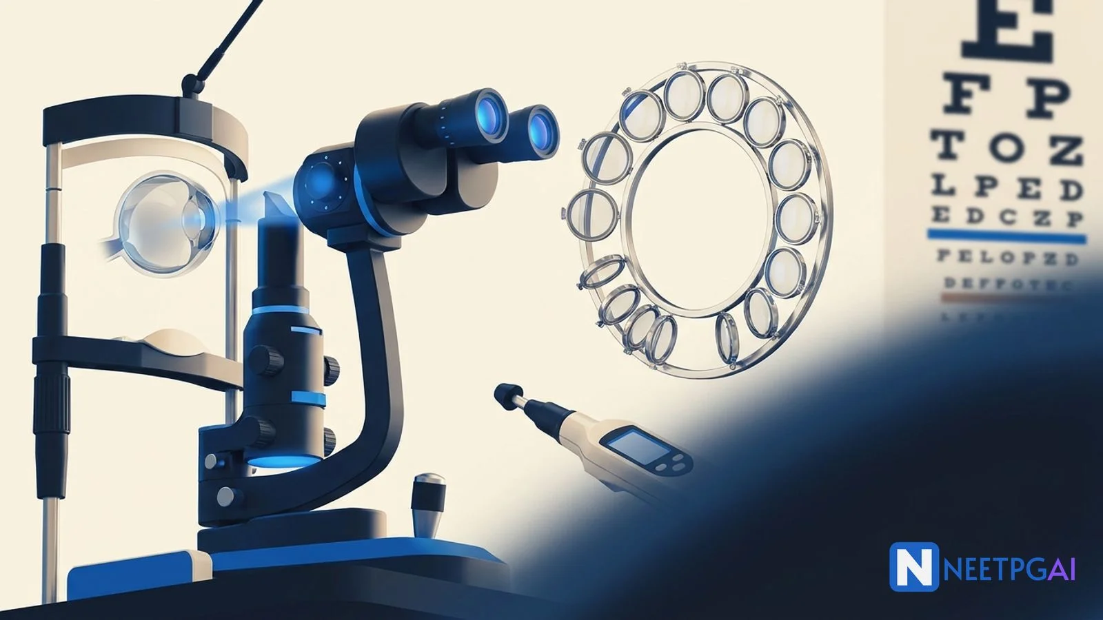
Ophthalmology contributes 8-12 questions to NEET PG and tests clinical pattern recognition rather than exhaustive textbook knowledge. The eight high-yield areas that return most frequently are:
- Glaucoma — open-angle vs angle-closure differentiation, IOP measurement (Goldmann applanation), anti-glaucoma drugs (timolol, latanoprost, pilocarpine), surgical options (trabeculectomy, iridotomy)
- Cataract — types (nuclear, cortical, PSC), surgery (phacoemulsification vs SICS), complications (posterior capsular opacification — Nd:YAG capsulotomy)
- Retinal diseases — diabetic retinopathy staging (4-2-1 rule for severe NPDR), ARMD (dry vs wet), retinal detachment (rhegmatogenous, tractional, exudative)
- Corneal diseases — ulcer types (bacterial vs fungal vs viral), keratoconus (Munson sign, Fleischer ring), corneal dystrophies
- Squint — esotropia vs exotropia, cover test and cover-uncover test, amblyopia management
- Refractive errors — myopia (concave lens), hypermetropia (convex lens), astigmatism (cylindrical lens), presbyopia (age 40+)
- Ocular emergencies — chemical injury (alkali worse than acid), CRAO (90-minute window), acute angle-closure crisis
- Neuro-ophthalmology — RAPD (Marcus Gunn pupil), Horner syndrome, Argyll Robertson pupil, visual field defects by lesion location
Ophthalmology is the subject where a single clinical sign often determines the answer. Unlike Medicine where you reason through a differential, ophthalmology MCQs frequently present a pathognomonic finding — Munson sign, cherry red spot, Argyll Robertson pupil — and expect you to name the diagnosis immediately. The good news: the number of such associations is finite, and NBE recycles them with remarkable consistency.
This guide covers the eight high-yield areas with the clinical facts NBE tests, the distinguishing features that separate correct from close-but-wrong options, and a practical study strategy. Pair it with the Ophthalmology subject hub and daily MCQ practice to convert reading into exam marks.
Glaucoma: types, measurement, and pharmacology
Glaucoma is a group of optic neuropathies characterized by progressive loss of retinal ganglion cells and their axons, resulting in characteristic optic disc cupping and visual field defects. It is the leading cause of irreversible blindness worldwide (WHO, 2020) and contributes 2-3 questions per NEET PG paper.
Primary open-angle glaucoma (POAG)
POAG is the most common type of glaucoma. The drainage angle is open, but aqueous outflow through the trabecular meshwork is reduced.
Clinical features: Bilateral, chronic, painless, insidious. Peripheral visual field loss progresses to tunnel vision. Central vision is preserved until late disease. IOP is elevated (typically 21-30 mmHg) but can be normal in normal-tension glaucoma.
Risk factors: Age above 40, family history, myopia, diabetes, thin central corneal thickness, African descent.
Visual field defects (in order of progression): Baring of blind spot, arcuate scotoma (Bjerrum scotoma), nasal step of Roenne, ring scotoma, tunnel vision, complete blindness.
Primary angle-closure glaucoma (PACG)
PACG occurs when the peripheral iris blocks the trabecular meshwork, preventing aqueous drainage.
Acute angle-closure crisis — a true ophthalmic emergency:
- Severe eye pain, headache, nausea/vomiting
- Blurred vision, halos around lights
- Corneal edema (steamy cornea)
- Fixed, mid-dilated pupil (vertically oval)
- Very high IOP (40-80 mmHg)
- Shallow anterior chamber
Precipitants: Dim lighting (pupil dilates), emotional stress, anticholinergic drugs (atropine), sympathomimetics (phenylephrine eye drops).
Emergency management:
- IV mannitol 20% (1-2 g/kg) — osmotic agent to lower IOP rapidly
- Topical timolol 0.5% — reduces aqueous production
- Topical pilocarpine 2% — constricts pupil, pulls iris away from angle (given after IOP drops below 40, otherwise sphincter is ischemic and unresponsive)
- Oral/IV acetazolamide — carbonic anhydrase inhibitor
- Definitive: laser peripheral iridotomy (LPI) — creates a hole in the peripheral iris to relieve pupillary block
IOP measurement
Goldmann applanation tonometry is the gold standard for IOP measurement. Based on the Imbert-Fick principle: the pressure inside a sphere equals the force needed to flatten a defined area of its surface. Measures the force to flatten a 3.06 mm diameter area of cornea. Affected by central corneal thickness (thicker cornea overestimates IOP, thinner underestimates).
Schiotz tonometry — indentation tonometry. Less accurate. Higher scale reading = lower IOP (inverse relationship — a common exam trap).
Anti-glaucoma drugs
| Drug class | Example | Mechanism | Key side effect |
|---|
| Beta-blockers | Timolol 0.5% | Reduces aqueous production | Bronchospasm (contraindicated in asthma), bradycardia |
| Prostaglandin analogs | Latanoprost, travoprost | Increases uveoscleral outflow | Iris pigmentation change, eyelash growth |
| Alpha-2 agonists | Brimonidine | Reduces production + increases outflow | Allergic conjunctivitis, drowsiness in children (contraindicated <2 years) |
| Carbonic anhydrase inhibitors | Dorzolamide (topical), acetazolamide (oral) | Reduces aqueous production | Metallic taste, metabolic acidosis (oral), hypokalemia |
| Miotics | Pilocarpine | Contracts ciliary muscle, opens trabecular meshwork | Miosis, accommodative spasm, brow ache |
| Osmotic agents | Mannitol (IV), glycerol (oral) | Reduces vitreous volume | Dehydration, renal overload |
First-line for POAG: Prostaglandin analogs (latanoprost) — once daily dosing, effective IOP reduction (25-30%), minimal systemic side effects.
Cataract: types, surgery, and complications
Cataract is opacification of the crystalline lens, causing progressive painless visual loss. It is the most common cause of treatable blindness globally and in India (National Blindness Survey, 2019). NEET PG tests cataract types, surgical techniques, and post-operative complications.
Types of cataract
| Type | Age group | Morphology | Key feature |
|---|
| Nuclear sclerosis | Age-related (most common) | Central yellowing, progressing to brown/black | Index myopia (second sight of aged — patient temporarily reads without glasses) |
| Cortical | Age-related | Spoke-like opacities from periphery (cuneiform) | Glare, difficulty in bright light |
| Posterior subcapsular (PSC) | Younger adults, steroids | Granular opacity on posterior capsule | Difficulty reading, glare in bright light; associated with systemic steroids and diabetes |
| Congenital | Birth/infancy | Lamellar (most common congenital type), total, polar | Requires urgent intervention to prevent amblyopia |
Maturity classification:
- Immature — partially opaque, iris shadow present on oblique illumination
- Mature — completely opaque, no iris shadow, pearly white lens
- Hypermature — cortex liquefies. Morgagnian cataract (brown nucleus sinks in liquefied cortex). Risk of phacolytic glaucoma (lens proteins leak, block trabecular meshwork) and phacomorphic glaucoma (swollen lens pushes iris forward).
Surgical techniques
Phacoemulsification (phaco):
- Incision: 2-3 mm (clear corneal or scleral tunnel)
- Technique: ultrasonic probe fragments and aspirates the lens nucleus
- IOL: foldable IOL inserted through the same small incision
- Advantages: faster recovery (1-2 weeks), minimal astigmatism, sutureless
- Best for: grade I-III nuclear sclerosis, PSC, cortical cataracts
Small incision cataract surgery (SICS/manual SICS):
- Incision: 6-7 mm scleral tunnel (self-sealing)
- Technique: nucleus delivered manually as a whole piece
- IOL: rigid PMMA IOL (or foldable)
- Advantages: does not require expensive phaco machine, effective for hard nuclei
- Best for: grade IV-V nuclear sclerosis, high-volume settings in developing countries
ECCE (extracapsular cataract extraction):
- Incision: 10-12 mm
- Technique: nucleus expressed as a whole, cortex aspirated
- Requires sutures, slower recovery
- Largely replaced by phaco and SICS
Post-operative complications
- Posterior capsular opacification (PCO) — most common late complication (20-40% within 2-5 years). Residual lens epithelial cells proliferate on the posterior capsule. Treatment: Nd:YAG laser capsulotomy (an exam favorite).
- Endophthalmitis — most devastating complication. Acute (within 6 weeks): Staphylococcus epidermidis (most common), S. aureus, Pseudomonas. Presents with pain, vision loss, hypopyon. Treatment: intravitreal antibiotics (vancomycin + ceftazidime) with or without vitrectomy (per ENDOPHTHALMITIS VITRECTOMY STUDY — vitrectomy beneficial if VA is light perception only).
- Cystoid macular edema (Irvine-Gass syndrome) — 4-12 weeks post-surgery. Treatment: topical NSAIDs, steroids.
Retinal diseases: diabetic retinopathy, ARMD, and detachment
Retinal diseases contribute 2-3 questions per NEET PG paper. The focus is on diabetic retinopathy staging, age-related macular degeneration classification, and retinal detachment types.
Diabetic retinopathy (DR)
DR is the leading cause of blindness in the working-age population (20-65 years) globally. The pathophysiology involves pericyte loss, basement membrane thickening, microaneurysm formation, and progressive retinal ischemia.
Staging:
| Stage | Findings | Management |
|---|
| Mild NPDR | Microaneurysms only | Observation, optimize glycemic control (HbA1c <7%) |
| Moderate NPDR | Microaneurysms + dot-blot hemorrhages + hard exudates + cotton wool spots | Observation, 6-monthly follow-up |
| Severe NPDR | 4-2-1 rule: hemorrhages in 4 quadrants, venous beading in 2 quadrants, IRMA in 1 quadrant | Close monitoring (3-monthly); consider PRP if high-risk features |
| PDR | Neovascularization of disc (NVD) or elsewhere (NVE), vitreous hemorrhage, tractional retinal detachment | Pan-retinal photocoagulation (PRP); anti-VEGF (bevacizumab, ranibizumab) |
| Diabetic macular edema | Can occur at any NPDR/PDR stage; macular thickening, hard exudates at macula | Anti-VEGF intravitreal injections (first-line); focal laser photocoagulation |
The 4-2-1 rule for severe NPDR is a perennial NEET PG favorite. Any one of the three criteria is sufficient for the diagnosis.
Age-related macular degeneration (ARMD)
| Type | Features | Management |
|---|
| Dry ARMD (non-exudative) | Drusen (yellow deposits under RPE), RPE atrophy, geographic atrophy | AREDS2 formula (vitamins C, E, zinc, lutein, zeaxanthin); no definitive treatment |
| Wet ARMD (exudative/neovascular) | Choroidal neovascularization (CNV), subretinal fluid, hemorrhage, rapid vision loss | Anti-VEGF intravitreal injections (ranibizumab, aflibercept, bevacizumab) — gold standard |
Amsler grid — used for self-monitoring of central visual field. Metamorphopsia (distortion of straight lines) is the earliest symptom of wet ARMD.
Retinal detachment
| Type | Cause | Mechanism | Key feature |
|---|
| Rhegmatogenous | Retinal break (tear/hole) | Vitreous fluid enters subretinal space through the break | Most common type; associated with myopia, trauma, lattice degeneration |
| Tractional | Fibrovascular membranes pull retina | Membranes from PDR, PVR pull retina off RPE | Does not involve retinal break; common in advanced diabetic eye disease |
| Exudative (serous) | Fluid accumulates without break or traction | Inflammation (Vogt-Koyanagi-Harada), tumors (choroidal melanoma), hypertensive retinopathy | Shifts with posture (fluid moves to dependent area) |
Symptoms of rhegmatogenous RD: Flashes of light (photopsia), floaters, curtain-like progressive visual field loss.
Treatment: Scleral buckling, pneumatic retinopexy, pars plana vitrectomy (PPV). The choice depends on the location and extent of the detachment.
Corneal diseases: ulcers, keratoconus, and dystrophies
Corneal pathology generates 1-2 questions per NEET PG paper. The focus is on differentiating bacterial from fungal ulcers and recognizing keratoconus signs.
Corneal ulcer differentiation
| Feature | Bacterial ulcer | Fungal ulcer | Viral (HSV) |
|---|
| Onset | Acute, rapid progression | Insidious, slow progression | Recurrent episodes |
| Pain | Severe | Moderate | Moderate, with reduced sensation |
| Margins | Well-defined, sharp | Feathery, irregular, satellite lesions | Dendritic (branching) pattern |
| Hypopyon | Common (sterile or infected) | Common (thick, may be endothelial plaque) | Uncommon |
| Risk factors | Contact lens wear, corneal trauma, dry eye | Vegetable matter trauma (agricultural workers), steroid use | Prior HSV infection, stress, immunosuppression |
| Organism | Pseudomonas (green discharge), Staphylococcus, Streptococcus | Aspergillus, Fusarium | HSV type 1 |
| Treatment | Fortified cefazolin + tobramycin hourly | Natamycin 5% (first-line in India), voriconazole | Acyclovir (topical and oral) |
Satellite lesions with feathery margins are pathognomonic for fungal keratitis. Dendritic ulcer staining with fluorescein in a branching pattern is pathognomonic for HSV keratitis.
Keratoconus
Keratoconus is progressive non-inflammatory thinning and protrusion of the cornea, producing an irregular conical shape.
Clinical signs:
- Munson sign — V-shaped indentation of the lower lid on downgaze (caused by the cone pushing the lid)
- Fleischer ring — iron deposition ring (hemosiderin) at the base of the cone (seen with cobalt blue filter)
- Vogt striae — fine vertical stress lines in Descemet membrane
- Rizzuti sign — conical reflection of light on the nasal cornea when light is shone from the temporal side
- Scissor reflex — on retinoscopy
Investigation: Corneal topography (Placido disc-based or Scheimpflug imaging) shows inferior steepening — the earliest diagnostic sign.
Management: Rigid gas permeable (RGP) contact lenses for visual rehabilitation. Corneal collagen cross-linking (C3R) with riboflavin and UV-A light halts progression (strengthens corneal collagen bonds). Penetrating keratoplasty for advanced disease.
Corneal dystrophies
| Dystrophy | Layer | Deposit | Key feature |
|---|
| Granular | Stroma | Hyaline | Bread-crumb-like opacities; stains red with Masson trichrome |
| Lattice | Stroma | Amyloid | Branching lattice lines; stains with Congo red (apple-green birefringence) |
| Macular | Stroma | Glycosaminoglycans (GAG) | Diffuse haze, extends to limbus; stains with Alcian blue/colloidal iron; most severe, only one that reduces corneal sensation |
| Fuchs endothelial | Endothelium | Guttata (collagen excrescences on Descemet membrane) | Morning corneal edema (clears during day), beaten-metal appearance |
Squint: types, tests, and management
Squint (strabismus) is misalignment of the visual axes. NEET PG tests classification, diagnostic tests, and the critical concept of amblyopia.
Classification
- Esotropia (convergent squint) — eye turns inward. Most common squint in children. Accommodative esotropia is associated with hypermetropia — corrected with plus lenses.
- Exotropia (divergent squint) — eye turns outward. More common in adults.
- Hypertropia/hypotropia — vertical misalignment.
Diagnostic tests
Cover test (cover-uncover): Cover the fixing eye and observe the squinting eye. If the uncovered eye moves to take up fixation, a manifest squint (tropia) is present. Direction of movement indicates the type: if the eye moves outward to fixate, it was esotropic.
Alternate cover test: Rapidly alternates the cover between eyes, preventing binocular fusion. Detects both manifest and latent squint (phoria). Greater total deviation than cover-uncover test.
Hirschberg test: Corneal light reflex test. The examiner holds a penlight at 33 cm and observes the position of the light reflex on each cornea. Each mm of decentration equals approximately 7 degrees of deviation (Hirschberg ratio). Quick screening test.
Amblyopia
Amblyopia is reduced best-corrected visual acuity in one or both eyes without detectable organic disease. Caused by abnormal visual experience during the critical period of visual development (birth to 7-8 years).
Types: Strabismic (most common), anisometropic (unequal refractive errors), stimulus deprivation (cataract, ptosis).
Treatment: Correct the underlying cause + occlusion therapy (patching the better eye to force the amblyopic eye to develop). Effective only during the critical period. After age 7-8, amblyopia becomes largely irreversible.
Refractive errors: optics essentials for NEET PG
Refractive error questions test basic optics — the type of lens used for correction and the optical principles behind each error.
| Error | Defect | Focal point | Corrective lens | Key association |
|---|
| Myopia | Eye too long or cornea too curved | In front of retina | Concave (minus) lens | Risk factor for retinal detachment, glaucoma |
| Hypermetropia | Eye too short or cornea too flat | Behind retina | Convex (plus) lens | Associated with angle-closure glaucoma (shallow anterior chamber) |
| Astigmatism | Unequal curvature of cornea/lens | Two focal points | Cylindrical lens | Regular (correctable) vs irregular (keratoconus — RGP lens) |
| Presbyopia | Age-related loss of accommodation | Near point recedes beyond 25 cm | Convex (plus) lens for near vision | Begins at 40 years; amplitude of accommodation decreases with age |
Accommodation: The ciliary muscle contracts, the zonules relax, and the lens becomes more convex (increases refractive power) for near vision. The near point is the closest point of clear vision — recedes from 10 cm at age 20 to 33 cm at age 45 to 100 cm at age 60.
Ocular emergencies: chemical injury and CRAO
Ocular emergencies require immediate recognition and management. NBE tests the priorities in each scenario.
Chemical injury
Alkali burns are worse than acid burns — the most important principle in ocular chemical injury.
- Alkali (lime/calcium hydroxide, ammonia, lye) — penetrates deep due to saponification of cell membranes. Causes liquefactive necrosis. Reaches anterior chamber rapidly. Prognosis is worse.
- Acid (sulfuric, hydrochloric) — causes coagulative necrosis. The coagulated tissue acts as a barrier preventing deeper penetration (except hydrofluoric acid, which penetrates like alkali).
Emergency management: Immediate copious irrigation with normal saline or clean water for at least 30 minutes. Check pH of conjunctival sac — continue irrigation until pH is 7.0-7.4. Do NOT wait for specialized solutions. Irrigation is the single most important step and takes priority over everything, including detailed examination.
Central retinal artery occlusion (CRAO)
CRAO is painless sudden loss of vision due to occlusion of the central retinal artery. A true ophthalmic emergency with a treatment window of 90-120 minutes before irreversible retinal damage.
Fundoscopy: Pale/whitish retina (edema from ischemia) with a cherry red spot at the macula (choroidal circulation visible through the thin foveal retina, which has no inner retinal layers). Cattle-trucking (segmentation of blood column in retinal arterioles).
Causes: Atherosclerosis, embolism (carotid plaque, cardiac source), giant cell arteritis (temporal arteritis — always check ESR in elderly patients with CRAO).
Emergency management: Digital massage of the globe (intermittent pressure to dislodge embolus), anterior chamber paracentesis (sudden IOP reduction may move the embolus distally), inhalation of carbogen (95% O2 + 5% CO2 to dilate retinal arterioles). Intra-arterial thrombolysis in specialized centers.
Neuro-ophthalmology: pupils and visual fields
Neuro-ophthalmology questions test pupillary abnormalities and visual field defect localization. These are pattern-recognition questions — learn the associations and the marks are yours.
Pupillary abnormalities
| Condition | Pupil findings | Cause | Key fact |
|---|
| Relative afferent pupillary defect (RAPD / Marcus Gunn pupil) | Swinging flashlight test: affected pupil dilates when light is swung from normal to affected eye | Optic nerve disease (optic neuritis, CRAO, glaucoma) | Tests afferent pathway; detected only by swinging flashlight test |
| Argyll Robertson pupil | Small, irregular, bilateral. Accommodates but does NOT react to light ("prostitute pupil" — accommodates but does not react) | Neurosyphilis | Light-near dissociation; dorsal midbrain lesion |
| Horner syndrome | Miosis + ptosis + anhidrosis (ipsilateral) | Sympathetic chain disruption (Pancoast tumor, carotid dissection, lateral medullary syndrome) | Cocaine test confirms; hydroxyamphetamine differentiates pre- and post-ganglionic |
| Third nerve palsy | Dilated pupil + ptosis + eye "down and out" | PComm aneurysm (pupil-involving), diabetes (pupil-sparing) | Pupil-involving third nerve palsy = aneurysm until proven otherwise = emergency |
| Adie tonic pupil | Dilated pupil, slow/absent light reaction, tonic near response | Post-viral damage to ciliary ganglion | Dilute pilocarpine (0.125%) constricts Adie pupil (denervation supersensitivity) but not normal pupil |
Visual field defects by lesion location
| Lesion site | Visual field defect |
|---|
| Optic nerve | Monocular scotoma or total monocular blindness |
| Optic chiasm (central) | Bitemporal hemianopia (pituitary adenoma) |
| Optic tract | Contralateral homonymous hemianopia |
| Temporal lobe (Meyer loop) | Contralateral superior homonymous quadrantanopia ("pie in the sky") |
| Parietal lobe | Contralateral inferior homonymous quadrantanopia ("pie on the floor") |
| Occipital lobe | Contralateral homonymous hemianopia with macular sparing |
Macular sparing occurs in occipital lobe lesions because the macular cortex has a dual blood supply from both the posterior cerebral artery and the middle cerebral artery.
Study strategy: converting ophthalmology into exam marks
Phase 1: Foundation (1 week)
Cover the eight areas using A.K. Khurana's Comprehensive Ophthalmology. Spend one day on each major topic. Build one-page summaries for: anti-glaucoma drugs table, DR staging, pupil abnormalities, and visual field defects by lesion location. Solve 15 ophthalmology MCQs daily.
Phase 2: MCQ drilling (1 week)
Increase to 25-30 mixed ophthalmology MCQs daily. Focus on clinical vignettes — ophthalmology questions increasingly present a patient scenario rather than a direct factual recall question. Build a spaced repetition deck of corneal signs, pupil abnormalities, and drug side effects.
For targeted MCQ practice, use the Ophthalmology subject page. For a broader study plan, see the Surgery high-yield topics guide which covers related surgical ophthalmology principles.
Sources and references
- A.K. Khurana, Comprehensive Ophthalmology, 7th Edition, 2019 — standard Indian ophthalmology reference for NEET PG.
- Jack J. Kanski, Clinical Ophthalmology: A Systematic Approach, 9th Edition, 2020 — gold-standard clinical reference.
- Parson, Diseases of the Eye, 22nd Edition, 2015 — concise reference widely used in Indian undergraduate and PG education.
- National Blindness and Visual Impairment Survey India, 2015-2019 — epidemiological data on cataract, glaucoma, and blindness prevalence.
- American Academy of Ophthalmology (AAO), Preferred Practice Patterns, 2024 — evidence-based clinical guidelines for glaucoma, DR, and ARMD management.
Frequently asked questions
How many ophthalmology questions appear in NEET PG?
Ophthalmology contributes 8-12 questions in NEET PG (2021-2024 analysis). Glaucoma and cataract together account for 3-4 questions most years, followed by retinal diseases (2-3), corneal pathology (1-2), and neuro-ophthalmology (1-2). The subject rewards visual learners who can associate clinical descriptions with specific diagnoses.
Which ophthalmology topics are tested most frequently?
Glaucoma (types, drugs, angle anatomy) and cataract (types, surgical techniques) dominate. Retinal diseases — diabetic retinopathy staging, retinal detachment types, and ARMD — appear with high frequency. Corneal ulcers (bacterial vs fungal differentiation) and neuro-ophthalmology (pupil abnormalities, visual field defects) are consistent contributors.
What is the best textbook for ophthalmology NEET PG preparation?
Comprehensive Ophthalmology by A.K. Khurana is the standard for Indian PG entrance exams. Supplement with Parson's Diseases of the Eye for clinical depth. For rapid revision, Khurana's MCQ book or a high-yield guide is more time-efficient than cover-to-cover reading.
How do I differentiate open-angle from angle-closure glaucoma in MCQs?
Open-angle glaucoma (POAG) is painless, chronic, bilateral, with gradual visual field loss. Angle-closure glaucoma (PACG) presents acutely with severe eye pain, halos, fixed mid-dilated pupil, and very high IOP. POAG is managed with topical drugs; acute PACG requires emergency laser peripheral iridotomy.
What are the key differences between phacoemulsification and SICS?
Phacoemulsification uses ultrasonic energy through a 2-3 mm incision. SICS uses a 6-7 mm scleral tunnel for manual nucleus delivery. Phaco has faster recovery and less astigmatism. SICS is preferred in hard cataracts and resource-limited settings.
How is diabetic retinopathy staged in NEET PG?
NPDR stages: mild (microaneurysms only), moderate (microaneurysms + hemorrhages + exudates), severe (4-2-1 rule). PDR: neovascularization. Treatment ranges from observation to PRP for PDR and anti-VEGF for macular edema.
What is the most common cause of preventable blindness in India?
Cataract accounts for 50-80% of avoidable blindness in India (National Blindness Survey, 2019). India performs over 6 million cataract surgeries annually.
What are the ocular emergencies tested in NEET PG?
Chemical injury (alkali worse than acid), CRAO (90-minute window, cherry red spot), acute angle-closure glaucoma (severe pain, mid-dilated pupil), orbital cellulitis, and retinal detachment. Each has a distinct emergency protocol that NBE tests.
Start your ophthalmology prep today. Open the Ophthalmology subject page and solve your first 15 MCQs — the clinical signs you drill now are the answers you will retrieve on exam day. Want unlimited AI-powered ophthalmology MCQs with detailed explanations? Explore NEETPGAI Pro.
Written by: NEETPGAI Editorial Team
Reviewed by: NEETPGAI Medical Advisory Board
Last reviewed: April 2026
This article is reviewed by qualified medical professionals for clinical accuracy and exam relevance. For corrections or updates, contact the editorial team.