Version 1.0 — Published September 2026
Quick Answer
Tetralogy of Fallot with an acute tet spell is a paediatric cardiology emergency and a high-yield NEET PG scenario. A 3-year-old with known TOF who becomes suddenly agitated, deeply cyanotic during feeding, then obtunded, needs an 8-step approach:
- Recognise the pattern — sudden intense cyanosis, agitation then lethargy, disappearance of the previously audible pulmonary ejection murmur = tet spell
- Knee-chest position — flex hips and knees onto abdomen (mimics squatting; raises systemic vascular resistance)
- Calm the child — minimise handling, allow the parent to hold, dim lights
- 100 percent oxygen by mask
- Morphine 0.1 mg/kg IV or IM — breaks the reflex, reduces catecholamine drive, relaxes the infundibulum
- IV crystalloid bolus 10-20 mL/kg — raises preload and pulmonary blood flow
- Refractory — phenylephrine (raises SVR), esmolol or propranolol IV (reduces infundibular spasm), ketamine, sodium bicarbonate for severe acidosis
- Do NOT give — vasodilators, digoxin acutely, isoproterenol (worsen R-to-L shunt); if refractory, emergency modified BT-Thomas shunt
The case
A 3-year-old boy known to have Tetralogy of Fallot on routine cardiology follow-up at a tertiary paediatric cardiac centre, awaiting scheduled primary complete repair in the next 4 weeks, is brought to the emergency room by his parents in acute distress.
The episode occurred while he was being fed his morning meal. He became suddenly agitated, refused the spoon, then cried inconsolably for approximately 2-3 minutes. During the crying his parents noticed intense cyanosis develop — his lips, tongue and fingertips turned deep purple, and his breathing became rapid and gasping. After approximately 3 minutes of the spell he became limp and floppy and his eyes rolled up briefly. His mother, who had been counselled about the manoeuvre at the last cardiology visit, immediately applied the knee-chest position — flexing his knees and hips onto his abdomen while holding him. Over the next 4 minutes his colour began to return, he took several deep breaths, and gradually he became responsive but drowsy and lethargic.
He was rushed to the paediatric ER by autorickshaw over a 25-minute journey. On arrival — heart rate 145, respiratory rate 40, SpO2 82 percent on room air (baseline saturation at last outpatient visit was 88 percent), capillary refill 3 seconds, warm but shut-down peripheries.
Past medical history — Tetralogy of Fallot diagnosed at 4 months of age after a routine well-baby examination detected a systolic murmur; echocardiography confirmed a large perimembranous VSD, an overriding aorta with 50 percent override, moderate infundibular pulmonary stenosis with a peak gradient of 65 mmHg, and secondary right ventricular hypertrophy. Started on oral propranolol 1 mg/kg/dose three times daily at diagnosis; developed 2 previous mild tet spells that responded to knee-chest positioning at home. Iron supplementation was prescribed for relative anaemia. Feeding tolerated well. Growth on the 25th centile for weight and 40th centile for height. Developmental history — walked at 15 months, first meaningful words at 18 months, currently in an anganwadi.
Family and social history — parents non-consanguineous; no known family history of congenital heart disease or genetic syndrome; father is a shopkeeper, mother a homemaker; they live in a semi-urban district and travel 90 km to the cardiac centre for follow-up. Immunisations up to date including additional inactivated influenza and pneumococcal vaccine.
On examination in the ER — the child is sleepy but arousable, does not cry vigorously, mild central cyanosis persists. Height 92 cm, weight 12 kg. Temperature 37.2. Heart rate 145 regular. Blood pressure 92/54. Respiratory rate 40. SpO2 82 percent on room air, 88 percent after high-flow oxygen by mask. Peripheral cyanosis with grade II digital clubbing of fingers and toes.
Cardiovascular — visible right parasternal heave; palpable systolic thrill at the upper left sternal border; on auscultation the murmur of pulmonary stenosis is now noticeably softer than at the last outpatient review — a grade 2/6 ejection systolic murmur in the pulmonary area; S2 single (P2 is absent or very soft because of the severe pulmonary stenosis); no diastolic murmur; no continuous murmur to suggest a persistent BT shunt or PDA.
Respiratory — tachypnoea, symmetrical air entry, no crackles.
Abdomen — soft, liver edge 1 cm below the costal margin (normal for age), no ascites.
CNS — arousable, no focal neurological deficit, no meningism.
Working diagnosis: acute tet spell in a known Tetralogy of Fallot patient with recovery in progress; assess for residual acidosis, hydration and readiness for scheduled surgical repair.
Initial assessment and time-critical management
The single most important principle is that a tet spell is a self-perpetuating vicious cycle — infundibular spasm reduces pulmonary blood flow, which worsens hypoxaemia and acidosis, which lowers systemic vascular resistance further, which increases right-to-left shunting — and each minute of hypoxaemia carries a risk of hypoxic brain injury, seizure, cerebrovascular event and death.
A — Airway: patent, spontaneous breathing.
B — Breathing: RR 40, SpO2 82 percent on room air, improved to 88 percent on high-flow oxygen — reflects fixed right-to-left shunting through the VSD.
C — Circulation: HR 145, BP 92/54 (age-appropriate for a 3-year-old), CRT 3 seconds, warm shut-down peripheries.
D — Disability: drowsy but arousable, no focal deficit.
E — Exposure: afebrile, no rash.
Emergency measures already begun by triage: knee-chest position (mother continues to hold), 100 percent oxygen by non-rebreather mask, cardiac monitor and pulse oximetry, IV access (a 24G cannula placed after two attempts).
Given persistent low saturation, drowsiness and history of a witnessed prolonged spell, immediate pharmacological measures are added.
- Morphine 1.2 mg IV (0.1 mg/kg) slow push — breaks the reflex spasm, reduces catecholamine drive, relaxes the infundibulum, sedates the child
- IV crystalloid bolus — Ringer lactate 200 mL (approximately 15 mL/kg) over 20 minutes — increases preload and pulmonary blood flow
- Propranolol on the home schedule maintained; consider IV esmolol infusion at 25-50 microgram/kg/min if refractory
- Sodium bicarbonate available at bedside if severe metabolic acidosis (pH under 7.20)
- Phenylephrine drawn up as a backup vasopressor to raise systemic vascular resistance if the spell continues despite the above
Contraindicated and dangerous drugs: do not give systemic vasodilators, acute digoxin, or isoproterenol — each worsens right-to-left shunting.
Tier 1 investigations (immediate)
- CBC — Hb 18 g/dL, haematocrit 55 percent (compensatory secondary polycythaemia from chronic hypoxaemia), WBC 12,000 with normal differential, platelets 320,000
- ABG on room air — pH 7.30, pCO2 32 mmHg, pO2 45 mmHg, HCO3 15 mmol/L, lactate 4 mmol/L (partly compensated metabolic acidosis, moderate hypoxaemia)
- Blood glucose — 78 mg/dL
- Basic metabolic panel — Na 138, K 4.0, Cl 105, HCO3 15, urea 22, creatinine 0.4 (normal for age)
- Coagulation — mildly prolonged (chronic hypoxia can produce a mixed haemostatic derangement)
- ECG — right axis deviation (plus 150 degrees), tall R waves in V1 with a strain pattern, deep S waves in V6 — features of right ventricular hypertrophy
- Chest X-ray — normal cardiac silhouette (approximately 55 percent cardiothoracic ratio in this age), boot-shaped heart (coeur en sabot) with upturned apex from the RVH, concavity of the pulmonary artery segment, right-sided aortic arch in about 25 percent of TOF cases (not this patient), pulmonary vascular markings decreased
- Bedside echocardiogram (reviewed against the last outpatient study) — large perimembranous VSD, 50 percent aortic override, severe infundibular pulmonary stenosis with a resting gradient of 75 mmHg (worse than the previous 65 mmHg), preserved biventricular function, no pericardial effusion, no BT shunt (surgery planned but not yet done)
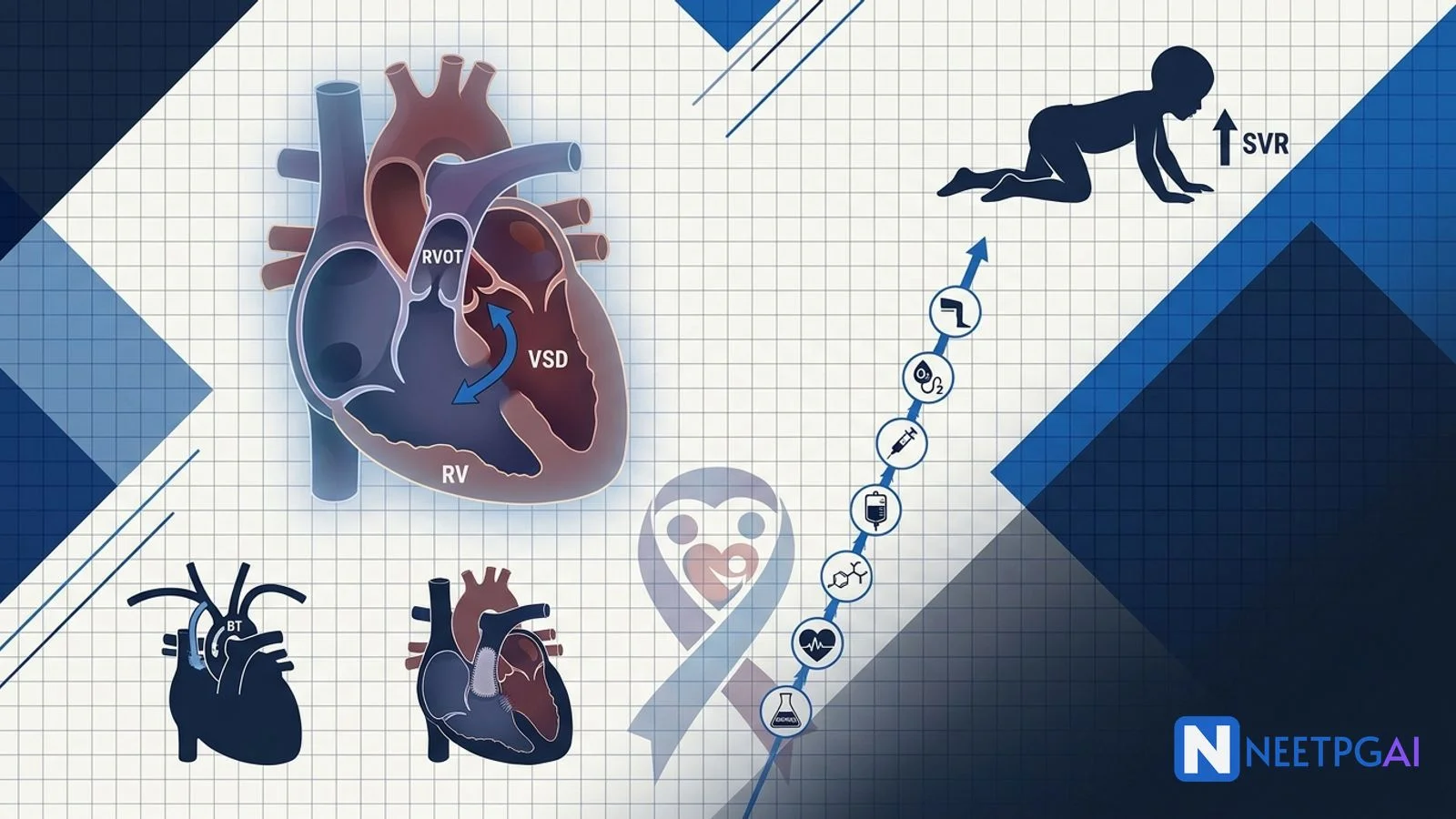
The diagnostic and management workflow
TOF pathophysiology — the four components
- Ventricular septal defect (VSD) — large, perimembranous, malalignment type
- Overriding aorta — dextroposed, straddling the septum; the aorta receives blood from both ventricles
- Right ventricular outflow tract obstruction — infundibular (subvalvular muscular) stenosis, valvular pulmonary stenosis, or both; the level and severity of RVOT obstruction determines the degree of cyanosis
- Right ventricular hypertrophy — secondary consequence of chronic pressure loading
Embryological basis: anterior and cephalad deviation of the infundibular (conal) septum during truncal septation, producing malalignment.
TOF is the commonest cyanotic congenital heart disease beyond infancy and represents 5-10 percent of all congenital heart defects. It is associated with 22q11.2 deletion (DiGeorge) syndrome in approximately 15 percent of cases — a chromosomal microarray is offered routinely at diagnosis.
Tet spell pathophysiology
Acute worsening of RVOT obstruction from infundibular muscular spasm shifts blood flow away from the pulmonary circulation and increases the right-to-left shunt across the VSD. Cyanosis intensifies suddenly; the previously audible ejection systolic murmur of pulmonary stenosis paradoxically becomes softer or disappears (less blood traverses the obstructed RVOT).
Triggers — crying, feeding, defaecation straining, fever, morning waking, dehydration, general anaesthetic induction, catecholamine surges.
Age — typical between 2 months and 4 years; declines after 4 years when children learn to squat spontaneously.
Squatting mechanism — mechanical kinking of the femoral and iliac arteries at the squatted position raises peripheral systemic vascular resistance, reduces the pressure gradient driving right-to-left shunting, and increases pulmonary blood flow.
Emergency management step-by-step
| Step | Action | Mechanism |
|---|
| 1 | Knee-chest position | Mimics squatting; raises SVR |
| 2 | Calm the child, minimise handling, allow parent to hold | Reduces catecholamine drive |
| 3 | 100 percent oxygen by non-rebreather | Pulmonary vasodilation modest but standard |
| 4 | IV or IO access | Delivery route for drugs and fluid |
| 5 | Morphine 0.1 mg/kg IV or IM | Breaks reflex, reduces catecholamines, relaxes infundibulum |
| 6 | IV crystalloid bolus 10-20 mL/kg | Raises preload and pulmonary blood flow |
| 7 | Sodium bicarbonate if pH under 7.20 | Corrects metabolic acidosis |
| 8 (refractory) | Phenylephrine 5-20 microgram/kg IV | Raises SVR |
| 8 (refractory) | Esmolol infusion or propranolol IV | Reduces infundibular spasm, slows HR |
| 8 (refractory) | Ketamine IV | Raises SVR and provides sedation |
| 9 (truly refractory) | Emergency modified BT-Thomas shunt | Palliative surgical shunt |
Do NOT give — systemic vasodilators (worsen R-to-L shunt), acute digoxin (positive inotropy worsens infundibular spasm), isoproterenol (increases infundibular contractility).
Long-term management
- Prophylactic oral propranolol 1-2 mg/kg/dose three times daily reduces the frequency of tet spells
- Iron therapy for relative or absolute iron deficiency in the setting of chronic cyanosis and compensatory erythrocytosis
- Adequate hydration and avoidance of dehydration
- Prompt treatment of fever and intercurrent illness
- Timely surgical repair at an experienced paediatric cardiac centre
Surgical strategy
- Primary complete repair at 3-6 months of age is preferred at experienced centres, avoiding the historical two-stage palliative approach in most patients
- Two-stage approach — a modified Blalock-Taussig-Thomas shunt (Gore-Tex tube from the subclavian to the pulmonary artery) is placed as a palliative bridge in neonates and young infants who are too small or unstable for complete repair, or as an emergency for refractory tet spells; complete repair is performed 6-12 months later
- Complete repair — patch closure of the VSD, resection of the obstructive infundibular muscle, pulmonary valvotomy or valve-sparing repair; a transannular patch is used when the pulmonary annulus is severely hypoplastic (widens the outflow but leaves the patient with lifelong pulmonary regurgitation)
Post-repair long-term issues
- Chronic pulmonary regurgitation from the transannular patch causes right ventricular dilatation over years and eventually right ventricular failure; managed by percutaneous transcatheter (Melody, SAPIEN) or surgical pulmonary valve replacement typically in adulthood
- Residual VSD or pulmonary stenosis
- RVOT aneurysm
- Ventricular tachycardia and sudden cardiac death — small but real long-term risk
- Atrial arrhythmias
- Endocarditis — prophylaxis in selected high-risk cases (per revised AHA guidance)
- Lifelong adult congenital heart disease follow-up is mandatory
Diagnosis
Acute hypercyanotic (tet) spell in a known 3-year-old boy with Tetralogy of Fallot awaiting scheduled complete surgical repair, with successful pre-hospital knee-chest positioning by the mother, moderate residual metabolic acidosis and hypoxaemia on arrival, and mild echocardiographic progression of the infundibular gradient — for admission, continued oxygen, IV morphine and fluid bolus, correction of acidosis, expedited surgical planning (advance scheduled complete repair, or interim BT-Thomas shunt if unstable), and reinforced caregiver education on trigger avoidance, propranolol adherence and knee-chest positioning.
Complications
Untreated or under-treated TOF
- Cyanotic spells with hypoxic brain injury, seizure, cerebrovascular event
- Cerebral abscess from paradoxical embolisation of septic material across the R-to-L shunt (classically streptococcal)
- Stroke from paradoxical embolism or from the hyperviscosity of severe erythrocytosis
- Coagulopathy — mixed picture of platelet dysfunction, prolonged bleeding time, hypofibrinogenaemia and reduced clotting factors
- Infective endocarditis on the VSD margin, RVOT muscle, or pulmonary valve
- Failure to thrive from work of breathing, feeding difficulty, and chronic hypoxaemia
- Squatting-related exercise limitation
- Death — mortality without surgical repair is high; the majority of unrepaired patients did not survive to adulthood in the pre-surgical era
Post-repair (long-term)
- Pulmonary regurgitation and right ventricular dilatation
- Ventricular tachycardia and sudden cardiac death
- Residual VSD or RVOT gradient
- Endocarditis
India-specific considerations
- TOF is one of the commonest cyanotic congenital heart defects in Indian paediatric cardiology practice
- Delayed diagnosis is common — antenatal fetal echocardiography is not universally available in the public sector, and many cases are diagnosed only after infants present with cyanosis, a murmur or a spell
- Universal newborn pulse oximetry screening at delivery is now recommended and is being scaled across tertiary maternity units — a saturation under 95 percent, or a right-hand to foot difference of more than 3 percent, prompts echocardiography
- RBSK (Rashtriya Bal Swasthya Karyakram) provides school-age screening and referral for congenital heart disease under the National Health Mission
- PMJAY (Ayushman Bharat) covers paediatric cardiac surgery at empanelled tertiary centres — Sri Sathya Sai Institute of Higher Medical Sciences, AIIMS, PGIMER, Narayana Health, Fortis, Apollo, CMC Vellore and state-run children's hospitals lead volume
- Access remains uneven — many families travel long distances, wait months to years for elective surgery, and face out-of-pocket costs beyond PMJAY caps
- NGO support — organisations such as Genesis Foundation, Being Human, and hospital-linked charities subsidise TOF surgery for financially constrained families
- Anganwadi and ASHA-worker education on recognising cyanotic spells, knee-chest positioning and prompt referral is a growing public-health priority
How NEET PG tests TOF
Recurring patterns:
Pattern 1 — The four components of TOF — perimembranous VSD, overriding aorta, RVOT obstruction, RVH.
Pattern 2 — Commonest cyanotic congenital heart disease beyond infancy — Tetralogy of Fallot.
Pattern 3 — Embryological basis — anterior cephalad deviation of the infundibular (conal) septum.
Pattern 4 — Chest X-ray sign — boot-shaped heart (coeur en sabot).
Pattern 5 — Genetic association — 22q11.2 deletion (DiGeorge) syndrome in approximately 15 percent.
Pattern 6 — Tet spell mechanism — infundibular muscular spasm increases R-to-L shunting through the VSD; the pulmonary ejection systolic murmur softens or disappears during a spell.
Pattern 7 — First-line management of tet spell — knee-chest position, oxygen, morphine, IV fluid bolus; refractory — phenylephrine, propranolol/esmolol, ketamine, sodium bicarbonate.
Pattern 8 — Contraindicated drugs — vasodilators, acute digoxin, isoproterenol.
Pattern 9 — Squatting — kinks femoral and iliac arteries, raises SVR, reduces R-to-L shunting; older children learn instinctively.
Pattern 10 — Complete surgical repair — preferred between 3 and 6 months at experienced centres; transannular patch causes long-term pulmonary regurgitation.
Pattern 11 — Modified Blalock-Taussig-Thomas shunt — Gore-Tex conduit from subclavian artery to pulmonary artery; palliative bridge or emergency operation.
Pattern 12 — Long-term complications — pulmonary regurgitation, RV dilatation and failure, ventricular tachycardia, sudden cardiac death, endocarditis.
High-yield one-liners:
- The four components — VSD, overriding aorta, RVOT obstruction (infundibular plus/minus valvular pulmonary stenosis), RVH
- Boot-shaped heart on X-ray = coeur en sabot
- Right-sided aortic arch in approximately 25 percent of TOF cases
- Prophylactic oral propranolol reduces tet-spell frequency
- Knee-chest positioning mimics squatting
- Morphine relaxes the infundibulum; do not give vasodilators
- Cerebral abscess (classically streptococcal) is a hallmark complication of unrepaired cyanotic CHD
- Compensatory secondary erythrocytosis (Hb often 18-22 g/dL)
- Modified BT-Thomas shunt = Gore-Tex conduit, subclavian artery to pulmonary artery
- Complete repair preferred at 3-6 months at experienced centres
- 22q11.2 deletion in about 15 percent — chromosomal microarray at diagnosis
Frequently Asked Questions
What are the four components of Tetralogy of Fallot and their embryological basis?
Tetralogy of Fallot has four anatomical components — a large perimembranous ventricular septal defect (VSD), an overriding (dextroposed) aorta straddling the septum, right ventricular outflow tract obstruction (which may be infundibular muscular, valvular pulmonary stenosis, or both), and right ventricular hypertrophy as a secondary consequence of chronic pressure loading. The embryological basis is anterior and cephalad deviation of the infundibular (conal) septum during septation of the truncus arteriosus, producing malalignment between the outlet septum and the muscular ventricular septum. This single developmental error explains the VSD, the overriding aorta, and the subvalvular pulmonary stenosis. TOF is the commonest cyanotic congenital heart disease beyond infancy and is associated with 22q11.2 deletion (DiGeorge) syndrome in approximately 15 percent of cases; a chromosomal microarray is offered routinely at diagnosis.
What is a Tet spell and what is its pathophysiological mechanism?
A tet spell (hypercyanotic spell) is an acute worsening of right ventricular outflow tract obstruction, usually driven by spasm of the infundibular muscle, which shifts blood flow away from the pulmonary circulation and increases right-to-left shunting across the VSD. Cyanosis intensifies suddenly, systemic oxygen saturation plummets, the child becomes agitated or lethargic, and the previously audible ejection systolic murmur of pulmonary stenosis paradoxically becomes softer or disappears (less blood traversing the obstructed RVOT). Triggers include crying, feeding, defaecation straining, fever, morning waking, dehydration, and any stimulus that acutely decreases systemic vascular resistance or increases catecholamine drive. Once systemic acidosis develops from hypoxaemia, systemic vascular resistance falls further, driving a vicious cycle of worsening shunt. Untreated severe spells lead to loss of consciousness, seizure, cerebrovascular event, or death. Older children instinctively squat, which mechanically kinks the femoral arteries and raises systemic vascular resistance, breaking the shunt cycle.
What is the step-by-step management of an acute Tet spell?
Acute Tet spell management works to increase systemic vascular resistance, decrease infundibular spasm, and increase pulmonary blood flow. Step 1: knee-chest position — flex the child's hips and knees onto the abdomen, or place the infant over the shoulder in a fetal position; this raises SVR. Step 2: calm the child and minimise handling, dim lights, allow the parent to hold. Step 3: 100 percent oxygen by mask (pulmonary vasodilatory effect is modest but standard). Step 4: intravenous or intraosseous access. Step 5: morphine 0.1 mg/kg IV or IM — breaks the reflex spasm, reduces catecholamine drive and relaxes the infundibulum. Step 6: intravenous crystalloid bolus 10-20 mL/kg — increases preload and pulmonary blood flow. Step 7: sodium bicarbonate for severe metabolic acidosis (pH under 7.20). Step 8 (refractory): phenylephrine 5-20 microgram/kg IV to raise SVR, or a beta blocker (esmolol infusion, or propranolol IV) to reduce infundibular spasm and slow the heart rate to allow better right ventricular filling; ketamine IV raises SVR and provides sedation for procedures. Contraindicated drugs are systemic vasodilators, digoxin acutely, and isoproterenol — each worsens the right-to-left shunt. If truly refractory, an emergency modified Blalock-Taussig-Thomas shunt is placed.
Why is squatting protective in Tetralogy of Fallot and why is it uncommon in infants?
Squatting is a compensatory manoeuvre that older children with TOF learn to relieve exertional cyanosis. Mechanical flexion of the hips and knees at the squatting position kinks the femoral and iliac arteries, sharply increasing peripheral systemic vascular resistance; this reduces the pressure gradient driving right-to-left shunting across the VSD, forcing more deoxygenated blood forward through the obstructed right ventricular outflow tract into the pulmonary circulation where it can be oxygenated. Squatting also increases venous return from the lower limbs, augmenting right ventricular preload and pulmonary blood flow. Infants and toddlers under 2 years cannot squat because of motor immaturity — they present with tet spells instead, which is why the emergency knee-chest position (in which a caregiver flexes the child's hips and knees onto the abdomen) mimics the haemodynamic effect of squatting. Once children walk and can squat spontaneously, tet spells become less frequent.
What is the current preferred surgical strategy for TOF and its long-term issues?
The current strategy at experienced centres is primary complete repair between 3 and 6 months of age, avoiding the historical two-stage palliative approach in most patients. Complete repair involves patch closure of the VSD, resection of the obstructive infundibular muscle, pulmonary valvotomy or valve-sparing repair, and, when the pulmonary annulus is severely hypoplastic, a transannular patch that widens the outflow but leaves the patient with lifelong pulmonary regurgitation. A modified Blalock-Taussig-Thomas shunt (Gore-Tex tube from the subclavian to the pulmonary artery) is placed as a palliative bridge in neonates and young infants who are too small or unstable for complete repair, or as an emergency operation for refractory tet spells. Long-term issues after complete repair include chronic pulmonary regurgitation causing right ventricular dilatation and failure (managed by percutaneous or surgical pulmonary valve replacement typically in adulthood), residual VSD, right ventricular outflow tract aneurysm, ventricular arrhythmias with a small but real risk of sudden cardiac death, atrial arrhythmias, and endocarditis. Lifelong adult congenital heart disease follow-up is essential.
This content is for educational purposes for NEET PG exam preparation. It is not a substitute for professional medical advice, diagnosis, or treatment. Clinical information has been reviewed by qualified medical professionals.
Written by: NEETPGAI Editorial Team
Reviewed by: Pending SME Review
Last reviewed: September 2026