Quick Answer
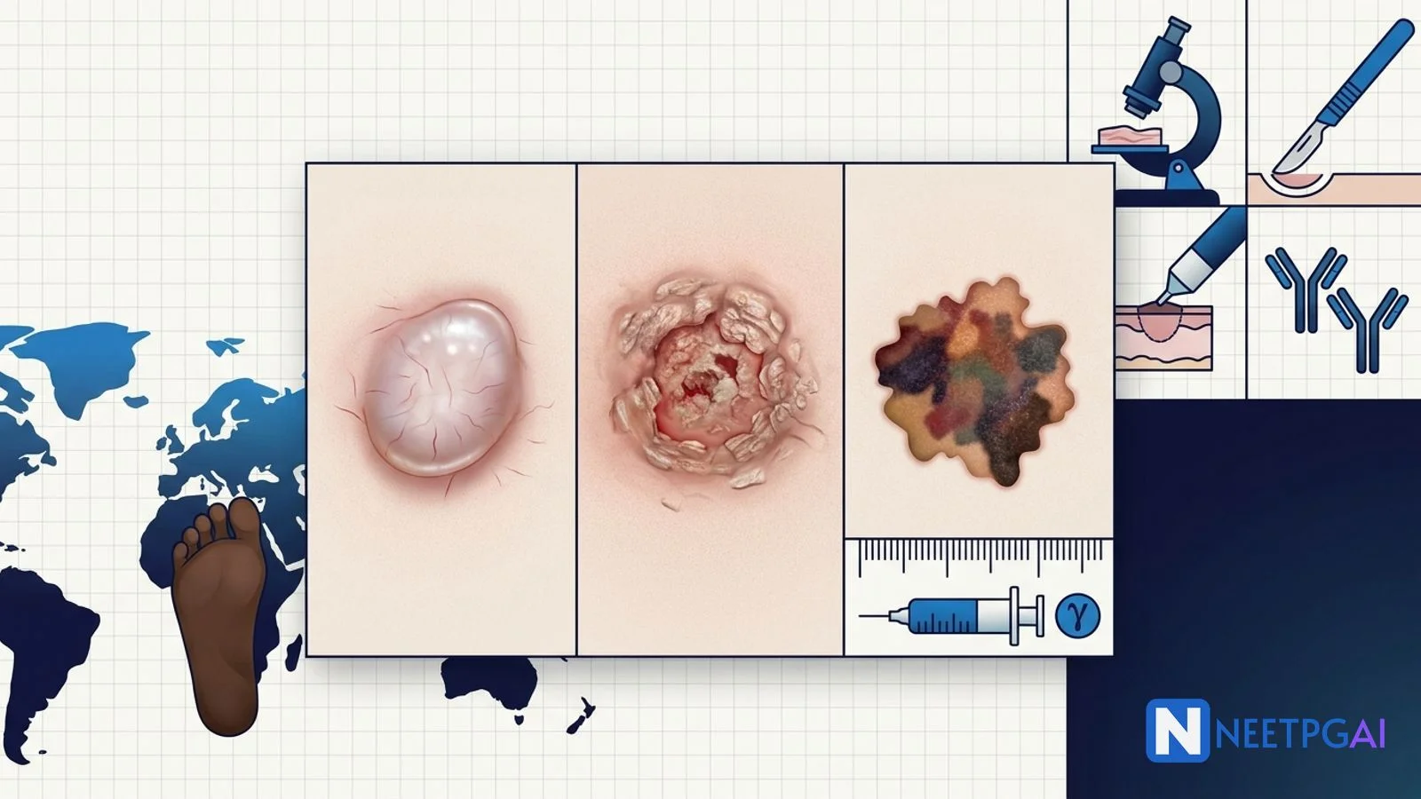
Skin cancer is a 1 to 2 question dermatology and oncology topic on NEET PG with distinct Western versus Indian epidemiology. Lock these:
- BCC — commonest skin cancer overall; pearly telangiectatic nodule on sun-exposed head and neck; rarely metastasises
- SCC — chronic sun-damaged skin, scars, immunosuppression; can metastasise 3-5 percent (up to 20 percent lip/ear/immunosuppressed)
- Actinic keratosis — SCC precursor; 1-10 percent per year risk of transformation
- Melanoma — 5 percent of skin cancers but 75 percent of deaths; ABCDE and Ugly Duckling sign
- Breslow thickness — the single strongest melanoma prognostic factor
- Wide excision margins — 0.5 cm for in situ, 1 cm for less than 1 mm, 1-2 cm for 1-2 mm, 2 cm for greater than 2 mm Breslow
- SLNB indication — 0.8 mm Breslow or greater, or high-risk thin tumours
- India context — acral lentiginous melanoma common on palm, sole, nail bed; often advanced presentation
Skin cancer sits at the intersection of dermatology, plastic surgery and medical oncology on NEET PG. The West-dominated epidemiology — driven by fair skin, high UV exposure and effective early-detection campaigns — contrasts sharply with the Indian picture, where melanotic protection from UV means lower absolute incidence but higher acral lentiginous variants and later presentation. This deep dive walks through basal cell carcinoma (BCC), squamous cell carcinoma (SCC), actinic keratosis, and cutaneous melanoma — with staging, treatment, and the newer immunotherapy landscape.
Pair this with the head and neck cancer guide for the SCC overlap on lip and skin-of-nose primaries.
Overview and risk factors
Skin cancers divide into non-melanoma (BCC and SCC — 95 percent of cases) and melanoma (5 percent of cases, 75 percent of deaths). Common risk factors:
- Cumulative UV exposure — UVB (280-320 nm) drives thymidine-dimer DNA damage; UVA (320-400 nm) causes indirect oxidative damage
- Fair skin (Fitzpatrick I-II), red or blonde hair, freckling, inability to tan
- Sunburn history — intermittent intense sunburn correlates with melanoma; chronic exposure with SCC
- Immunosuppression — solid organ transplant recipients (SCC risk up 65-100 fold), HIV, chemotherapy
- Prior radiation therapy
- Chronic wounds — Marjolin ulcer arising in burn scars, chronic osteomyelitis sinuses, venous ulcers (usually SCC)
- Xeroderma pigmentosum — autosomal recessive nucleotide-excision repair deficiency, multiple childhood skin cancers
- Gorlin (basal cell naevus) syndrome — PTCH1 mutation, multiple BCCs, jaw keratocysts, palmar/plantar pits, medulloblastoma
- Chemical carcinogens — arsenic (contaminated groundwater in West Bengal, Bihar, Bangladesh), tar, soot
- Human papillomavirus — SCC in immunosuppressed and anogenital sites
Basal cell carcinoma (BCC)
The commonest skin cancer overall — 70 percent of skin cancers, though under-reported in India.
Clinical subtypes
- Nodular (most common) — pearly, translucent papule or nodule with fine surface telangiectasia, rolled edge and central ulcer (rodent ulcer); sun-exposed head, neck, upper trunk
- Superficial — red, scaly, well-demarcated patch that mimics eczema, psoriasis or Bowen disease; trunk more than head
- Morpheaform (sclerosing, infiltrative) — scar-like, indistinct margins, deeply invasive; higher recurrence
- Pigmented — resembles nodular but with brown/black pigment; must be distinguished from melanoma
Behaviour and treatment
BCC grows slowly, invades locally, and rarely metastasises (less than 0.1 percent). Local destruction of eye, nose, ear or bone is the main morbidity.
Treatment options:
- Surgical excision with 4 mm margins for low-risk primary BCC
- Mohs micrographic surgery — tissue-sparing, high-cure technique for face, cosmetically sensitive sites, high-risk histology (morpheaform), recurrent tumours
- Electrodesiccation and curettage — low-risk lesions on the trunk, not for hairy areas or high-risk sites
- Topical imiquimod 5 percent cream or 5-fluorouracil — superficial BCC only
- Photodynamic therapy — superficial BCC
- Radiotherapy — elderly patients unsuitable for surgery, adjuvant for perineural invasion
- Hedgehog pathway inhibitors — vismodegib and sonidegib for locally advanced or metastatic disease
Squamous cell carcinoma (SCC)
The second commonest skin cancer, about 20 percent of skin cancers.
Presentation
Scaly, hyperkeratotic papule or plaque, often ulcerated, on sun-exposed sites (bald scalp, ear pinna, lip, dorsum of hand, forearm) or arising in a scar or chronic wound (Marjolin ulcer). Faster growth than BCC. Bowen disease is intraepidermal SCC in situ — a well-demarcated erythematous scaly patch.
Metastatic risk
- Overall 3-5 percent
- High-risk sites — lip and ear approach 15-20 percent metastasis
- Immunosuppressed transplant recipients — up to 20 percent
- Regional cervical or axillary lymph nodes are the commonest first site
Treatment
- Surgical excision with 4-6 mm margins for low-risk; 6-10 mm for high-risk
- Mohs surgery for face and high-risk sites
- Sentinel lymph node biopsy for high-risk primary SCC (thick tumour, immunosuppression, high-risk site)
- Adjuvant radiotherapy for perineural invasion, positive margins, extensive nodal disease
- Cemiplimab and pembrolizumab — PD1 checkpoint inhibitors for locally advanced or metastatic cutaneous SCC (approved after clinical trials showed durable responses)
Actinic keratosis (AK)
- Rough, scaly erythematous macule or thin papule on chronically sun-damaged skin (bald scalp, face, dorsum of hand)
- Precursor to SCC — approximately 0.5-10 percent per year risk of individual transformation, though the field-cancerisation concept means multiple lesions signify wider risk
- Treatment: cryotherapy (single lesions), topical 5-fluorouracil, imiquimod, ingenol mebutate, diclofenac gel for field treatment, photodynamic therapy
- Sunscreen, wide-brim hats, avoidance of peak UV, and follow-up surveillance are essential
Cutaneous melanoma
5 percent of skin cancers but 75 percent of skin-cancer deaths. Global incidence continues to rise.
Subtypes
| Subtype | Frequency | Site | Notes |
|---|
| Superficial spreading melanoma | 70 percent — commonest | Trunk (men), legs (women) | Long radial growth phase before vertical invasion |
| Nodular melanoma | 15-20 percent | Any site | Vertical growth phase from the outset; aggressive |
| Lentigo maligna melanoma | 5-10 percent | Face of elderly | Slow-growing on chronic sun damage; long in-situ phase (lentigo maligna) |
| Acral lentiginous melanoma | 5-10 percent West; higher in Indian, African, East Asian populations | Palms, soles, subungual (nail bed) | Often diagnosed late; not sun-related; commonest melanoma in Indians |
| Amelanotic melanoma | Any subtype without pigment | Any site | Pink or red; easily missed |
ABCDE and Ugly Duckling sign
- Asymmetry — one half does not mirror the other
- Border irregularity — notched, scalloped, blurred
- Colour variegation — two or more shades of brown, black, red, white or blue
- Diameter greater than 6 mm (pencil-eraser rule)
- Evolving — change in size, shape, colour, elevation, or new symptoms (bleeding, itch, ulceration)
The Ugly Duckling sign — a lesion that looks different from a patient's other naevi — is often more sensitive than ABCDE alone.
Diagnosis
Excisional biopsy with a narrow (1-2 mm) margin is preferred; incisional or punch biopsy is acceptable for very large lesions or cosmetically sensitive sites. Shave biopsy is discouraged because it may transect the tumour and prevent accurate Breslow measurement.
Breslow thickness and staging
Breslow thickness is measured vertically in millimetres from the top of the granular layer to the deepest invasive cell — the single strongest independent prognostic factor.
| Breslow thickness | Approximate 5-year survival | Wide excision margin |
|---|
| Melanoma in situ | Approaches 100 percent | 0.5 cm |
| Less than 1 mm | 90-95 percent | 1 cm |
| 1-2 mm | 80 percent | 1-2 cm |
| 2-4 mm | 65 percent | 2 cm |
| Greater than 4 mm | 40-50 percent | 2 cm |
AJCC 8 TNM staging integrates Breslow thickness, ulceration (upstages within each T), sentinel lymph node status, and distant metastases with LDH.
Sentinel lymph node biopsy (SLNB)
- Offered for primary melanoma with Breslow 0.8 mm or greater, or thinner tumours with ulceration or high mitotic rate
- Not routinely offered for in situ or thin (less than 0.8 mm) non-ulcerated melanoma — sub-1 percent yield
- A positive SLNB upstages to stage III and prompts consideration of adjuvant systemic therapy
- Completion lymph node dissection after a positive SLNB is no longer routine (MSLT-II, DeCOG) — replaced by nodal ultrasound surveillance for most patients
Systemic therapy for advanced melanoma
- BRAF plus MEK inhibitors for BRAF V600 mutant disease (40-50 percent of cutaneous melanomas) — dabrafenib plus trametinib, encorafenib plus binimetinib, vemurafenib plus cobimetinib
- PD1 inhibitors — nivolumab, pembrolizumab (monotherapy)
- PD1 plus CTLA4 inhibitors — nivolumab plus ipilimumab (higher response and higher toxicity)
- PD1 plus LAG3 — nivolumab plus relatlimab
- Adjuvant therapy for resected stage III and IV disease — checkpoint inhibitors or BRAF/MEK inhibitors
- Intralesional oncolytic virus — talimogene laherparepvec (T-VEC) for injectable cutaneous, subcutaneous and superficial nodal disease
India-specific considerations
- Lower absolute incidence of BCC, SCC and melanoma than Western populations because of melanin photoprotection
- Acral lentiginous melanoma is the commonest melanoma subtype in Indians (palms, soles, subungual) — non-UV related and often diagnosed late
- Delayed presentation — many patients present at advanced stages because of low awareness, misdiagnosis as fungal infection or trauma-related change, and limited dermatology access in peripheral centres
- Arsenic-linked SCC — endemic areas of West Bengal, Bihar, Bangladesh with contaminated groundwater have an increased burden of arsenical keratoses and SCC
- Immunosuppression drivers — solid organ transplant, HIV and prolonged systemic steroid or immunosuppressant use raise skin cancer risk
- Xeroderma pigmentosum is well described in Indian consanguineous populations
- Sun-protection counselling — despite lower absolute UV-driven cancer incidence, sunscreen use, wide-brim hats and long sleeves are increasingly promoted for melasma, photoageing and dermatology-clinic populations
- Access to immunotherapy — pembrolizumab, nivolumab and cemiplimab are available at tertiary centres and select PMJAY-empanelled institutions, though cost and continuity remain barriers
- Regional cancer centres — Tata Memorial Mumbai, AIIMS New Delhi, RGCIRC Delhi, and state cancer institutes coordinate multidisciplinary melanoma management
NEET PG MCQ traps
- Commonest skin cancer overall — BCC
- Commonest melanoma subtype overall — superficial spreading
- Commonest melanoma subtype in Indians — acral lentiginous
- Rodent ulcer — nodular BCC (pearly, telangiectatic, rolled edge, central ulceration)
- Marjolin ulcer — SCC in a chronic wound, burn scar or sinus tract
- Gorlin syndrome — multiple BCCs, jaw keratocysts, palmoplantar pits, medulloblastoma; PTCH1 mutation
- Xeroderma pigmentosum — nucleotide excision repair defect; multiple childhood skin cancers
- Erythroplasia of Queyrat — SCC in situ on glans penis
- Bowen disease — SCC in situ (scaly erythematous patch)
- Highest prognostic factor for melanoma — Breslow thickness
- Historical prognostic marker replaced by Breslow — Clark level
- Wide excision margin for less than 1 mm melanoma — 1 cm
- Wide excision margin for greater than 2 mm melanoma — 2 cm
- SLNB threshold — Breslow 0.8 mm or greater (or thinner if ulcerated/high mitotic rate)
- BRAF mutation frequency in melanoma — approximately 40-50 percent, most commonly V600E
- Ipilimumab target — CTLA-4
- Nivolumab and pembrolizumab target — PD-1
- Cemiplimab indication — locally advanced or metastatic cutaneous SCC
- T-VEC — oncolytic herpes simplex virus for injectable melanoma
- Hedgehog inhibitors — vismodegib and sonidegib for advanced BCC
- Mohs surgery preferred sites — face, cosmetically sensitive areas, recurrent tumours, morpheaform BCC
- Ugly Duckling sign — a naevus that looks different from a patient's other naevi
Frequently asked questions
What are the classic risk factors for non-melanoma and melanoma skin cancer?
The dominant modifiable risk factor is cumulative ultraviolet exposure — UVB (280-320 nm) drives thymidine-dimer DNA damage, and UVA (320-400 nm) contributes indirectly through reactive oxygen species. Intense intermittent sunburns in childhood correlate strongly with melanoma; chronic occupational sun exposure correlates with squamous cell carcinoma. Fair skin (Fitzpatrick I-II), red or blonde hair, light eyes, freckling and inability to tan raise all three cancer risks. Immunosuppression after solid organ transplantation raises SCC risk 65-100 fold and BCC risk 10 fold. Prior therapeutic radiation, chronic wounds and burn scars (Marjolin ulcer for SCC), xeroderma pigmentosum, basal cell naevus (Gorlin) syndrome, arsenic exposure (contaminated groundwater in West Bengal), tar and PUVA all raise risk. Melanoma additionally has a strong genetic component — CDKN2A and CDK4 germline mutations, large congenital naevi, and dysplastic naevus syndrome.
How does the ABCDE rule help distinguish melanoma from a benign naevus?
The ABCDE mnemonic is a screening tool designed for clinical dermoscopy or naked-eye evaluation. A — Asymmetry: one half of the lesion does not mirror the other. B — Border irregularity, notching, scalloping or blurring rather than a smooth round margin. C — Colour variegation with two or more shades (brown, black, red, white, blue). D — Diameter greater than 6 mm (the pencil-eraser rule), although early melanomas can be smaller. E — Evolving change in size, shape, colour, elevation or symptoms (bleeding, itching, ulceration) over weeks to months. The complementary Ugly Duckling sign recognises that a person's naevi tend to look alike; a naevus that looks different from its neighbours deserves biopsy. NEET PG typically tests the mnemonic verbatim and expects candidates to recall that E stands for Evolving.
Why is Breslow thickness the most important prognostic factor in melanoma?
Breslow thickness is the vertical measurement in millimetres from the top of the granular layer of the epidermis to the deepest invasive melanoma cell. It is the single strongest independent predictor of survival and of metastatic risk, and it drives both wide excision margins and sentinel lymph node biopsy decisions in the AJCC 8th edition TNM staging. Approximate 5-year survival by thickness: melanoma in situ approaches 100 percent, less than 1 mm around 90-95 percent, 1-2 mm around 80 percent, 2-4 mm around 65 percent and greater than 4 mm 40-50 percent. Ulceration on histology upstages within each T category (T1a versus T1b for example). Clark level (anatomical depth by dermal layer) is now largely historical — Breslow superseded it because millimetre depth reproducibly integrates the biological aggressiveness of tumours across differing skin sites.
When is sentinel lymph node biopsy indicated in cutaneous melanoma?
Sentinel lymph node biopsy is a staging procedure that identifies clinically occult nodal disease in the drainage basin of the primary melanoma using lymphoscintigraphy with radiolabelled colloid and blue dye. Current NCCN and international guidelines offer SLNB to patients with a primary melanoma of 0.8 mm Breslow thickness or greater, or thinner tumours with high-risk features (ulceration, high mitotic rate). SLNB is not routinely offered for melanoma in situ or thin (less than 0.8 mm) non-ulcerated tumours because the sub-1 percent positive-node yield does not justify the procedure. A positive SLNB upstages the patient to stage III and prompts consideration of adjuvant systemic therapy (nivolumab, pembrolizumab, or dabrafenib-trametinib for BRAF V600 mutant disease). Complete lymphadenectomy after a positive SLNB is no longer routine — the MSLT-II and DeCOG trials showed no melanoma-specific survival benefit over nodal ultrasound surveillance.
What is the treatment landscape for advanced and metastatic melanoma?
The treatment of advanced melanoma has been transformed in the last decade. Approximately 40-50 percent of cutaneous melanomas harbour a BRAF V600 mutation (most commonly V600E), which enables targeted therapy with combined BRAF and MEK inhibitors — dabrafenib plus trametinib, encorafenib plus binimetinib, or vemurafenib plus cobimetinib — with rapid response but frequent secondary resistance. Immune checkpoint inhibitors are the second pillar — anti-PD1 monotherapy (nivolumab, pembrolizumab), combined anti-PD1 plus anti-CTLA4 (nivolumab plus ipilimumab, higher response but greater toxicity) and the newer anti-LAG3 combination (nivolumab plus relatlimab). Intralesional oncolytic virus talimogene laherparepvec (T-VEC) is approved for cutaneous, subcutaneous and superficial nodal metastases. Adjuvant checkpoint inhibitor therapy for resected stage III and stage IV disease is now standard. Access and cost remain the dominant barriers in India, though some agents are available at tertiary oncology centres and under compassionate-use or PMJAY-linked arrangements.
This content is for educational purposes for NEET PG exam preparation. It is not a substitute for professional medical advice, diagnosis, or treatment. Clinical information has been reviewed by qualified medical professionals.
Written by: NEETPGAI Editorial Team
Reviewed by: Pending SME Review
Last reviewed: September 2026