Version 1.0 — Published July 2026
Quick Answer
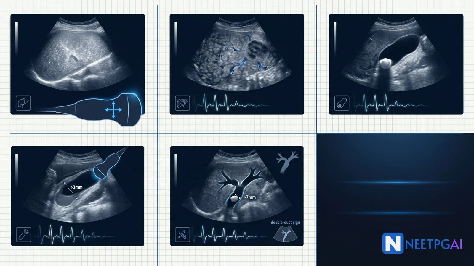
Abdominal ultrasound of the liver and gallbladder contributes 3-5 image-based MCQs per NEET PG paper across radiology, medicine, surgery, and gastroenterology. Five patterns recur reliably year after year:
- Hepatic steatosis (fatty liver) — increased liver echogenicity relative to renal cortex + posterior beam attenuation + poor intrahepatic vessel visualisation; graded 1-3; India NAFLD prevalence 25-32 percent
- Hepatocellular carcinoma on cirrhosis — heterogeneous mosaic-pattern nodule with peripheral hypoechoic halo + intralesional Doppler flow on a nodular cirrhotic liver with splenomegaly, portal hypertension, and ascites; LI-RADS on multi-phase CT/MRI
- Cholelithiasis — hyperechoic mobile stone in gallbladder lumen with posterior acoustic shadowing; five Fs risk profile; north-Indian belt prevalence 6-15 percent
- Acute cholecystitis — gallbladder wall thickening over 3 mm + pericholecystic fluid + sonographic Murphy sign + gallbladder distension over 5 cm + gallstones; complications include emphysematous, gangrenous, perforation, mucocele, and Mirizzi syndrome
- CBD stone with obstructive pattern — dilated CBD over 7 mm (over 10 mm post-cholecystectomy or in elderly) + intraluminal stone with shadow + dilated intrahepatic radicles; double duct sign suggests periampullary cancer
Locking these 5 patterns plus 5-8 additional PYQ ultrasound images (hepatic haemangioma, hydatid cyst, liver abscess, gallbladder polyp, gallbladder carcinoma, porcelain gallbladder) over 2-3 weeks moves accuracy from 40 to 85 percent in hepatobiliary imaging MCQs.
Why hepatobiliary ultrasound MCQs are high-yield for NEET PG
Abdominal ultrasound is the most-used initial imaging modality in Indian clinical practice — cheap, radiation-free, bedside-portable, and diagnostic in most hepatobiliary conditions. NEET PG, INI-CET, and FMGE feature ultrasound-based image MCQs in 3-5 questions per paper, often paired with a classical vignette (right upper quadrant pain in a 40-year-old fertile woman, jaundice with weight loss in an elderly man, incidental finding on a routine health check) to test pattern recognition, cause identification, and next-step imaging or management.
The management decision often hinges on distinguishing benign from malignant lesions (hepatic haemangioma vs HCC), acute from chronic pathology (acute cholecystitis vs chronic cholecystitis), and recognising complications that need urgent intervention (emphysematous cholecystitis, Mirizzi syndrome, gallbladder perforation). Ultrasound measurement cut-offs (wall thickness 3 mm, CBD 7 mm, gallbladder length under 10 cm and transverse under 5 cm) are repeatedly tested.
Drilling these 5 patterns plus 5-8 additional PYQ hepatobiliary images over 2-3 weeks moves accuracy from 40 to 85 percent.
Foundational approach before the MCQs
Systematic hepatobiliary ultrasound read
| Step | What to look at | Key findings |
|---|
| Liver echotexture | Compare with adjacent right renal cortex; look for coarse texture, nodularity | Fatty liver, cirrhosis, chronic hepatitis |
| Liver contour and lobar ratio | Smooth vs nodular; caudate-to-right-lobe ratio | Cirrhosis if nodular contour + hypertrophied caudate |
| Focal lesions | Number, size, echogenicity, halo, calcification, Doppler | Cyst, haemangioma, FNH, adenoma, HCC, metastasis |
| Gallbladder wall | Thickness on anterior wall (normal under 3 mm), layering, hyperaemia | Acute cholecystitis, adenomyomatosis |
| Gallbladder lumen | Stones (mobile, shadow), sludge, polyps (fixed, no shadow) | Cholelithiasis, sludge, polyp, carcinoma |
| CBD | Diameter at porta hepatis (normal up to 7 mm; up to 10 mm post-cholecystectomy) | Obstruction, stone, mass |
| Intrahepatic biliary radicles | Dilated parallel to portal veins (parallel channel sign) | Downstream obstruction |
| Pancreas | Head bulk, duct diameter (normal under 3 mm) | Periampullary cancer, chronic pancreatitis |
| Portal vein and splenic size | Diameter (normal under 13 mm), collaterals, splenomegaly | Portal hypertension |
| Ascites and pleural effusion | Free anechoic fluid in Morison pouch, pelvis, subphrenic | Cirrhosis, malignancy |
Key measurement pearls (memorise cold)
| Structure | Normal cut-off | Abnormal significance |
|---|
| Gallbladder wall thickness | Under 3 mm | Acute cholecystitis if over 3 mm with other signs |
| Gallbladder length | Under 10 cm longitudinal | Distended if over 10 cm |
| Gallbladder transverse | Under 5 cm | Hydrops if over 5 cm |
| CBD adult under 60 | Under 7 mm | Obstruction if over 7 mm |
| CBD elderly / post-cholecystectomy | Up to 10 mm (+1 mm per decade over 60) | Age-adjusted allowance |
| Pancreatic duct | Under 3 mm | Chronic pancreatitis, obstruction if over 3 mm |
| Portal vein diameter | Under 13 mm | Portal hypertension if over 13 mm |
| Spleen length | Under 12 cm | Splenomegaly if over 12 cm |
| Liver span (mid-clavicular) | Under 15 cm | Hepatomegaly if over 15 cm |
MCQ 1: 48-year-old T2DM woman with elevated ALT and echogenic liver on routine health check
USG image description: [Grayscale abdominal ultrasound of a 48-year-old woman using a 3.5 MHz curvilinear probe in the right subcostal view. The liver parenchyma is diffusely hyperechoic (brighter than the adjacent right renal cortex) with a marked hepatorenal echogenicity contrast. There is posterior beam attenuation with reduced visualisation of the diaphragm and posterior right lobe. The intrahepatic vessels (portal and hepatic veins) are poorly visualised due to the increased parenchymal echogenicity. Liver span 16.5 cm (hepatomegaly). Gallbladder appears normal with thin wall (2.1 mm) and no stones. Portal vein diameter 10 mm. Spleen 10.5 cm. No ascites. No focal lesions in the liver.]
Clinical vignette: A 48-year-old homemaker from Delhi presents for a routine executive health check-up. She has T2DM for 6 years on metformin, hypertension on telmisartan, and dyslipidaemia. BMI 31 kg/m2, waist circumference 96 cm, fasting glucose 132 mg/dL, HbA1c 7.4 percent, LDL 138 mg/dL, triglycerides 218 mg/dL, HDL 38 mg/dL. Liver function tests — ALT 78 U/L (elevated), AST 46 U/L, GGT 68 U/L, alkaline phosphatase 112 U/L, bilirubin 0.8 mg/dL, albumin 4.2 g/dL, INR 1.0. HBsAg negative, anti-HCV negative. No history of alcohol consumption (she is a lifelong teetotaller). Ceruloplasmin normal, ferritin normal.
Options:
- (a) Grade 2 hepatic steatosis (moderate NAFLD) — lifestyle intervention plus metabolic control
- (b) Hepatic cirrhosis — Child-Pugh assessment plus HCC surveillance
- (c) Autoimmune hepatitis — ANA plus anti-smooth muscle antibody plus IgG
- (d) Wilson disease — 24-hour urinary copper plus slit-lamp for KF ring
Correct answer: (a) Grade 2 hepatic steatosis (moderate NAFLD) — lifestyle intervention plus metabolic control
Reasoning: The ultrasound shows classical moderate (Grade 2) hepatic steatosis — diffusely hyperechoic liver relative to renal cortex, posterior beam attenuation, and reduced visualisation of intrahepatic vessels and diaphragm. The clinical picture is textbook NAFLD (non-alcoholic fatty liver disease) — a middle-aged obese T2DM woman with dyslipidaemia (all criteria for metabolic syndrome), mildly elevated ALT (ALT typically exceeds AST in NAFLD; AST over ALT with ratio over 2 suggests alcoholic liver disease), and negative viral, autoimmune, and metabolic aetiologies.
India is at the epicentre of the NAFLD epidemic — recent studies estimate NAFLD prevalence in urban Indian adults at 25-32 percent, driven by high refined-carbohydrate diet, sedentary lifestyle, and Asian Indians developing central adiposity and insulin resistance at lower BMI thresholds than Western populations. Management is lifestyle intervention — 7-10 percent weight loss reverses steatosis; Mediterranean diet, reduced refined carbohydrates, regular aerobic exercise 150 minutes per week, tight glycaemic control, statins for dyslipidaemia. Pioglitazone or vitamin E may be considered in biopsy-proven NASH. FibroScan (transient elastography) for liver stiffness assessment identifies fibrosis stage; F3-F4 fibrosis requires HCC surveillance every 6 months.
Cirrhosis would show nodular liver contour, splenomegaly, portal hypertension features — absent here. Autoimmune hepatitis typically has AST over ALT, positive autoantibodies, elevated IgG. Wilson disease is unlikely at 48 years without KF ring or extrapyramidal features.
Teaching pearl — ultrasound grading of hepatic steatosis:
| Grade | Echogenicity | Vessel visualisation | Diaphragm | Attenuation |
|---|
| Grade 1 (mild) | Slight increase over renal cortex | Normal | Normal | None |
| Grade 2 (moderate) | Clearly hyperechoic | Reduced | Reduced | Mild |
| Grade 3 (severe) | Markedly hyperechoic | Absent | Absent | Marked, posterior right lobe obscured |
MCQ 2: 62-year-old chronic HBV cirrhotic man with a 3 cm liver nodule on surveillance ultrasound
USG image description: [Grayscale and colour Doppler abdominal ultrasound of a 62-year-old man on 6-monthly surveillance ultrasound for known compensated cirrhosis. The liver shows a coarse echotexture, nodular surface contour, hypertrophied caudate lobe, and atrophic right lobe. In segment VII, there is a 3.2 cm heterogeneous mixed-echogenicity nodule with a peripheral hypoechoic halo (thin rim) and an internal mosaic pattern of alternating hypoechoic and hyperechoic areas. Colour Doppler shows intralesional vascularity with a basket pattern and high-velocity arterial flow within the nodule. The spleen is enlarged at 15 cm, portal vein diameter is 14 mm (portal hypertension), and there are recanalised paraumbilical vein collaterals at the falciform ligament. Small volume ascites in Morison pouch. No portal vein thrombus visible on this view.]
Clinical vignette: A 62-year-old retired schoolteacher with chronic HBV cirrhosis (Child-Pugh A6, MELD 9) diagnosed 8 years ago is on 6-monthly surveillance with ultrasound and alpha-fetoprotein per AASLD and INASL guidelines. He is on entecavir 0.5 mg daily for viral suppression (HBV DNA under 20 IU/mL for 4 years). Previous surveillance ultrasounds have shown stable cirrhotic changes without focal lesions. Today's ultrasound shows the new 3.2 cm segment VII nodule described above. Serum AFP has risen from 8 ng/mL 6 months ago to 340 ng/mL today. Liver function — bilirubin 1.4 mg/dL, albumin 3.5 g/dL, INR 1.2, ALT 42, AST 48, platelets 92,000. He is asymptomatic — no weight loss, no abdominal pain, no jaundice.
Options:
- (a) Hepatocellular carcinoma — multi-phase contrast-enhanced CT or MRI for LI-RADS classification
- (b) Focal nodular hyperplasia — contrast-enhanced ultrasound for spoke-wheel pattern
- (c) Hepatic haemangioma — no further imaging, reassure
- (d) Regenerative nodule of cirrhosis — repeat ultrasound in 3 months
Correct answer: (a) Hepatocellular carcinoma — multi-phase contrast-enhanced CT or MRI for LI-RADS classification
Reasoning: The ultrasound shows classical hepatocellular carcinoma (HCC) on a background of cirrhosis — a heterogeneous mosaic-pattern nodule (variable internal echogenicity reflecting necrosis, haemorrhage, and fibrous septae) with a peripheral hypoechoic halo (fibrous pseudocapsule) and intralesional Doppler flow (arterial neovascularisation). The background liver shows all cirrhotic features — nodular contour, hypertrophied caudate, atrophic right lobe, splenomegaly, portal hypertension, and ascites. The AFP jump from 8 to 340 ng/mL over 6 months (any AFP over 200 ng/mL in a cirrhotic with a nodule strongly supports HCC), the appropriate risk background (chronic HBV cirrhosis), and the size (over 1 cm on screening ultrasound triggers formal diagnostic imaging) all fit HCC.
Next step — multi-phase contrast-enhanced CT (arterial + portal venous + delayed phases) or MRI with hepatocyte-specific contrast (gadoxetate — Primovist) for LI-RADS (Liver Imaging Reporting And Data System) classification. LI-RADS 5 (definitive HCC, no biopsy needed) requires the combination of arterial phase hyperenhancement (APHE) + washout in portal venous or delayed phase + enhancing capsule appearance in a nodule over 1 cm in a cirrhotic. Biopsy is reserved for indeterminate LI-RADS 3-4 lesions or if targeted therapy trials require histology.
Focal nodular hyperplasia has a central scar with spoke-wheel arterial flow — occurs in young women, not cirrhotic men. Haemangioma is homogeneously hyperechoic with posterior enhancement, no Doppler flow, in a non-cirrhotic liver. A regenerative nodule of cirrhosis is typically under 1 cm, iso-echoic to background, without a halo or intralesional Doppler flow.
Teaching pearl — HCC screening and diagnosis in cirrhotics:
- Screening — 6-monthly ultrasound plus AFP in all cirrhotics regardless of aetiology (Child A-B; Child C only if transplant candidate)
- Nodule under 1 cm — repeat ultrasound in 3 months
- Nodule 1-2 cm — one dynamic contrast-enhanced imaging (CT or MRI); LI-RADS 5 pattern = HCC; indeterminate = second modality
- Nodule over 2 cm — same LI-RADS pathway
- Portal vein tumour thrombus — expansile hypoechoic filling defect with arterial flow within = pathognomonic HCC (vs bland thrombus with no flow)
- Management — BCLC staging drives treatment; very early HCC (BCLC 0) = ablation; early (BCLC A) = resection or transplant; intermediate (BCLC B) = TACE; advanced (BCLC C) = systemic therapy (sorafenib, lenvatinib, atezolizumab-bevacizumab); terminal (BCLC D) = best supportive care
MCQ 3: 42-year-old obese multiparous woman with intermittent right upper quadrant pain after fatty meals
USG image description: [Grayscale abdominal ultrasound of a 42-year-old multiparous woman using a 3.5 MHz curvilinear probe in the right subcostal view with the patient in left lateral decubitus position. The gallbladder is well-distended (measurements 8.5 cm longitudinal and 3.2 cm transverse — normal size). The wall is thin and smooth at 2.4 mm (normal). Within the lumen, there are three hyperechoic (bright) foci ranging 8-14 mm in size along the dependent wall of the gallbladder. Each shows strong posterior acoustic shadowing clearly separating them from adjacent bowel gas artefact. On repositioning the patient from supine to left lateral decubitus, the foci mobilise and roll to the new dependent position confirming their intraluminal free nature. There is no pericholecystic fluid, no wall thickening, no Murphy sign on scanning. The CBD is 5 mm (normal). Intrahepatic biliary radicles are not dilated. The liver, pancreas, and spleen are unremarkable.]
Clinical vignette: A 42-year-old multiparous (para 3) homemaker from Punjab presents to the surgical outpatient with a 6-month history of intermittent right upper quadrant pain, typically 1-2 hours after fatty meals (paratha, samosa, sweets). The pain is dull, lasts 30-60 minutes, radiates to the right scapula, and is associated with mild nausea. No fever, no jaundice, no weight loss, no altered bowels. She has been prescribed ranitidine and antispasmodics for suspected gastritis with partial relief. BMI 32 kg/m2. Family history — mother had gallstones removed at age 50. Examination — mild right upper quadrant tenderness with no guarding, no Murphy sign, no palpable gallbladder, no jaundice. Labs — CBC normal, LFT normal (bilirubin 0.6, ALT 22, ALP 88), amylase and lipase normal.
Options:
- (a) Symptomatic cholelithiasis — elective laparoscopic cholecystectomy
- (b) Acute cholecystitis — IV antibiotics plus urgent cholecystectomy
- (c) Gallbladder polyp — surveillance ultrasound every 6 months
- (d) Gallbladder sludge — ursodeoxycholic acid trial
Correct answer: (a) Symptomatic cholelithiasis — elective laparoscopic cholecystectomy
Reasoning: The ultrasound shows classical cholelithiasis — multiple hyperechoic intraluminal foci with strong posterior acoustic shadowing and gravitational mobility on repositioning (both features distinguish stones from polyps or sludge and from bowel gas artefact). The absence of gallbladder wall thickening, pericholecystic fluid, and sonographic Murphy sign rules out acute cholecystitis. The clinical picture is textbook biliary colic — postprandial right upper quadrant pain after fatty meals, radiation to the right scapula, transient duration under 6 hours, and no signs of infection or obstruction.
She fits all classical risk factors — the five Fs (Female + Forty + Fat + Fertile — three pregnancies + Fair — northern India Punjab has higher gallstone prevalence than southern India). Genetic polymorphisms of the ABCG5/ABCG8 cholesterol transporter contribute to the north Indian belt cholesterol-stone predilection. Management — she is symptomatic and has gallstones, which is a Class I indication for elective laparoscopic cholecystectomy (gold standard, day-care surgery, 4-port or single-incision technique, less than 1 percent bile duct injury rate in experienced hands). Asymptomatic gallstones generally do NOT need prophylactic cholecystectomy except in specific scenarios — stones over 3 cm (high gallbladder cancer risk), porcelain gallbladder (high cancer risk), children with sickle cell disease undergoing splenectomy, morbid obesity undergoing bariatric surgery, and pre-transplant patients.
Acute cholecystitis would show wall thickening over 3 mm, pericholecystic fluid, sonographic Murphy, fever, elevated WBC — absent here. Polyps are fixed to the wall, do not shadow, do not mobilise. Sludge appears as low-level dependent echoes without shadowing.
Teaching pearl — indications for cholecystectomy in asymptomatic cholelithiasis:
| Indication | Reason |
|---|
| Stones over 3 cm | Increased gallbladder cancer risk |
| Porcelain gallbladder | Very high gallbladder cancer risk (traditionally 25 percent, revised down to 6 percent) |
| Polyp over 10 mm coexisting with stones | High neoplastic risk |
| Sickle cell disease undergoing splenectomy | Increased pigment stones and cholecystitis risk |
| Morbid obesity undergoing bariatric surgery | Rapid weight loss increases stone formation |
| Pre-solid-organ-transplant candidate | Post-transplant immunosuppression complicates emergency surgery |
| Long-term TPN, ileal disease/resection | Increased pigment stone risk |
| Cystic fibrosis | Increased cholelithiasis risk |
| Children with cholelithiasis | Symptomatic risk over adult lifetime |
MCQ 4: 68-year-old diabetic man with fever, right upper quadrant pain, and thick-walled gallbladder with air in the wall
USG image description: [Grayscale abdominal ultrasound of a 68-year-old diabetic man presenting with 3-day history of fever and right upper quadrant pain. The gallbladder is distended at 11 cm longitudinal and 5.4 cm transverse. The wall is markedly thickened at 6.8 mm (measured on the anterior wall) with a striated appearance (alternating hyperechoic and hypoechoic layers from oedema). There is a rim of pericholecystic fluid (anechoic collection surrounding the gallbladder). Within the gallbladder lumen, an impacted stone is visible at the neck with posterior shadowing. Along the anterior gallbladder wall, there are high-amplitude echoes with dirty posterior shadowing and ring-down artefact consistent with gas (air) within the gallbladder wall. Sonographic Murphy sign is strongly positive with maximal tenderness on direct transducer pressure over the gallbladder. Colour Doppler shows hyperaemia of the wall.]
Clinical vignette: A 68-year-old male retired shopkeeper with poorly controlled T2DM (HbA1c 10.2 percent, on premixed insulin) is brought to the emergency by his family with a 3-day history of severe right upper quadrant pain radiating to the right shoulder, high-grade fever with rigors, vomiting, and reduced oral intake. He had similar milder episodes over the past 2 months, self-managed with painkillers. On examination — temperature 39.4 C, HR 118 (tachycardic), BP 96/62 (borderline hypotensive), RR 24. Marked right upper quadrant tenderness with guarding and Murphy sign, no jaundice, no palpable mass. Labs — WBC 22,800 with 92 percent neutrophils, CRP 340 mg/L, bilirubin 1.8 mg/dL, ALT 96, ALP 210, creatinine 1.6 (baseline 1.0 — acute kidney injury), lactate 3.2 mmol/L, glucose 340 mg/dL, HbA1c 10.2. Blood cultures sent. He is drowsy but arousable.
Options:
- (a) Emphysematous cholecystitis — urgent laparoscopic or open cholecystectomy plus broad-spectrum IV antibiotics
- (b) Uncomplicated acute cholecystitis — IV antibiotics plus elective cholecystectomy after 6 weeks
- (c) Chronic cholecystitis — elective laparoscopic cholecystectomy
- (d) Adenomyomatosis — MRCP for evaluation
Correct answer: (a) Emphysematous cholecystitis — urgent laparoscopic or open cholecystectomy plus broad-spectrum IV antibiotics
Reasoning: The ultrasound shows classical emphysematous cholecystitis — features of acute cholecystitis (thick striated wall over 6 mm, pericholecystic fluid, distended gallbladder over 5 cm transverse, impacted neck stone, positive sonographic Murphy) PLUS high-amplitude echoes with dirty shadowing and ring-down artefact along the gallbladder wall representing gas within the wall itself — the pathognomonic finding of emphysematous cholecystitis. The clinical picture is textbook — an elderly diabetic (or immunocompromised) patient with sepsis and organ dysfunction (tachycardia, hypotension, elevated lactate, AKI, altered sensorium) from a gas-forming organism infection — commonly Clostridium perfringens, E. coli, Klebsiella, or anaerobes.
Emphysematous cholecystitis has 20-25 percent mortality (10 times higher than uncomplicated acute cholecystitis) and requires urgent surgical intervention — laparoscopic or open cholecystectomy (open often required for severe disease) with concurrent broad-spectrum IV antibiotics covering Gram-negatives and anaerobes (piperacillin-tazobactam or meropenem plus metronidazole; add vancomycin if MRSA suspected). Percutaneous cholecystostomy is a bridge if the patient is unfit for immediate surgery — decompresses the gallbladder and controls sepsis, with interval cholecystectomy in 6-8 weeks. In this patient with sepsis and AKI, cholecystostomy while stabilising haemodynamics is a reasonable bridge to definitive surgery.
Uncomplicated acute cholecystitis without gas would be managed with early (within 72 hours) laparoscopic cholecystectomy — but this patient has emphysematous features requiring more urgent surgical thinking. Chronic cholecystitis is a stable outpatient diagnosis without sepsis. Adenomyomatosis shows Rokitansky-Aschoff sinuses with comet-tail artefact — not sepsis.
Teaching pearl — complications of acute cholecystitis:
| Complication | Key ultrasound finding | Management |
|---|
| Emphysematous cholecystitis | Gas in wall (ring-down, dirty shadow); diabetic elderly | Urgent surgery + broad-spectrum antibiotics |
| Gangrenous cholecystitis | Asymmetric wall thickening, intraluminal membranes (sloughed mucosa), absent wall Doppler | Urgent surgery |
| Perforation | Focal wall defect, pericholecystic collection, free peritoneal fluid | Urgent surgery |
| Mucocele | Distended gallbladder over 5 cm transverse without inflammation | Cholecystectomy |
| Empyema | Distended gallbladder with echogenic pus, wall thickening | Urgent drainage or cholecystectomy |
| Mirizzi syndrome | Stone impacted in cystic duct or neck causing extrinsic CBD compression with obstructive jaundice | MRCP for classification; surgical or endoscopic management |
| Gallstone ileus | Air in biliary tree, dilated small bowel, ectopic gallstone in RIF (Rigler triad on X-ray) | Enterolithotomy; interval cholecystectomy |
MCQ 5: 55-year-old man with painless jaundice, weight loss, and dilated CBD with intraluminal shadowing focus
USG image description: [Grayscale abdominal ultrasound of a 55-year-old man presenting with painless progressive jaundice and 8 kg weight loss over 3 months. The common bile duct is dilated at 14 mm (normal under 7 mm) throughout its length. Within the distal CBD near the ampulla, there is a hyperechoic (bright) focus measuring 9 mm with strong posterior acoustic shadowing consistent with a CBD stone. The intrahepatic biliary radicles are markedly dilated running parallel to the portal vein branches producing the classical parallel channel sign (a dilated biliary radicle alongside a portal vein branch, when the biliary radicle should not normally be visible at this calibre). The gallbladder is distended (12 cm longitudinal, 6 cm transverse) with multiple stones and sludge in the dependent lumen. The pancreatic duct is not dilated (2 mm, normal). The pancreatic head is not enlarged. Liver echotexture is normal. No focal liver lesions. No ascites.]
Clinical vignette: A 55-year-old male businessman presents with a 3-week history of progressive jaundice, dark urine, pale stools, pruritus, and 8 kg weight loss over 3 months. No abdominal pain except mild right upper quadrant discomfort. No fever. No history of gallstones previously known. Past medical history — hypertension controlled on amlodipine. On examination — deep icterus, no pallor, no lymphadenopathy. Palpable non-tender distended gallbladder in the right upper quadrant (Courvoisier sign positive — an enlarged palpable non-tender gallbladder in a jaundiced patient suggests malignant obstruction rather than stone disease; but note that in choledocholithiasis with a proximal stone allowing gradual downstream obstruction, gallbladder distension can also occur — Courvoisier is a rule with exceptions). No hepatomegaly. No ascites. Labs — bilirubin 14.8 mg/dL (predominantly direct at 11.2), ALP 620 IU/L, GGT 480 IU/L, ALT 92, AST 78, INR 1.1, albumin 3.6, CA 19-9 42 U/mL (mildly elevated).
Options:
- (a) Choledocholithiasis (CBD stone) — MRCP followed by ERCP with stone extraction and later laparoscopic cholecystectomy
- (b) Periampullary carcinoma with double duct sign — contrast-enhanced CT with pancreatic protocol
- (c) Primary sclerosing cholangitis — MRCP for beaded appearance and ANCA
- (d) Klatskin tumour (hilar cholangiocarcinoma) — MRCP for Bismuth-Corlette classification
Correct answer: (a) Choledocholithiasis (CBD stone) — MRCP followed by ERCP with stone extraction and later laparoscopic cholecystectomy
Reasoning: The ultrasound shows classical choledocholithiasis — a dilated CBD over 7 mm (14 mm here) with an intraluminal hyperechoic focus with posterior shadowing in the distal CBD, plus dilated intrahepatic biliary radicles (parallel channel sign), plus gallbladder distension with multiple stones (source of the migratory CBD stone). Ultrasound sensitivity for CBD stones is only 30-70 percent (overlying bowel gas obscures the distal CBD), so a positive ultrasound finding is quite specific.
The pancreatic duct is NOT dilated (2 mm, normal) — this rules out the double duct sign which would be simultaneously dilated CBD (over 7 mm) AND pancreatic duct (over 3 mm), a highly specific finding for periampullary carcinoma (pancreatic head adenocarcinoma or ampullary carcinoma). Painless progressive jaundice with weight loss classically raises suspicion for pancreatic head cancer, but the direct visualisation of a stone with shadowing in the CBD and absence of pancreatic duct dilatation and pancreatic head mass point strongly to choledocholithiasis in this case.
Next step — MRCP (magnetic resonance cholangiopancreatography) is the non-invasive gold standard for CBD stone visualisation (95 percent sensitivity), followed by therapeutic ERCP (endoscopic retrograde cholangiopancreatography) with sphincterotomy and stone extraction using balloon or basket, plus later elective laparoscopic cholecystectomy (usually within 6 weeks of ERCP, once cholangitis is treated) to remove the source gallbladder. In patients with cholangitis (Charcot triad — fever, right upper quadrant pain, jaundice; or Reynold pentad adding hypotension and altered mental status), urgent ERCP within 24-48 hours is life-saving after antibiotic and fluid resuscitation. Endoscopic ultrasound (EUS) is an alternative for CBD stone visualisation with slightly higher sensitivity than MRCP.
Teaching pearl — differentiating causes of dilated CBD:
| Cause | CBD dilated? | Pancreatic duct? | Gallbladder | Key clue |
|---|
| Choledocholithiasis | Yes | No | Distended with stones | Direct stone visualisation + shadowing |
| Periampullary carcinoma | Yes | Yes (double duct) | Distended non-tender (Courvoisier) | Weight loss, painless jaundice, no stone in CBD |
| Chronic pancreatitis with head involvement | Yes | Yes | Variable | Pancreatic calcifications, atrophy, ductal beading |
| Klatskin tumour (hilar cholangiocarcinoma) | Intrahepatic dilated, CBD often normal | No | Collapsed (obstructed proximally) | Intrahepatic biliary dilation with normal CBD |
| Ampullary cancer | Yes | Yes | Distended | Endoscopy shows ampullary mass; biopsy diagnostic |
| Post-cholecystectomy CBD stone | Yes | No | Absent | History; CBD often chronically dilated to 10 mm post-cholecystectomy |
| Age-related CBD dilatation | Up to 10 mm (over 60, +1 mm per decade) | No | Normal | Asymptomatic incidental finding |
| Post-cholecystectomy without stone | Up to 10 mm | No | Absent | Compensatory dilatation, no obstruction |
Common pitfalls in hepatobiliary ultrasound MCQs
Five frequent error patterns appear in NEET PG dissection of hepatobiliary ultrasound images.
Pitfall 1: Missing the hepatorenal contrast in fatty liver
Fatty liver is diagnosed by comparing liver echogenicity with the adjacent right renal cortex — the liver should be slightly brighter than or equal to the cortex; in steatosis, it is much brighter. Do not judge fatty liver by absolute liver brightness alone (the ultrasound gain setting affects this); always cross-check against the renal cortex, and correlate with posterior beam attenuation and vessel visualisation.
Pitfall 2: Confusing gallstones with polyps or sludge
The three findings inside a gallbladder differ in a few key features. Stones — hyperechoic, mobile with gravity, strong posterior acoustic shadowing. Polyps — hyperechoic, fixed to the wall (do NOT move on repositioning), NO posterior shadowing. Sludge — low-level echoes without shadowing, gravitationally layered, mobile with slow settling. Polyps over 10 mm, polyps with rapid growth, and polyps in patients over 50 with gallstones warrant cholecystectomy for gallbladder cancer risk. Stones need cholecystectomy only if symptomatic.
Pitfall 3: Diagnosing acute cholecystitis without sonographic Murphy sign
Wall thickening over 3 mm and gallbladder distension are seen in many conditions — hepatitis, hypoalbuminaemia, heart failure, cirrhosis, HIV cholangiopathy, adenomyomatosis. The sonographic Murphy sign (maximal tenderness with the transducer directly over the gallbladder) is the single most specific finding for acute cholecystitis (85-90 percent positive predictive value when combined with stones and wall thickening). It is more reliable than clinical Murphy sign because the ultrasound directly localises the gallbladder rather than assuming its position.
Pitfall 4: Missing the double duct sign
The double duct sign — simultaneously dilated CBD (over 7 mm) AND pancreatic duct (over 3 mm) — is highly specific for periampullary pathology, most commonly pancreatic head or ampullary carcinoma. In every jaundice ultrasound, look at BOTH ducts. A dilated CBD alone with a normal pancreatic duct suggests a proximal cause (CBD stone, cholangiocarcinoma above the ampulla, benign stricture). Both ducts dilated together points to the ampulla or pancreatic head.
Pitfall 5: Forgetting age-adjusted CBD cut-offs
The normal CBD is up to 7 mm in adults under 60. Add 1 mm per decade over 60 (so 9 mm at age 80 is normal). Post-cholecystectomy CBD may dilate up to 10-12 mm as compensatory reservoir function. Do not diagnose obstruction in an asymptomatic elderly patient with a 9 mm CBD if the intrahepatic radicles are not dilated and no cause is visible. Also, do not miss obstruction in a young patient with a 7.5 mm CBD if intrahepatic radicles are dilated.
How to study hepatobiliary ultrasound for NEET PG
- Memorise the 5 patterns in this article cold — fatty liver, HCC on cirrhosis, cholelithiasis, acute cholecystitis, CBD stone
- Review 5-8 additional PYQ images — hepatic haemangioma (homogeneously hyperechoic, no Doppler), simple cyst (anechoic, thin wall, posterior enhancement), hydatid cyst (WHO classification CE 1-5), liver abscess (thick-walled hypoechoic collection), gallbladder polyp (fixed, no shadow), gallbladder carcinoma (irregular wall thickening, mass invading liver), porcelain gallbladder (curvilinear echogenic wall calcification with shadowing)
- Learn the measurement cut-offs cold — GB wall 3 mm, GB length 10 cm, GB transverse 5 cm, CBD 7 mm (+1 mm per decade over 60), pancreatic duct 3 mm, portal vein 13 mm, spleen 12 cm, liver span 15 cm
- Pair each pattern with its clinical vignette — obese T2DM woman with fatty liver, HBV cirrhotic with HCC surveillance nodule, forty fertile female with biliary colic, elderly diabetic with sepsis and emphysematous cholecystitis, painless jaundice with weight loss and double duct sign
- Learn the LI-RADS 5 criteria for HCC diagnosis in cirrhosis (APHE + washout + capsule; nodule over 1 cm)
- Practice reading images in a stepwise algorithm — liver echotexture → focal lesion → gallbladder wall → gallbladder lumen → CBD → intrahepatic radicles → pancreatic duct → portal vein → spleen → ascites
- Use spaced repetition — 1d, 3d, 7d, 14d, 30d review of the same 30-40 high-yield hepatobiliary images
- Practice in the question bank — NEETPGAI offers a tagged hepatobiliary imaging set; do 20-30 questions per day for 2-3 weeks
Key takeaways
- Abdominal ultrasound of the liver and gallbladder contributes 3-5 image MCQs per NEET PG paper
- Fatty liver — hepatorenal echogenicity contrast + posterior attenuation + reduced vessel visualisation; graded 1-3; India NAFLD prevalence 25-32 percent
- HCC on cirrhosis — mosaic-pattern nodule with peripheral halo and intralesional Doppler flow on nodular cirrhotic liver; LI-RADS 5 for definitive diagnosis
- Cholelithiasis — mobile hyperechoic stone with posterior shadowing; five Fs risk; north-Indian belt prevalence 6-15 percent
- Acute cholecystitis — wall over 3 mm + pericholecystic fluid + sonographic Murphy + distension + stones; complications include emphysematous, gangrenous, perforation, Mirizzi
- CBD stone — dilated CBD over 7 mm + intraluminal shadow + dilated intrahepatic radicles; MRCP then ERCP then interval cholecystectomy
- Double duct sign — periampullary carcinoma until proven otherwise
- Learn measurement cut-offs cold; they are directly tested
Frequently Asked Questions
How is hepatic steatosis graded on ultrasound and why does India have such a high burden?
Hepatic steatosis (fatty liver) is graded on ultrasound using the comparison of liver echogenicity with adjacent right renal cortex and the visualisation of intrahepatic vessels and the diaphragm. Grade 1 (mild) shows a slight diffuse increase in liver echogenicity compared with renal cortex, with normal visualisation of diaphragm and intrahepatic vessels. Grade 2 (moderate) shows more prominent hepatorenal contrast, mild posterior beam attenuation, and slightly reduced visualisation of intrahepatic vessels and diaphragm. Grade 3 (severe) shows marked hepatorenal contrast, significant posterior beam attenuation, and poor or absent visualisation of intrahepatic vessels, diaphragm, and posterior right lobe. Ultrasound sensitivity for fatty liver drops below 20 percent fat content; MRI proton density fat fraction (PDFF) and controlled attenuation parameter (CAP) on FibroScan are more quantitative. India is at the epicentre of the NAFLD/NASH epidemic — recent community-based studies estimate NAFLD prevalence in urban Indian adults at 25-32 percent, driven by high refined-carbohydrate diet, sedentary lifestyle, central adiposity in Asian Indians at lower BMI thresholds than Western populations, and insulin resistance. NASH progresses to cirrhosis in 15-25 percent over 10-20 years and is now a leading cause of hepatocellular carcinoma in non-alcoholic non-viral background. NEET PG tests the hepatorenal echogenicity contrast as the diagnostic hallmark, the 3-grade classification, and the association with metabolic syndrome and T2DM.
What are the ultrasound features of hepatocellular carcinoma on a background of cirrhosis?
Hepatocellular carcinoma (HCC) on a cirrhotic background shows a heterogeneous nodule with variable echogenicity — small HCC under 3 cm is often hypoechoic, larger HCC becomes heterogeneous or mixed echogenicity with a mosaic pattern reflecting internal necrosis, haemorrhage, and fibrous septae. A peripheral hypoechoic halo (corresponding to the fibrous pseudocapsule or compressed liver parenchyma) is characteristic. Colour Doppler shows intralesional vessels with a basket pattern of vascularity and high-velocity arterial signals. The background cirrhotic liver shows a nodular contour, coarse echotexture, hypertrophied caudate lobe, atrophic right lobe, splenomegaly over 12 cm, portal vein diameter over 13 mm, portosystemic collaterals (recanalised paraumbilical vein, splenorenal shunt, oesophageal varices), and ascites. Portal vein tumour thrombus (expansile hypoechoic filling defect with arterial flow within it — pathognomonic for HCC over bland thrombus) may be seen. Ultrasound is the recommended screening modality every 6 months for cirrhotic patients per AASLD and INASL guidelines; a nodule over 1 cm on screening ultrasound triggers multi-phase contrast-enhanced CT or MRI for LI-RADS classification (arterial phase hyperenhancement + washout + capsule appearance = LI-RADS 5 = definitive HCC, no biopsy needed). Serum alpha-fetoprotein over 200 ng/mL in a cirrhotic with a nodule supports HCC but is neither sensitive (30-40 percent of HCC are AFP-normal) nor specific. NEET PG tests the mosaic-pattern-plus-halo appearance, the background cirrhotic features, the LI-RADS 5 imaging criteria, and the 6-monthly ultrasound screening in cirrhotics.
What are the ultrasound features of cholelithiasis and who is at highest risk in India?
Cholelithiasis on ultrasound shows one or more hyperechoic (bright) foci within the gallbladder lumen with strong posterior acoustic shadowing and gravitational mobility on repositioning the patient from supine to left lateral decubitus. Mobility distinguishes stones from polyps and adherent sludge; posterior acoustic shadowing distinguishes stones from air artefact. Wall-echo-shadow (WES) sign — a curvilinear echogenic line (gallbladder wall) + a thin hypoechoic line (bile) + a strongly echogenic line with dense posterior shadowing (stone) — indicates a contracted gallbladder full of stones. Ultrasound sensitivity for stones over 2 mm is above 95 percent. Sludge appears as low-level echoes without shadowing that layer dependently and move slowly with repositioning; it is a precursor to stones and can obstruct on its own. India has a north-south gradient in gallstone prevalence — the northern belt (Uttar Pradesh, Bihar, Punjab, Haryana, Delhi) has a prevalence of 6-15 percent in adults, one of the highest in Asia, while southern India has lower prevalence 2-6 percent. Risk factors summarised as the classical five Fs — Female (2-3x male risk), Forty (over 40), Fat (BMI over 30), Fertile (pregnancy, oral contraceptives), Fair (northern Indian belt has higher prevalence than southern; genetic polymorphisms of ABCG5/G8 cholesterol transporters). Additional risks — rapid weight loss, TPN, ileal disease or resection (bile salt malabsorption), haemolytic anaemia (pigment stones), and cystic fibrosis. Gallbladder cancer prevalence is also disproportionately high in the north Indian belt and is strongly associated with chronic cholelithiasis over 3 cm stones plus porcelain gallbladder plus gallbladder polyps over 10 mm. NEET PG tests the mobility-plus-shadowing diagnostic criteria, the WES sign, the five Fs, and the north-Indian belt epidemiology.
How is acute cholecystitis diagnosed on ultrasound and what are the complications?
Acute cholecystitis on ultrasound requires a constellation of findings — (1) gallstones (present in 90-95 percent — acalculous cholecystitis is the 5-10 percent), (2) gallbladder wall thickening over 3 mm (measured on the anterior wall to avoid posterior wall through-transmission artefact), (3) pericholecystic fluid (anechoic rim around the gallbladder wall), (4) sonographic Murphy sign (maximal tenderness with the transducer directly over the visualised gallbladder — 85-90 percent positive predictive value, more reliable than clinical Murphy sign as it is anatomically localised), (5) gallbladder distension over 5 cm in transverse diameter or over 10 cm in longitudinal (Courvoisier not applicable here — that is distal biliary obstruction). Additional supporting findings — hyperaemic wall on colour Doppler, striated wall from oedema, and impacted stone in the neck. Sonographic Murphy is the single highest-yield sign; a positive Murphy plus stones plus wall thickening = 92 percent specificity. Complications tested on NEET PG include (a) emphysematous cholecystitis — gas in the gallbladder wall or lumen appearing as high-amplitude echoes with dirty shadowing or ring-down artefact; classically in diabetic elderly men, high mortality, needs urgent cholecystectomy; (b) gangrenous cholecystitis — asymmetric wall thickening, intraluminal membranes (sloughed mucosa), pericholecystic abscess, absent wall vascularity on Doppler; (c) perforation — pericholecystic fluid collection or free intraperitoneal fluid, wall defect; (d) mucocele — hydropic distension over 5 cm without inflammation, from chronic cystic duct obstruction; (e) empyema — pus in the gallbladder with wall thickening; (f) Mirizzi syndrome — extrinsic compression of the common hepatic duct by a stone impacted in the cystic duct or gallbladder neck, causing obstructive jaundice — five types on Csendes classification. Management is cholecystectomy (laparoscopic within 72 hours preferred over interval cholecystectomy after 6 weeks); percutaneous cholecystostomy for critically ill or high surgical risk. NEET PG tests the wall-thickening 3 mm cut-off, the sonographic Murphy sign, and the emphysematous vs gangrenous vs Mirizzi complication list.
What is the significance of common bile duct dilation and the double duct sign?
The normal common bile duct (CBD) diameter measured at the porta hepatis is up to 7 mm in adults under 60, up to 10 mm in adults over 60 (with an additional 1 mm allowed per decade above 60), and up to 10-12 mm post-cholecystectomy (compensatory dilatation from the loss of the gallbladder's reservoir function). CBD dilatation over these cut-offs suggests distal biliary obstruction, and the aetiology depends on the site and imaging pattern. Choledocholithiasis (CBD stone) is the commonest cause — direct ultrasound visualisation of a hyperechoic stone with posterior shadowing within the CBD (sensitivity 30-70 percent, lower than gallbladder stones due to overlying bowel gas), often with a dilated CBD over 7 mm and dilated intrahepatic biliary radicles (parallel channel sign — dilated bile duct parallel to portal vein branch). Distal CBD stones are best seen on endoscopic ultrasound (EUS) or MRCP. The double duct sign — simultaneous dilatation of both the CBD (over 7 mm) AND the pancreatic duct (over 3 mm) — is a highly specific ultrasound and CT finding for periampullary pathology, most commonly (a) ampullary or peri-ampullary carcinoma (adenocarcinoma of the ampulla of Vater), (b) pancreatic head carcinoma (ductal adenocarcinoma), (c) chronic pancreatitis with head involvement, and (d) rarely, chronic granulomatous disease of the ampulla. The double duct sign in a jaundiced patient with weight loss = presumed pancreatic head or periampullary cancer until proven otherwise — proceed to contrast-enhanced CT abdomen with pancreatic protocol, EUS with fine-needle aspiration, and CA 19-9. Painless obstructive jaundice with a palpable non-tender gallbladder (Courvoisier sign) also points away from stone disease (which produces a scarred fibrotic small gallbladder) and toward malignant obstruction. NEET PG tests the 7 mm CBD cut-off, the post-cholecystectomy allowance, the double duct sign association with periampullary cancer, and the Courvoisier sign interpretation.
This content is for educational purposes for NEET PG exam preparation. It is not a substitute for professional medical advice, diagnosis, or treatment. Clinical information has been reviewed by qualified medical professionals.
Written by: NEETPGAI Editorial Team
Reviewed by: Pending SME Review
Last reviewed: July 2026