Version 1.0 — Published July 2026
Quick Answer
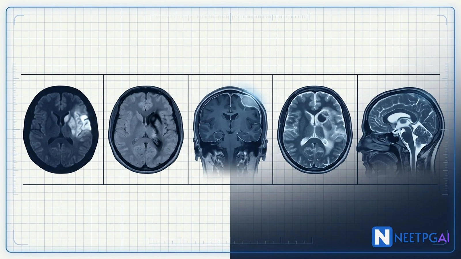
MRI brain emergencies contribute 4-6 image-based MCQs per NEET PG paper across radiology, medicine, neurology, and neurosurgery. Five patterns recur reliably year after year:
- Acute ischemic stroke — DWI bright, ADC dark (cytotoxic oedema within minutes); DWI-FLAIR mismatch selects wake-up stroke patients for thrombolysis under 4.5 hours; MCA territory shows insular ribbon loss
- Intracerebral haemorrhage — SWI (T2*) shows blooming; hypertensive bleed in basal ganglia, thalamus, pons, cerebellum; amyloid angiopathy in elderly lobar; T1 bright at day 3-7 (methaemoglobin)
- Meningioma — dural-based extra-axial mass with dural tail, avid homogeneous enhancement, hyperostosis of overlying bone; CSF cleft confirms extra-axial location
- High-grade glioma vs metastases — GBM heterogeneous ring, central necrosis, crosses corpus callosum ("butterfly glioma"); metastases multiple at grey-white junction with disproportionate oedema
- Acute hydrocephalus — dilated ventricles with transependymal CSF flow (periventricular T2-FLAIR bright), sulcal effacement, tonsillar herniation risk; NPH has Hakim triad (ataxia, incontinence, dementia)
Locking these 5 patterns plus 10-15 additional PYQ images (venous sinus thrombosis, cerebral abscess, MS plaques, herpes encephalitis, PRES) over 2-3 weeks moves accuracy from 40 to 80 percent in neuroradiology MCQs.
Why neuroradiology MCQs are high-yield for NEET PG
Neuroradiology cuts across radiology, medicine, neurology, neurosurgery, and emergency medicine. NEET PG, INI-CET, and FMGE feature MRI brain images in 4-6 questions per paper, almost always paired with a classical clinical vignette (sudden onset hemiparesis, thunderclap headache, seizure, altered sensorium, gait disturbance) to test pattern recognition, sequence understanding, and management decisions. Many of these presentations are true emergencies where the imaging drives the immediate care algorithm — thrombolysis versus thrombectomy versus emergent surgical decompression versus VP shunt.
Drilling these 5 patterns plus 10-15 additional PYQ images over 2-3 weeks moves accuracy from 40 to 80 percent.
Foundational approach before the MCQs
Systematic MRI brain read
| Step | What to look at | Common abnormalities |
|---|
| Vignette anchor | Age, sex, symptom onset (sudden vs gradual), key finding | Sudden = stroke or haemorrhage; gradual = tumour or hydrocephalus; fever = infection |
| Sequence recognition | T1 (grey dark, CSF dark), T2 (CSF bright), FLAIR (CSF nulled — WM lesions bright), DWI-ADC, SWI, post-contrast T1 | Choose sequence to answer question |
| Location | Intra-axial vs extra-axial; grey vs white matter; supratentorial vs infratentorial | Extra-axial = meningioma, schwannoma; grey-white junction = metastases; deep grey = hypertensive bleed |
| Signal pattern | T1/T2 signal, enhancement, restricted diffusion | Blood evolution, tumour vs infection vs demyelination |
| Mass effect | Sulcal effacement, midline shift, herniation | Emergency triggers |
| Ventricular size | Evans index over 0.3; transependymal flow | Hydrocephalus |
Sequence-by-sequence mental shortlist
| Sequence | Signal principle | Best used for |
|---|
| T1 | Fat bright, CSF dark, WM slightly brighter than GM | Anatomy, subacute blood (methaemoglobin bright), fat, gadolinium enhancement |
| T2 | CSF bright, fluid bright, WM darker than GM | Oedema, cysts, most pathology bright |
| FLAIR | T2 with CSF nulled | Periventricular WM lesions, MS plaques, transependymal flow, subarachnoid haemorrhage |
| DWI-ADC | Cytotoxic oedema — DWI bright, ADC dark | Acute infarct, epidermoid, abscess, hypercellular tumour |
| SWI (T2)* | Paramagnetic blooming | Haemorrhage, microbleeds, calcification, venous thrombosis |
| Post-contrast T1 | Enhancement where BBB disrupted | Tumours, infection, inflammation, meningeal disease |
MCQ 1: 62-year-old man with sudden right hemiparesis on waking
Image description: [Axial MRI brain of a 62-year-old man brought to the emergency department after being found on the floor of his bedroom at 6 am, with a witnessed last-normal time of 11 pm the previous night. The DWI shows a well-defined wedge-shaped area of hyperintense (bright) restricted diffusion in the left middle cerebral artery territory — involving the left insular cortex (insular ribbon loss), left frontal-parietal cortex, and left basal ganglia. The corresponding ADC map shows matching hypointense (dark) signal in the same region — confirming true cytotoxic oedema (restricted diffusion) rather than T2 shine-through. The FLAIR image at the same level shows the affected cortex is still isointense compared to the contralateral side — the FLAIR has not yet become bright, indicating a DWI-FLAIR mismatch. No haemorrhage on SWI. A paired MR angiogram shows a left M1 segment occlusion of the middle cerebral artery.]
Clinical vignette: A 62-year-old hypertensive diabetic gentleman, retired schoolteacher, is brought by his wife to the stroke centre emergency department at 6:30 am. He was last seen normal at 11 pm when he retired to bed after his usual evening walk. He was found on the bedroom floor at 6 am, unable to move his right side, with slurred speech. NIHSS score on arrival 14. BP 172/98, HR 82, RR 18, saturation 96 percent on room air. Blood glucose 168 mg/dL. He is otherwise medically stable. The stroke team activates the wake-up stroke pathway. CT brain is unremarkable (no established infarct, no haemorrhage). MRI brain with MR angiography is completed within 30 minutes.
Options:
- (a) Hyperacute left MCA territory ischemic stroke — thrombolysis eligible via DWI-FLAIR mismatch criteria
- (b) Established left MCA infarct beyond thrombolysis window — supportive care only
- (c) Intracerebral haemorrhage — reverse anticoagulation and admit
- (d) Transient ischemic attack — start aspirin and discharge with follow-up
Correct answer: (a) Hyperacute left MCA territory ischemic stroke — thrombolysis eligible via DWI-FLAIR mismatch criteria
Reasoning: The DWI bright / ADC dark pattern is diagnostic of cytotoxic oedema from acute ischemic infarction. The wedge-shaped territorial distribution with insular ribbon loss localises to the left MCA territory, confirmed by the MRA showing M1 occlusion. The critical decision-driving feature is the DWI-FLAIR mismatch — the FLAIR has not yet become bright in the same region — which validated by the WAKE-UP trial (2018) and the DEFUSE-3 / DAWN extended-window criteria indicates the infarct is less than 4.5 hours old, even though the last-known-well time was much earlier. This patient is eligible for intravenous thrombolysis (tenecteplase or alteplase) and endovascular thrombectomy given the large-vessel occlusion.
Established infarct beyond thrombolysis window would show FLAIR hyperintensity matching the DWI. ICH would show blooming on SWI and haematoma signal on T1/T2. TIA shows no DWI restriction (by definition tissue is not infarcted).
Teaching pearl — acute stroke imaging protocol:
- DWI-ADC couplet is the earliest and most sensitive sequence for acute infarct — positive within 3-30 minutes of arterial occlusion; DWI signal persists 7-14 days before pseudonormalising
- DWI-FLAIR mismatch selects wake-up stroke patients for thrombolysis under 4.5 hours (WAKE-UP protocol)
- Extended window thrombectomy — DAWN trial (24 hours) and DEFUSE-3 (16 hours) allow endovascular therapy for large-vessel occlusion using perfusion mismatch (MR or CT perfusion) — core less than 70 mL with mismatch ratio over 1.8
- Territorial patterns — MCA (insular ribbon, frontal-parietal), ACA (medial frontal, paracentral lobule), PCA (occipital, thalamus); lacunar (small under 15 mm subcortical dots in basal ganglia, thalamus, pons, internal capsule)
- DWI-positive but ADC-normal or ADC-bright — think T2 shine-through (not true restriction), old infarct, or vasogenic oedema
MCQ 2: 58-year-old hypertensive with sudden severe headache and left hemiparesis
Image description: [Axial MRI brain of a 58-year-old hypertensive gentleman presenting with sudden severe headache and left hemiparesis 4 hours before imaging. The SWI (susceptibility-weighted image) shows a large hypointense (dark) mass with marked blooming artefact in the right putamen and posterior limb of the internal capsule, measuring 3.5 cm in largest diameter, with the dark signal appearing disproportionately larger than the actual haematoma from the paramagnetic blooming of deoxyhaemoglobin. The T1 image shows the lesion as isointense to slightly hyperintense (early bright signal at the periphery — early methaemoglobin conversion). The T2 image shows the lesion as markedly hypointense (deoxyhaemoglobin dark). Surrounding vasogenic oedema is present as T2 bright signal in the adjacent white matter. Mild mass effect with 3 mm midline shift. No intraventricular extension. SWI also shows multiple small microbleeds scattered in the basal ganglia, thalami, and pons bilaterally, consistent with chronic hypertensive small-vessel disease.]
Clinical vignette: A 58-year-old gentleman with a 20-year history of poorly controlled hypertension (last recorded BP 178/108, on irregular telmisartan), type 2 diabetes, and current smoker, was watching television when he developed a sudden severe occipital headache followed within minutes by left-sided weakness and difficulty speaking. His son brought him to the emergency 45 minutes after symptom onset. BP on arrival 208/118, HR 88, GCS E3M5V4 = 12. NIHSS 12 (left hemiparesis, left hemianaesthesia, dysarthria, mild left hemi-neglect). CT brain (non-contrast) done first shows a hyperdense (bright) collection in the right basal ganglia measuring 3.5 cm — confirming intracerebral haemorrhage. MRI brain is done for further characterisation and to identify any underlying vascular malformation.
Options:
- (a) Hypertensive intracerebral haemorrhage — right putaminal bleed
- (b) Cerebral amyloid angiopathy — lobar cortical haemorrhage
- (c) Haemorrhagic transformation of ischemic infarct
- (d) Arteriovenous malformation rupture
Correct answer: (a) Hypertensive intracerebral haemorrhage — right putaminal bleed
Reasoning: The location (basal ganglia, specifically putamen) is the classical site of hypertensive intracerebral haemorrhage. Hypertensive bleeds preferentially involve deep grey structures fed by penetrating perforator vessels — putamen (most common, 35-40 percent), thalamus (15-20 percent), pons (10 percent), cerebellum (10 percent), and lobar (10-15 percent). The lipohyalinosis and Charcot-Bouchard microaneurysms in these tiny perforators (lenticulostriate, thalamoperforator, paramedian pontine) rupture under sustained high pressure. Additional supporting features are the multiple microbleeds on SWI in a deep distribution (basal ganglia, thalami, pons) — the signature of chronic hypertensive small-vessel disease — and the young-to-middle-aged (under 65) patient profile with sustained uncontrolled hypertension.
Amyloid angiopathy occurs in elderly patients over 65 with lobar cortical-subcortical haemorrhages (frontal, parietal, occipital) and microbleeds in a cortical-subcortical distribution — not deep. Haemorrhagic transformation of infarct would show a wedge-shaped area of infarction on prior DWI with secondary haemorrhage; the vignette does not describe this. AVM rupture typically has a nidus visible on angiography and often occurs in younger patients.
Teaching pearl — MRI blood signal evolution:
| Stage | Time | Component | T1 | T2 | SWI |
|---|
| Hyperacute | Under 6 hours | Oxyhaemoglobin | Iso to slightly hypo | Bright | Iso (no blooming) |
| Acute | 6 hours to 3 days | Deoxyhaemoglobin | Iso | Dark | Marked blooming |
| Early subacute | 3-7 days | Intracellular methaemoglobin | Bright | Dark | Blooming |
| Late subacute | 1-4 weeks | Extracellular methaemoglobin | Bright | Bright | Blooming |
| Chronic | Over 4 weeks | Haemosiderin | Iso | Dark rim | Persistent blooming |
- SWI (T2) is the most sensitive sequence* for microbleeds — a scattered pattern of tiny dark dots reveals underlying small-vessel disease (hypertensive if deep; amyloid if lobar; DAI if traumatic)
- ICH management — acute BP control (target SBP 140-160 in first 24 hours, INTERACT-2 trial), reverse anticoagulation, neurosurgical consult for cerebellar bleed over 3 cm or intraventricular extension with hydrocephalus, and admit to neuro-ICU
- Investigate the cause — hypertension if deep and typical; consider AVM, aneurysm, tumour, or amyloid if atypical location, young patient, or lobar in elderly
MCQ 3: 55-year-old woman with 6 months of progressive left-sided headache and mild left arm weakness
Image description: [Axial post-contrast T1 MRI brain of a 55-year-old woman. The image shows a well-defined dural-based extra-axial mass attached to the right parietal convexity, measuring 3.2 x 2.8 cm. The mass demonstrates avid homogeneous enhancement on post-contrast T1. A characteristic dural tail sign is visible — the adjacent dura is thickened and enhancing, tapering away from the mass. There is a CSF cleft between the mass and the underlying brain, confirming its extra-axial location. The overlying calvarial bone shows hyperostosis (thickened, sclerotic bone). The underlying cortex is buckled inwards with mild mass effect but no invasion. T1 non-contrast shows the mass as isointense to grey matter. T2 shows the mass as mildly hyperintense. There is minimal surrounding vasogenic oedema. A small area of internal calcification is visible on the corresponding CT.]
Clinical vignette: A 55-year-old woman, a retired banker, presents to the neurology outpatient clinic with a 6-month history of progressive dull left-sided headache, worse in the morning on waking, sometimes accompanied by nausea. Over the past 2 months she has noticed mild weakness of her left arm when picking up her grandchildren and occasional word-finding difficulty. No seizures, no visual disturbance, no cranial nerve symptoms. On examination — alert, oriented, MMSE 29/30. Cranial nerves normal. Motor examination — mild 4+/5 power in left upper limb, otherwise normal. Deep tendon reflexes normal, plantars flexor. No sensory or cerebellar deficits. Fundus normal. BP 132/84. MRI brain is arranged.
Options:
- (a) Meningioma
- (b) Glioblastoma multiforme
- (c) Cerebral metastasis
- (d) Cerebral abscess
Correct answer: (a) Meningioma
Reasoning: The imaging is textbook meningioma. Six diagnostic features are present — (1) dural-based extra-axial location with a broad base against the convexity dura; (2) CSF cleft between the mass and the underlying brain (confirms extra-axial); (3) avid homogeneous enhancement on post-contrast T1; (4) dural tail sign — the adjacent dura is thickened and enhancing; (5) hyperostosis of the overlying calvarium — bone remodelling from chronic slow-growing pressure; (6) internal calcification (psammoma bodies). The age (over 40), female sex (2:1 female predominance), gradual progressive symptoms over 6 months, and slow growth pattern are all classical for meningioma.
GBM is an intra-axial mass with heterogeneous ring enhancement and central necrosis — completely different pattern. Metastases are grey-white junction, often multiple, with disproportionate oedema and thin ring. Cerebral abscess shows a smooth thin uniformly enhancing ring with strong central DWI restriction (pus is viscous) and dark T2 rim (paramagnetic free radicals) with surrounding oedema.
Teaching pearl — meningioma epidemiology, subtypes, and management:
- Most common primary intracranial tumour in adults (30-35 percent of all primary brain tumours); most common benign intracranial tumour
- Peak age 40-70 years; female-to-male ratio 2:1 (except spinal meningiomas 4:1)
- Common locations — parasagittal (25 percent, most common), convexity (20 percent), sphenoid ridge (20 percent), olfactory groove (10 percent), tuberculum sellae (10 percent), cerebellopontine angle (10 percent), spinal (5 percent)
- Histological grading (WHO) — Grade I (benign, 80 percent, meningothelial / fibrous / transitional / psammomatous), Grade II (atypical, 15 percent), Grade III (anaplastic, 5 percent — malignant)
- Genetic associations — NF2 mutations (chromosome 22q loss) in sporadic and NF2-related meningiomas; hormonally responsive (progesterone receptor positive in 70 percent — often enlarge in pregnancy)
- Management — small asymptomatic incidental meningiomas can be observed with serial MRI at 6-12 month intervals; symptomatic, growing, or over 3 cm need surgical resection graded by Simpson classification (I complete resection with dural attachment and hyperostotic bone; V decompression only); stereotactic radiosurgery (Gamma Knife) for surgically inaccessible or residual tumours
MCQ 4: 68-year-old man with 4 weeks of progressive right hemiparesis, headache, and confusion
Image description: [Axial post-contrast T1 MRI brain of a 68-year-old man. The image shows a large heterogeneous mass centred on the splenium of the corpus callosum with bilateral extension into both hemispheres — the classical butterfly appearance. The mass demonstrates irregular thick ring enhancement with a central non-enhancing dark core (necrosis) measuring 4 cm in largest diameter. There is extensive surrounding vasogenic oedema as T2 bright signal in the periventricular and adjacent white matter, respecting the grey-white junction. Marked mass effect with effacement of the posterior horns of the lateral ventricles and 4 mm midline shift. DWI shows restricted diffusion in the enhancing rim (dark on ADC). MR spectroscopy over the enhancing rim shows a raised choline peak, reduced NAA peak, and a lactate-lipid peak. No other lesions visible. The paired axial FLAIR shows the same butterfly distribution with extensive white matter oedema.]
Clinical vignette: A 68-year-old retired gentleman, previously well, presents to the neurology emergency with 4 weeks of progressive right-sided weakness, worsening headache (worse on lying down, wakes him from sleep), and confusion — his wife reports he has become forgetful, calling their daughter by the wrong name, and has had two witnessed generalised seizures in the past week. No history of malignancy, no smoking, no weight loss, no fever. Examination — GCS 14, mildly disoriented to time. Motor — right hemiparesis 3/5. Right-sided upper motor neuron facial weakness. Papilloedema on fundoscopy. No lymphadenopathy, no palpable masses. CT chest-abdomen-pelvis unremarkable. Mammography (as he was thought to be female initially — corrected) not applicable. PET-CT body unremarkable.
Options:
- (a) Glioblastoma multiforme (GBM) — butterfly glioma
- (b) Cerebral metastasis from unknown primary
- (c) Primary CNS lymphoma
- (d) Cerebral tuberculoma with abscess
Correct answer: (a) Glioblastoma multiforme (GBM) — butterfly glioma
Reasoning: The imaging is diagnostic of GBM — a heterogeneous intra-axial mass with irregular thick ring enhancement, central necrosis, extensive surrounding vasogenic oedema, and crossing of the corpus callosum producing the butterfly glioma appearance. Butterfly glioma is nearly pathognomonic — the ability to cross the corpus callosum via the splenium or genu is a defining feature of GBM (also seen with primary CNS lymphoma and less commonly with rare demyelination). DWI restriction in the enhancing rim reflects hypercellularity. MR spectroscopy shows the classical GBM signature — raised choline (increased cell membrane turnover), reduced NAA (neuronal loss), and lactate-lipid peak (necrosis and anaerobic metabolism). The age (over 65), absence of a systemic primary despite full workup, and the pattern-recognition triad clinch GBM.
Metastases are usually multiple, at the grey-white junction, spherical with thin regular ring enhancement, and have a known or newly discovered systemic primary. Primary CNS lymphoma often crosses the corpus callosum too, but classically shows homogeneous non-necrotic enhancement, DWI restriction throughout the mass (not just the rim), lower ADC values, and dramatic response to steroids (vanishing tumour effect on repeat imaging — an important pitfall). Tuberculoma is usually smaller with a target sign (central caseation dark, peripheral rim enhancement) and often multiple in a patient from a TB-endemic area.
Teaching pearl — GBM management principles:
- GBM is WHO grade IV astrocytoma — the most common malignant primary brain tumour in adults; median age at diagnosis 65
- IDH-wildtype GBM (90 percent) has poor prognosis (median survival 12-15 months with standard therapy); IDH-mutant (previously secondary GBM) has better prognosis (24-30 months)
- MGMT promoter methylation predicts better response to temozolomide
- Stupp protocol — maximal safe surgical resection (aiming for gross-total resection when feasible) followed by concurrent radiotherapy (60 Gy in 30 fractions) with temozolomide, then adjuvant temozolomide for 6 cycles
- Tumour treating fields (TTFields) — alternating electric fields via scalp electrodes — added benefit in the EF-14 trial
- Bevacizumab for recurrent or progressive disease; no clear survival benefit but reduces oedema and steroid requirement
- Metastasis workup for a solitary ring-enhancing mass — CT chest-abdomen-pelvis, mammography, PET-CT, skin examination for melanoma; if all negative, consider primary CNS process
- Solitary metastasis vs GBM distinction — perfusion MRI (rCBV markedly elevated in high-grade glioma, less so in metastasis), spectroscopy (lipid-lactate peaks common to both but choline-NAA ratios differ), and DWI can help; ultimate answer often comes from stereotactic biopsy
MCQ 5: 78-year-old man with 4 months of gait unsteadiness, urinary urgency, and forgetfulness
Image description: [Axial and sagittal MRI brain of a 78-year-old gentleman. The axial FLAIR shows marked dilatation of both lateral ventricles with the frontal horns appearing rounded. The Evans index (maximal frontal horn width divided by biparietal internal diameter) measures 0.38 (greater than the 0.3 cut-off, confirming ventriculomegaly). The temporal horns are prominent at 4 mm (greater than the 2 mm cut-off). There is effacement of the cortical sulci over the high convexity but preservation of the sulci over the lateral surfaces — the disproportionately enlarged subarachnoid space hydrocephalus (DESH) pattern. The corpus callosum is thinned and bowed upwards. A flow void is visible in the aqueduct of Sylvius on sagittal T2 (hyperdynamic CSF flow — the aqueductal flow void sign). No transependymal periventricular hyperintensity is seen (this feature would suggest an acute obstructive component). No mass lesion, no aqueductal stenosis, no colloid cyst. A prior CT scan from the same patient 2 years ago showed normal-sized ventricles.]
Clinical vignette: A 78-year-old retired railway official is brought to the neurology outpatient clinic by his daughter with a 4-month history of progressive gait unsteadiness, urinary urgency, and forgetfulness. The gait is described by the daughter as "shuffling and wide-based, as if his feet are stuck to the floor". He has fallen twice in the past month. He now needs prompting to use the toilet and has had 3 episodes of incontinence. His memory for recent events (breakfast items, morning conversations) is patchy but distant memory is preserved. No headache, no visual disturbance, no seizures, no focal weakness. Past history — hypertension controlled, mild BPH, no diabetes, no stroke. Examination — MMSE 22/30 (attention and recent memory affected). Gait — magnetic wide-based short-stepped shuffling with reduced arm swing. Motor and reflexes normal. No cerebellar signs. Fundus normal. Postural BP normal. Prostate mildly enlarged.
Options:
- (a) Normal pressure hydrocephalus (NPH) — VP shunt candidate
- (b) Alzheimer disease
- (c) Aqueductal stenosis with obstructive hydrocephalus
- (d) Parkinson disease
Correct answer: (a) Normal pressure hydrocephalus (NPH) — VP shunt candidate
Reasoning: The clinical triad of gait ataxia (wide-based magnetic gait — the earliest and most reversible sign), urinary incontinence, and cognitive decline is the Hakim triad of normal pressure hydrocephalus (NPH) — a specific subtype of communicating hydrocephalus in elderly patients. The MRI features are classical — ventriculomegaly out of proportion to sulcal atrophy (Evans index 0.38), the disproportionately enlarged subarachnoid space hydrocephalus (DESH) pattern (tight high-convexity sulci with wide Sylvian fissures), thinning and upward bowing of the corpus callosum, and the aqueductal flow void on T2 sagittal reflecting hyperdynamic CSF flow. The absence of transependymal periventricular hyperintensity is consistent with the chronic communicating nature of NPH (no acute obstruction).
Alzheimer disease shows diffuse cortical atrophy with proportionately enlarged ventricles (Evans index generally under 0.3) and hippocampal atrophy — sulci are widened, not effaced. Aqueductal stenosis produces obstructive hydrocephalus with lateral and third ventricle dilatation but normal-sized fourth ventricle, plus transependymal flow if acute. Parkinson disease has bradykinesia, resting tremor, rigidity, and characteristic MRI is often normal (or shows substantia nigra hyperechogenicity on transcranial ultrasound and dopamine transporter reduction on DAT scan).
Teaching pearl — NPH management:
- CSF tap test (large-volume lumbar puncture removing 30-50 mL CSF) — gait improvement in 24-72 hours predicts good VP shunt response (sensitivity 26-62 percent, specificity 33-100 percent)
- Extended CSF drainage (external lumbar drain 300 mL / 3 days) has higher predictive value than the single tap test
- CSF pressure — is characteristically NORMAL on lumbar puncture (hence "normal pressure" hydrocephalus) — the pathology is impaired arachnoid granulation absorption, not raised pressure
- Ventriculoperitoneal (VP) shunt with programmable valve — gait improves in 60-80 percent of well-selected patients, cognition and continence less predictably
- Complications — shunt infection, shunt overdrainage (subdural haematoma), obstruction, and later Alzheimer-superimposed decline
- Idiopathic NPH vs secondary NPH — secondary follows subarachnoid haemorrhage (aneurysm rupture), meningitis, trauma, or prior brain surgery
Common pitfalls in MRI brain emergency MCQs
Five frequent error patterns appear in NEET PG dissection of neuroradiology image MCQs.
Pitfall 1: Missing DWI-FLAIR mismatch and stopping thrombolysis eligibility at last-known-well time
A patient with an unknown or beyond-4.5-hour last-known-well time can still be thrombolysis-eligible if the DWI-FLAIR mismatch shows DWI-positive but FLAIR-negative lesion — the FLAIR turnaround takes 3-6 hours, so DWI-positive/FLAIR-negative approximates a lesion under 4.5 hours old. NEET PG is increasingly testing wake-up stroke protocols; do not reflexively write off patients with "unknown onset time" as ineligible.
Pitfall 2: Confusing hypertensive vs amyloid bleed locations
Hypertensive bleeds are DEEP — putamen, thalamus, pons, cerebellum. Amyloid angiopathy bleeds are LOBAR — frontal, parietal, occipital cortical-subcortical. The microbleed distribution on SWI is the tell — deep microbleeds indicate hypertensive small-vessel disease; lobar microbleeds indicate amyloid. Amyloid is a disease of the elderly (over 65) and is often recurrent. This location distinction commonly determines the exam answer.
Pitfall 3: Missing the extra-axial vs intra-axial call
Meningioma is extra-axial — dural-based with a CSF cleft separating it from the brain, buckling the underlying cortex without invasion. GBM and metastases are intra-axial — within the brain parenchyma with no CSF cleft. Missing this call leads to the wrong differential list. The CSF cleft, dural tail, hyperostosis, and broad dural base are the four hallmarks that should reflexively trigger "meningioma".
Pitfall 4: Confusing GBM, metastases, primary CNS lymphoma, and abscess (the "ring-enhancing mass" differential)
Ring-enhancing mass differentials in adults include GBM, metastases, primary CNS lymphoma, abscess, tuberculoma, and demyelinating tumefactive MS. Key distinguishers — GBM (thick irregular ring, necrotic centre, crosses corpus callosum — butterfly, MRS raised choline and lactate-lipid); metastases (thin regular ring, multiple, grey-white junction, disproportionate oedema); PCNSL (homogeneous non-necrotic enhancement, DWI restriction throughout, immunosuppressed patient — HIV, transplant, steroid response); abscess (smooth thin uniformly enhancing ring, strong central DWI restriction because pus is viscous, dark T2 rim); tuberculoma (target sign, often multiple, TB-endemic population); tumefactive MS (open ring toward cortex, young patient, other MS features).
Pitfall 5: Missing NPH and treating it as Alzheimer disease
The elderly patient with gait ataxia, urinary incontinence, and cognitive decline is often labelled Alzheimer or vascular dementia and denied a treatable diagnosis. The magnetic wide-based gait as the first symptom (before cognitive decline) and the MRI showing ventriculomegaly out of proportion to sulcal atrophy plus DESH pattern should trigger the NPH workup. NPH is one of the few reversible dementias. A missed NPH is a missed VP shunt candidate — high-yield ethical and clinical NEET PG teaching point.
How to study neuroradiology for NEET PG
- Memorise the 5 patterns in this article cold — DWI-ADC couplet, blood signal evolution, meningioma hallmarks, butterfly GBM, NPH triad and DESH
- Review 10-15 additional PYQ images — cerebral venous sinus thrombosis (cord sign, empty delta), cerebral abscess (thin ring, DWI restriction), MS plaques (Dawson fingers, callosal fingers), herpes encephalitis (bitemporal, insular), PRES (posterior parieto-occipital vasogenic oedema), tuberculoma (target sign), cerebral toxoplasmosis (eccentric target sign, HIV), colloid cyst (bright T1 at foramen of Monro), and pituitary macroadenoma with apoplexy
- Pair images with classic vignettes — sudden onset means stroke or bleed; gradual means tumour or hydrocephalus; fever plus focal deficit means abscess or encephalitis
- Learn one sequence at a time — spend a day each on T1, T2, FLAIR, DWI-ADC, SWI, and post-contrast T1; understand what each shows and when to prefer it
- Use spaced repetition — 1d, 3d, 7d, 14d, 30d review of the same 25-30 high-yield MRI images
- Practice in the question bank — NEETPGAI offers a tagged neuroradiology set; do 20-30 questions per day for 2-3 weeks
Key takeaways
- Neuroradiology contributes 4-6 image MCQs per NEET PG paper across radiology, medicine, neurology, and neurosurgery
- Acute infarct — DWI bright / ADC dark within 3-30 minutes; DWI-FLAIR mismatch selects wake-up stroke for thrombolysis
- ICH — SWI blooming most sensitive; hypertensive deep (putamen/thalamus/pons/cerebellum), amyloid lobar (elderly)
- Meningioma — extra-axial with dural tail, homogeneous enhancement, hyperostosis, CSF cleft
- GBM vs metastases — butterfly (crosses corpus callosum), thick ring, necrosis, MRS raised choline-lactate for GBM; multiple grey-white junction with disproportionate oedema for metastases
- Hydrocephalus — transependymal flow acutely; NPH has Hakim triad plus DESH pattern, VP shunt candidate
- Pair images with classic vignettes for fast pattern recognition
Frequently Asked Questions
What is the diagnostic sequence for acute ischemic stroke on MRI and what does DWI-FLAIR mismatch mean?
MRI is the gold-standard modality for acute ischemic stroke because it can detect cytotoxic oedema within minutes of arterial occlusion, well before CT changes appear. The diagnostic sequence is diffusion-weighted imaging (DWI) with the apparent diffusion coefficient (ADC) map. In acute infarction, cytotoxic oedema restricts water motion inside dying neurons — this appears bright (hyperintense) on DWI and dark (hypointense) on ADC. The DWI signal appears within 3-30 minutes of vessel occlusion and persists for 7-14 days before pseudonormalising. The DWI-FLAIR mismatch refers to a DWI-positive but FLAIR-negative lesion, which indicates a hyperacute infarct less than 4.5 hours old (FLAIR takes 3-6 hours to become bright) — this pattern is used to select wake-up stroke patients for thrombolysis under the WAKE-UP trial protocol even when the exact stroke onset time is unknown. The infarct pattern also localises the vessel — MCA territory infarcts show insular ribbon loss and involve the frontal-parietal cortex, ACA involves the medial frontal and paracentral lobule, PCA involves the occipital lobe and posterior thalamus, and lacunar infarcts appear as small (under 15 mm) subcortical dot-like restrictions in the basal ganglia, thalamus, pons, or internal capsule. NEET PG tests DWI bright / ADC dark as the pathognomonic couplet and the 4.5 hour and 24 hour treatment windows.
How is intracerebral haemorrhage characterised on MRI and why is SWI the most sensitive sequence?
Intracerebral haemorrhage (ICH) is best characterised on gradient-echo T2 star (T2*) or susceptibility-weighted imaging (SWI), which are exquisitely sensitive to blood breakdown products because of their paramagnetic effect on the local magnetic field. SWI produces marked blooming artefact (dark signal disproportionately larger than the actual haemorrhage) from deoxyhaemoglobin, methaemoglobin, and haemosiderin. The signal evolution of blood on MRI is temporally predictable — hyperacute (under 6 hours) shows oxyhaemoglobin (T1 iso to slightly hypo, T2 bright); acute (6 hours to 3 days) shows deoxyhaemoglobin (T1 iso, T2 dark); early subacute (3-7 days) shows intracellular methaemoglobin (T1 bright, T2 dark — the classic bright T1 haematoma); late subacute (1-4 weeks) shows extracellular methaemoglobin (T1 bright, T2 bright); chronic (over 4 weeks) shows haemosiderin (T1 iso, T2 dark rim). Aetiology is location-driven — hypertensive haemorrhage occurs in the basal ganglia (most common, especially putamen), thalamus, pons, and cerebellum (deep grey structures with penetrating perforator vessels). Cerebral amyloid angiopathy causes lobar cortical-subcortical haemorrhages in elderly patients (over 65), often multiple and recurrent, with microbleeds on SWI. Trauma-related contusions are cortical (frontal and temporal poles). AV malformations and aneurysm rupture produce their own patterns. NEET PG tests hypertensive vs amyloid location, the T1 bright methaemoglobin at 3-7 days, and the SWI blooming for microbleed detection.
What are the classical MRI features of a meningioma and how are they distinguished from other extra-axial masses?
Meningioma is the most common primary intracranial tumour in adults (overall) and the most common benign intracranial tumour. On MRI, the classical features are a well-defined dural-based extra-axial mass with a broad base against the dura, typically iso to slightly hypointense on T1 and iso to slightly hyperintense on T2, with avid homogeneous enhancement on post-contrast T1. The dural tail sign is enhancement of the adjacent thickened dura mater tapering away from the mass — highly suggestive but not pathognomonic (seen in schwannoma, dural metastases, and granulomatous disease as well). Additional features include hyperostosis of the overlying calvarium (bone remodelling from chronic slow-growing pressure), calcification (psammoma bodies, especially in psammomatous variant), CSF cleft between the mass and the underlying brain confirming extra-axial location, and buckling of the underlying cortex without invasion. Common locations are parasagittal (25 percent), convexity (20 percent), sphenoid ridge (20 percent), olfactory groove, tuberculum sella, and cerebellopontine angle. Differentials of an extra-axial mass include vestibular schwannoma (CPA, ice-cream-cone shape into the internal auditory canal), dural metastases (multiple, often from breast or prostate primary), haemangiopericytoma (aggressive, often no hyperostosis, may erode bone), and dural-based lymphoma. Small asymptomatic incidental meningiomas can be observed with serial MRI; symptomatic or growing tumours need surgical resection (Simpson grade I-V). NEET PG tests the dural tail, hyperostosis, and homogeneous enhancement pattern.
How do high-grade gliomas differ from cerebral metastases on MRI and what is the significance of butterfly glioma?
High-grade gliomas (glioblastoma multiforme, WHO grade IV) and cerebral metastases are the two commonest solitary and multiple ring-enhancing intracranial masses in adults, and distinguishing them is a classic NEET PG image question. GBM appears as a heterogeneous intra-axial mass with irregular thick ring enhancement, central necrosis (non-enhancing dark core), surrounding vasogenic oedema (T2 bright white matter oedema respecting the grey-white junction), and mass effect. GBM classically crosses the corpus callosum producing the butterfly glioma appearance — a bilateral hemispheric lesion extending across the genu or splenium of the corpus callosum — nearly pathognomonic when seen. DWI shows restricted diffusion in the enhancing rim (from hypercellularity). MR spectroscopy shows raised choline, reduced NAA, and lactate lipid peaks. Cerebral metastases in contrast are typically multiple (in 70 percent of cases), located at the grey-white junction (arterial embolic distribution), spherical and well-circumscribed, with thin regular ring enhancement, and disproportionate surrounding vasogenic oedema out of proportion to the mass size (the small-lesion big-oedema pattern). Common primaries are lung (adenocarcinoma most common), breast, melanoma, renal cell, and gastrointestinal. A solitary metastasis can mimic GBM but the ring is usually thinner and more regular. Investigation of a solitary intracranial mass suspected to be metastatic includes CT chest-abdomen-pelvis, mammography, PET-CT, and dermatology examination. NEET PG tests butterfly glioma for GBM and multiple grey-white junction lesions with disproportionate oedema for metastases.
What are the MRI features of acute hydrocephalus and how are obstructive and communicating types distinguished?
Hydrocephalus is characterised on MRI by ventricular dilatation out of proportion to sulcal size, with three additional acute features — transependymal CSF flow (periventricular T2-FLAIR hyperintensity from CSF egress into the periventricular white matter under raised pressure), effacement of the cortical sulci and basal cisterns, and in severe cases descent of the cerebellar tonsils below the foramen magnum (tonsillar herniation). The Evans index (maximal frontal horn width divided by maximal biparietal internal diameter at the same level) greater than 0.3 confirms ventricular enlargement. The temporal horn width greater than 2 mm is another sensitive early sign. Obstructive (non-communicating) hydrocephalus results from CSF flow blockage within the ventricular system — aqueductal stenosis (dilated lateral and third ventricles with normal fourth ventricle), colloid cyst of the third ventricle (bright round mass at the foramen of Monro causing acute obstruction and sudden death risk), tumours (pineal tumour, brainstem glioma, posterior fossa tumour compressing the fourth ventricle), Chiari malformation, and haemorrhage clot. Communicating hydrocephalus results from impaired CSF absorption at the arachnoid villi — post-subarachnoid haemorrhage, post-meningitis, post-traumatic, and normal pressure hydrocephalus (NPH). NPH is a specific subtype in elderly patients with the classical Hakim triad of gait ataxia (wide-based magnetic gait — earliest and most reversible), urinary incontinence, and cognitive decline; MRI shows ventriculomegaly out of proportion to sulcal atrophy with disproportionately enlarged subarachnoid spaces (DESH) and CSF flow void in the aqueduct. Treatment of obstructive hydrocephalus is emergent external ventricular drainage or endoscopic third ventriculostomy; NPH is treated with ventriculoperitoneal shunt. NEET PG tests transependymal flow as the acute sign, Hakim triad for NPH, and the location of obstruction (aqueduct vs foramen of Monro vs fourth ventricle).
This content is for educational purposes for NEET PG exam preparation. It is not a substitute for professional medical advice, diagnosis, or treatment. Clinical information has been reviewed by qualified medical professionals.
Written by: NEETPGAI Editorial Team
Reviewed by: Pending SME Review
Last reviewed: July 2026