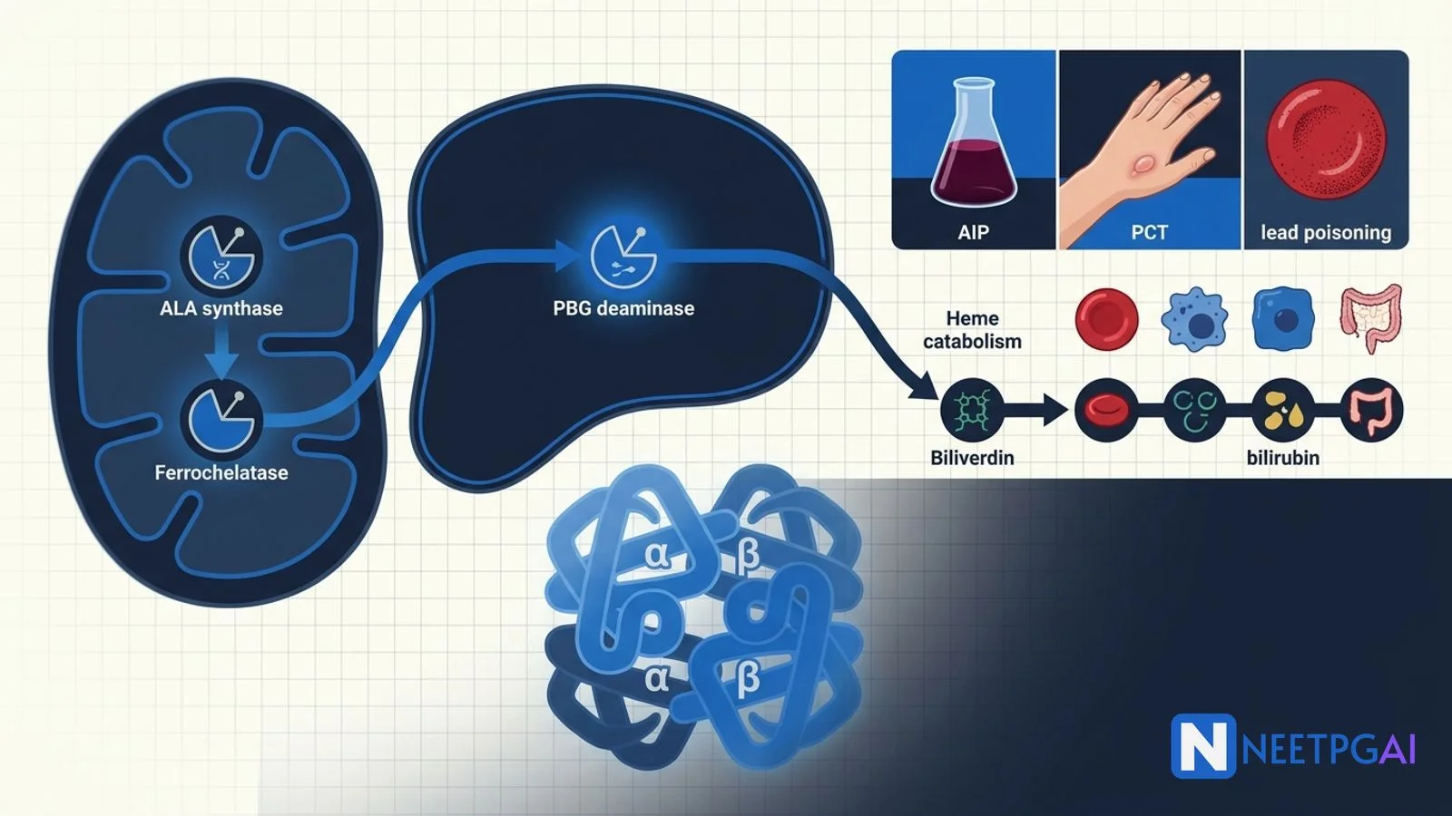
Quick Answer
Hemoglobin, heme, and bilirubin metabolism is a 3 to 4 question topic in NEET PG Biochemistry. Lock these:
- Hemoglobin — 4 globin + 4 heme; adult HbA α2β2, HbA2 α2δ2, HbF α2γ2.
- Heme synthesis rate-limiting — ALA synthase (mitochondria; requires B6; feedback-inhibited by heme).
- Acute intermittent porphyria — PBG deaminase deficiency; 5 P's; NO photosensitivity; hemin + carbohydrates.
- Porphyria cutanea tarda — uroporphyrinogen decarboxylase; commonest; cutaneous; alcohol + HCV.
- Lead poisoning — inhibits ALA dehydratase + ferrochelatase; basophilic stippling; wrist drop; chelate.
- Bilirubin — heme oxygenase → biliverdin → unconjugated → UGT1A1 conjugation.
- Crigler-Najjar I — complete UGT deficiency; kernicterus. Dubin-Johnson — MRP2 defect; black liver.
- G6PD deficiency — X-linked; primaquine, dapsone, sulfonamides trigger haemolysis; Heinz bodies.
Hemoglobin and heme metabolism is a high-yield NEET PG Biochemistry topic that spans haematology, pharmacology, and toxicology. The pathway is elegant — a two-compartment (mitochondrial and cytoplasmic) synthesis, drug-precipitated porphyric attacks, lead-inhibited enzymes, and the classical bilirubin transport disorders. India-specific relevance includes G6PD deficiency prevalence and drug-induced haemolysis with dapsone and primaquine. This NEETPGAI deep dive covers the pathway, disease mechanisms, and MCQ traps.
Pair this guide with the reproductive physiology deep dive for physiology-biochemistry integration.
Hemoglobin structure
Adult hemoglobin is a tetramer of four globin chains (2 alpha + 2 non-alpha) with a heme moiety in each chain (iron-protoporphyrin IX). Types:
| Hemoglobin | Structure | Percent (adult) | Function |
|---|
| HbA | α2β2 | 96 to 98% | Main adult |
| HbA2 | α2δ2 | 2 to 3% | Elevated in beta thalassaemia trait |
| HbF | α2γ2 | Less than 1% | Fetal; higher O2 affinity; elevated in sickle cell, beta thalassaemia |
| HbA1c | Glycated HbA | 4 to 6% (normal) | Marker of long-term glycaemic control |
Embryonic hemoglobins — Gower 1 (ζ2ε2), Gower 2 (α2ε2), Portland (ζ2γ2) — replaced by fetal HbF by the end of the first trimester.
Oxygen dissociation curve — sigmoid; right shift (higher P50, easier O2 unloading) with acidosis, hypercapnia, hyperthermia, and elevated 2,3-BPG (chronic hypoxia); left shift with alkalosis, hypocapnia, hypothermia, and HbF.
Heme synthesis pathway
Heme is synthesised in every nucleated cell but especially in the bone marrow (85 percent, for erythropoiesis) and liver (15 percent, for cytochromes). The pathway begins and ends in the mitochondrion with cytoplasmic intermediates.
| Step | Enzyme | Substrate → Product | Location |
|---|
| 1 (rate-limiting) | ALA synthase (needs B6) | Glycine + succinyl-CoA → 5-aminolaevulinic acid (ALA) | Mitochondrion |
| 2 | ALA dehydratase (needs Zn) | 2 ALA → porphobilinogen (PBG) | Cytoplasm |
| 3 | Porphobilinogen deaminase (hydroxymethylbilane synthase) | 4 PBG → hydroxymethylbilane | Cytoplasm |
| 4 | Uroporphyrinogen III synthase | HMB → uroporphyrinogen III | Cytoplasm |
| 5 | Uroporphyrinogen decarboxylase | Uroporphyrinogen III → coproporphyrinogen III | Cytoplasm |
| 6 | Coproporphyrinogen oxidase | Copro III → protoporphyrinogen IX | Mitochondrion |
| 7 | Protoporphyrinogen oxidase | Protoporphyrinogen IX → protoporphyrin IX | Mitochondrion |
| 8 | Ferrochelatase (adds Fe2+) | Protoporphyrin IX + Fe2+ → heme | Mitochondrion |
Regulation — hepatic ALAS1 is feedback-inhibited by heme. Drugs that induce cytochrome P450 (barbiturates, sulfonamides, alcohol, rifampicin, phenytoin, carbamazepine, ketoconazole) deplete free heme by pulling it into new cytochromes — releasing ALAS1 from inhibition, over-producing porphyrins, and precipitating acute porphyria attacks in susceptible individuals.
Erythroid ALAS2 — regulated by iron availability via iron-response elements on its mRNA; mutations cause X-linked sideroblastic anaemia.
Porphyrias — enzyme block table
Each porphyria corresponds to a block in the pathway. Two clinical clusters — acute (neurologic and abdominal) and cutaneous (photosensitive).
| Porphyria | Enzyme | Type | Diagnostic marker | Key clue |
|---|
| ALA dehydratase deficiency porphyria | ALAD | Acute | Urinary ALA | Very rare |
| Acute intermittent porphyria (AIP) | PBG deaminase | Acute (no skin) | Urinary PBG, ALA | 5 P's; port-wine urine on standing |
| Congenital erythropoietic porphyria (Gunther) | Uroporphyrinogen III synthase | Cutaneous (severe) | Urinary uroporphyrin I | Red teeth (erythrodontia), hypertrichosis (werewolf), severe photosensitivity |
| Porphyria cutanea tarda (PCT) | Uroporphyrinogen decarboxylase | Cutaneous (commonest) | Urinary uroporphyrin | Alcohol + HCV; skin blisters on sun-exposed areas |
| Hereditary coproporphyria | Coproporphyrinogen oxidase | Acute + cutaneous | Urinary + fecal copropophyrin | Mixed |
| Variegate porphyria | Protoporphyrinogen oxidase | Acute + cutaneous | Fecal protoporphyrin | South African, King George III association |
| Erythropoietic protoporphyria (EPP) | Ferrochelatase | Cutaneous (burning) | RBC protoporphyrin | Painful photosensitivity within minutes |
Acute intermittent porphyria (AIP) — high-yield
- Autosomal dominant PBG deaminase deficiency.
- The 5 P's — Pain (severe colicky abdominal pain, misdiagnosed as surgical abdomen), Polyneuropathy (motor; can progress to respiratory failure), Psychiatric (anxiety, agitation, hallucinations), Precipitants (drugs, alcohol, fasting, hormones, infection), Port-wine urine on standing (PBG oxidises in air).
- NO photosensitivity — the block is BEFORE the photosensitive porphyrinogens.
- Diagnosis — urinary PBG and ALA elevated; genetic testing.
- Treatment — high-carbohydrate diet (10 percent dextrose infusion — glucose suppresses ALAS1), IV hemin (or heme arginate) — replenishes heme pool, suppresses ALAS1. Avoid triggers. Newer — givosiran (RNAi against ALAS1, approved for recurrent AIP).
Porphyria cutanea tarda (PCT) — commonest porphyria
- Uroporphyrinogen decarboxylase deficiency (sporadic or familial).
- Triggers — alcohol, hepatitis C, HIV, iron overload, oestrogens.
- Presents with skin blisters, hyperpigmentation, hypertrichosis on sun-exposed areas.
- Diagnosis — elevated urinary uroporphyrin; iron studies (often high ferritin).
- Treatment — trigger avoidance (alcohol, oestrogens), phlebotomy to reduce iron, low-dose hydroxychloroquine.
Heme catabolism and bilirubin
Senescent red cells are phagocytosed by macrophages of the reticuloendothelial system (spleen, liver, bone marrow). Heme is degraded in a stepwise pathway:
- Heme oxygenase — cleaves heme, releasing iron (recycled via transferrin), carbon monoxide, and biliverdin (green).
- Biliverdin reductase — reduces biliverdin (green) to unconjugated bilirubin (yellow) using NADPH.
- Unconjugated (indirect) bilirubin — hydrophobic, transported bound to albumin to the liver. Elevated in haemolysis, Gilbert, Crigler-Najjar, neonatal jaundice.
- Hepatocyte uptake — by OATP1B1 and OATP1B3 (Rotor syndrome — defective).
- Conjugation — by UGT1A1 (UDP-glucuronyltransferase) to bilirubin diglucuronide (water-soluble). Neonates have low UGT1A1 → physiological jaundice.
- Biliary excretion — via MRP2 canalicular transporter (Dubin-Johnson — defective).
- Gut — bacterial beta-glucuronidase deconjugates → urobilinogen. Some reabsorbed into portal circulation (enterohepatic), excreted in urine as urobilin. The rest is oxidised to stercobilin (brown; faecal colour).
Bilirubin transport disorders
| Disorder | Defect | Bilirubin type | Clinical |
|---|
| Gilbert | Decreased UGT1A1 (promoter TA insertion) | Unconjugated (mild, less than 5 mg/dL) | Benign; unconjugated jaundice with fasting, stress, illness; 5 to 10% population |
| Crigler-Najjar type I | Complete absence of UGT1A1 | Unconjugated (over 20 mg/dL) | Kernicterus; phototherapy + liver transplant; no phenobarbital response |
| Crigler-Najjar type II | Partial UGT1A1 deficiency | Unconjugated (moderate) | Responds to phenobarbital (induces UGT) |
| Dubin-Johnson | MRP2 (canalicular transporter) defect | Conjugated | Grossly BLACK liver on biopsy; benign |
| Rotor | OATP1B1/1B3 uptake defect | Conjugated | Normal liver on biopsy; benign |
Physiological neonatal jaundice — appears after day 2, peaks day 4 to 5, resolves by day 10. Due to immature UGT, increased haemolysis, and enterohepatic recirculation. Treated with phototherapy. Rule out pathological jaundice (appears less than 24 hours, rises rapidly, direct bilirubin over 20 percent).
Kernicterus — bilirubin encephalopathy from unconjugated bilirubin crossing the immature blood-brain barrier and depositing in basal ganglia and brain stem. Preventable with prompt phototherapy and exchange transfusion.
Lead poisoning
Lead inhibits ALA dehydratase (early) and ferrochelatase (late), blocking heme synthesis and causing sideroblastic-like anaemia.
Clinical features:
- Haematologic — microcytic hypochromic anaemia with basophilic stippling (ribosomal RNA aggregation), ring sideroblasts on marrow.
- Neurologic — peripheral neuropathy (motor — wrist drop, foot drop), encephalopathy in children (irritability, developmental regression, seizures).
- Abdominal — colicky pain, constipation.
- Renal — Fanconi syndrome (proximal tubular dysfunction), chronic tubulointerstitial nephropathy.
- Musculoskeletal — gum lead line (Burton line) — bluish discoloration at the gingival margin from bacterial hydrogen sulphide reacting with lead.
- Bone — dense metaphyseal bands on paediatric long-bone radiograph.
Diagnosis — blood lead level (BLL); elevated free erythrocyte protoporphyrin (FEP) or zinc protoporphyrin; urinary ALA elevated.
Treatment — remove source. Chelation for BLL over 45 µg/dL in children — succimer (DMSA) oral; over 70 µg/dL or symptomatic — dimercaprol (BAL) IM + EDTA (calcium disodium edetate) IV.
Sideroblastic anaemia
Impaired heme synthesis with iron accumulation in mitochondria → ring sideroblasts on bone marrow Prussian blue stain (iron rings around the nucleus).
| Type | Cause | Comment |
|---|
| Hereditary | ALAS2 mutation (X-linked recessive) | Male predominance; responsive to pyridoxine (B6) |
| Acquired reversible | B6 deficiency, isoniazid, chloramphenicol, alcohol, lead, copper deficiency | Trigger avoidance |
| Acquired clonal | MDS with ring sideroblasts (MDS-RS) | Elderly; SF3B1 mutation; risk of AML transformation |
Peripheral smear — dimorphic (microcytic and normocytic populations).
G6PD deficiency — India context
Glucose-6-phosphate dehydrogenase is the rate-limiting enzyme of the pentose phosphate pathway. It generates NADPH, which regenerates reduced glutathione (GSH), the red cell's main antioxidant.
- X-linked recessive — males affected, females usually carriers (some symptomatic due to lyonisation).
- Prevalence in India — about 3 to 15 percent regionally, especially in tribal populations (Bhil, Vasava, Andaman islanders).
- Triggers — oxidant drugs (primaquine, dapsone, sulfonamides, nitrofurantoin, methylene blue, rasburicase), fava beans, infection.
- Clinical — acute intravascular haemolysis 1 to 3 days after trigger — jaundice, dark urine (haemoglobinuria), back pain, anaemia.
- Peripheral smear — Heinz bodies (denatured haemoglobin, seen with supravital stain), bite cells (splenic pitting), blister cells.
- Diagnosis — G6PD assay (do NOT test during acute haemolysis — reticulocytes have higher G6PD levels giving false-normal).
- Management — remove trigger; supportive care; transfusion if severe.
- Prevention — screen for G6PD before starting primaquine (radical cure of vivax malaria) or dapsone (leprosy, PJP prophylaxis).
NEET PG MCQ traps
- HbA — α2β2; HbA2 — α2δ2 (raised in beta thalassaemia trait); HbF — α2γ2.
- Right shift O2 curve — acidosis, high 2,3-BPG, hyperthermia, hypercapnia.
- HbF — higher O2 affinity than HbA (left shift).
- Rate-limiting heme step — ALA synthase (mitochondrial; needs B6).
- Enzyme blocks — ALA dehydratase and ferrochelatase inhibited by lead.
- AIP — PBG deaminase; 5 P's; NO photosensitivity; hemin + glucose treatment.
- PCT — commonest porphyria; uroporphyrinogen decarboxylase; alcohol + HCV; phlebotomy + hydroxychloroquine.
- Erythropoietic protoporphyria — ferrochelatase; painful photosensitivity within minutes.
- Gunther disease (CEP) — red teeth (erythrodontia), severe photosensitivity; uroporphyrinogen III synthase.
- Basophilic stippling — lead poisoning (also thalassaemia).
- Gum lead line (Burton) — lead poisoning.
- Ring sideroblasts — Prussian blue positive iron rings around nucleus.
- Bilirubin conjugation — UGT1A1 in hepatocyte.
- Gilbert — mild unconjugated; benign.
- Crigler-Najjar I — complete UGT deficiency; kernicterus; liver transplant.
- Crigler-Najjar II — phenobarbital responsive.
- Dubin-Johnson — MRP2 defect; conjugated; BLACK liver on biopsy.
- Rotor — OATP1B1/1B3 defect; conjugated; NORMAL liver on biopsy.
- Neonatal jaundice — immature UGT; phototherapy converts bilirubin to water-soluble isomers.
- G6PD deficiency — X-linked; Heinz bodies and bite cells; primaquine, dapsone, sulfonamides.
- G6PD assay — not during acute haemolysis (false-normal from reticulocytes).
- Heme oxygenase — heme → biliverdin + CO + Fe.
- Stercobilin — faecal brown; urobilin — urinary yellow.
Frequently asked questions
What is the rate-limiting step of heme synthesis and where does it occur?
The rate-limiting enzyme of heme synthesis is delta-aminolaevulinic acid synthase (ALA synthase, ALAS). It catalyses the condensation of glycine and succinyl-CoA to form 5-aminolaevulinic acid (ALA) in the mitochondrial matrix, using pyridoxal phosphate (vitamin B6) as cofactor. Two isoforms exist — ALAS1 in liver (feedback-inhibited by heme) and ALAS2 in erythroid cells (regulated by iron availability). Drugs like barbiturates, sulfonamides, alcohol, and rifampicin induce hepatic ALAS1 (via CYP450 induction that depletes heme) and can precipitate acute intermittent porphyria attacks in susceptible individuals.
How do you clinically differentiate the acute porphyrias?
Acute intermittent porphyria (AIP) — porphobilinogen deaminase deficiency; presents with the 5 P's — Pain (abdominal), Polyneuropathy (motor), Psychiatric symptoms (anxiety, hallucinations), Precipitants (drugs, alcohol, fasting), and Port-wine urine on standing (from PBG oxidation). NO photosensitivity because the block is BEFORE the photosensitive porphyrinogens. Porphyria cutanea tarda (PCT) — uroporphyrinogen decarboxylase; commonest porphyria; cutaneous photosensitivity, blisters on sun-exposed skin, alcohol and HCV triggers. Variegate porphyria and hereditary coproporphyria have BOTH acute attacks AND cutaneous features. Erythropoietic protoporphyria — ferrochelatase; burning photosensitivity.
How does lead poisoning affect heme synthesis?
Lead inhibits two enzymes in the heme synthesis pathway — ALA dehydratase (ALAD, converts ALA to porphobilinogen) and ferrochelatase (inserts Fe2+ into protoporphyrin IX to form heme). Result — accumulation of ALA and protoporphyrin IX, with reduced heme. Clinical picture — microcytic hypochromic anaemia with basophilic stippling (from ribosomal RNA aggregation), sideroblastic features (ring sideroblasts on marrow), abdominal colic, peripheral neuropathy (wrist drop, foot drop from motor demyelination), gum lead line (Burton line) from bacterial sulphide deposits, and childhood encephalopathy. Elevated blood lead level, free erythrocyte protoporphyrin (FEP), and urinary ALA confirm diagnosis. Chelate with succimer (DMSA), EDTA, or dimercaprol depending on severity.
How do you differentiate the hereditary hyperbilirubinaemias?
Gilbert syndrome — mild deficiency of UGT1A1; mild unconjugated hyperbilirubinaemia (less than 5 mg/dL) precipitated by fasting, stress, infection; benign, no treatment. Crigler-Najjar type I — complete absence of UGT1A1; severe unconjugated hyperbilirubinaemia at birth (over 20 mg/dL) with kernicterus; phototherapy and liver transplant. Crigler-Najjar type II — partial UGT1A1 deficiency; responds to phenobarbital (enzyme inducer). Dubin-Johnson — defective MRP2 (canalicular multidrug resistance protein 2); conjugated hyperbilirubinaemia; grossly BLACK liver on biopsy. Rotor syndrome — organic anion uptake defect (SLCO1B1, SLCO1B3); conjugated hyperbilirubinaemia; normal-appearing liver on biopsy (helpful discriminator from Dubin-Johnson).
Why is G6PD deficiency an important cause of drug-induced haemolysis in India?
Glucose-6-phosphate dehydrogenase (G6PD) is the rate-limiting enzyme of the hexose monophosphate shunt (pentose phosphate pathway) which generates NADPH. NADPH regenerates reduced glutathione (GSH) which protects red cells from oxidative stress. G6PD deficiency (X-linked recessive) is highly prevalent in India (about 3 to 15 percent regionally, especially tribal populations). Oxidant drugs — primaquine, dapsone, sulfonamides, nitrofurantoin, methylene blue — precipitate acute intravascular haemolysis with Heinz bodies (denatured haemoglobin), bite cells, jaundice, dark urine (haemoglobinuria), and back pain. Fava beans and infection are additional triggers. India-relevance is high because dapsone (leprosy) and primaquine (radical cure of vivax malaria) are widely used — screen for G6PD before starting.
This content is for educational purposes for NEET PG exam preparation. It is not a substitute for professional medical advice, diagnosis, or treatment. Clinical information has been reviewed by qualified medical professionals.
Written by: NEETPGAI Editorial Team
Reviewed by: Pending SME Review
Last reviewed: July 2026