Version 1.0 — Published August 2026
Quick Answer
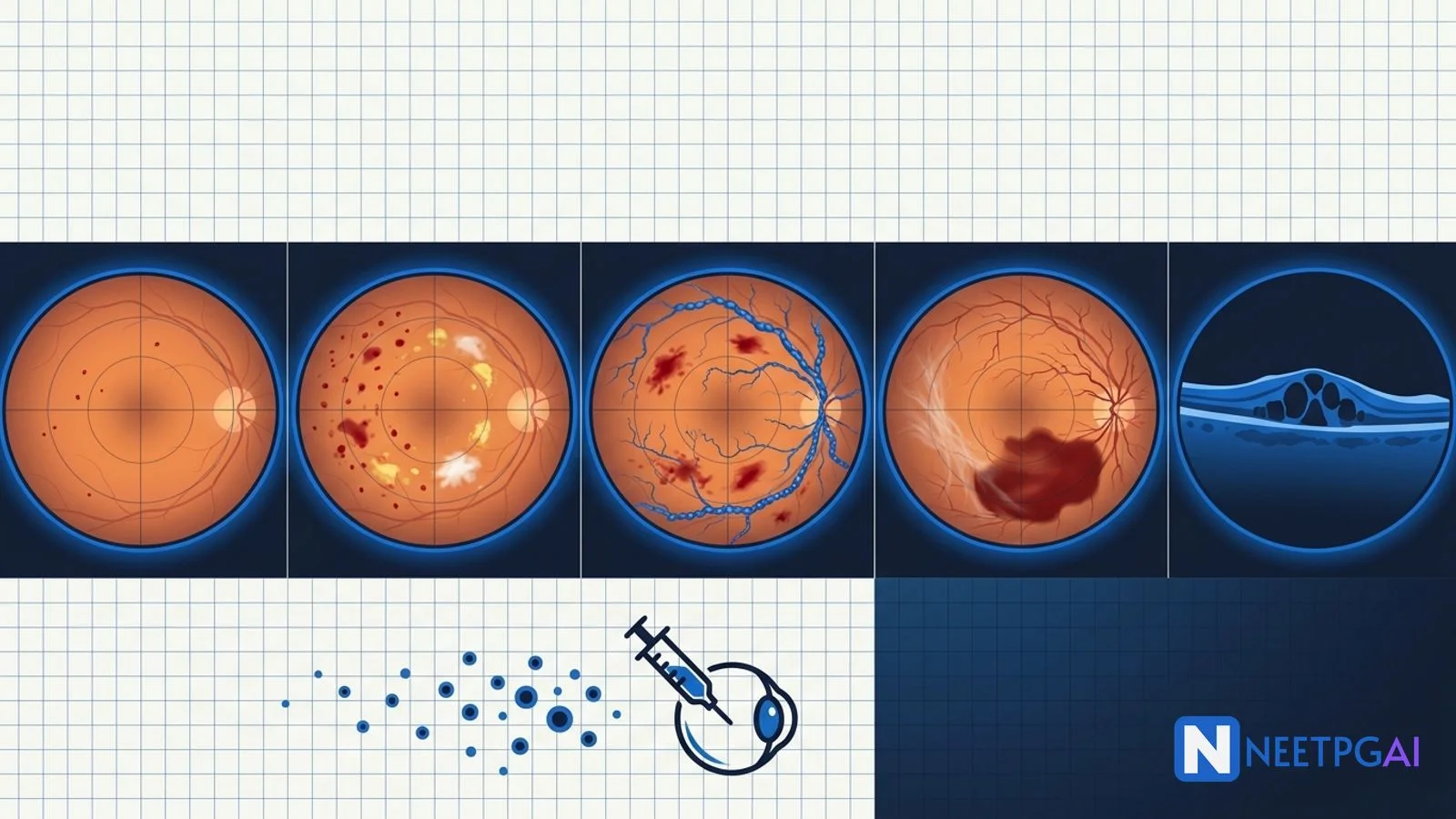
Diabetic retinopathy (DR) image MCQs contribute 3-5 questions per NEET PG paper across ophthalmology, medicine, and PSM. Five high-yield patterns recur reliably year after year:
- Mild NPDR — only microaneurysms (tiny round red dots at the posterior pole)
- Moderate NPDR — microaneurysms PLUS dot-and-blot haemorrhages, hard exudates, cotton wool spots — but not meeting the severe threshold
- Severe NPDR (4-2-1 rule) — intra-retinal haemorrhages in all 4 quadrants OR venous beading in 2 quadrants OR IRMA in 1 quadrant
- Proliferative DR (PDR) — neovascularization of disc (NVD) or elsewhere (NVE), with or without pre-retinal / vitreous haemorrhage
- Diabetic macular edema (DME) — retinal thickening at the macula; OCT is gold standard; centre-involving (CI-DME) needs prompt anti-VEGF; non-centre-involving may be observed or treated focally
Locking these 5 patterns plus 5-6 additional fundoscopy PYQ images (hypertensive retinopathy grades, central retinal vein occlusion, central retinal artery occlusion, papilloedema, age-related macular degeneration) over 1-2 weeks moves DR-MCQ accuracy from 45 to 85 percent.
Why diabetic retinopathy image MCQs are high-yield for NEET PG
Diabetic retinopathy is the leading cause of blindness in the working-age adult population globally and one of the highest-burden public-health complications of diabetes in India. NEET PG tests DR heavily because it cuts across ophthalmology (grading, treatment), internal medicine and endocrinology (glycaemic control, hypertension, dyslipidaemia), and PSM (NPCBVI, screening programmes, artificial intelligence-based fundus screening). The fundoscopy findings are stereotyped, photogenic, and diagnostic, making them ideal for image MCQs.
Drilling these 5 patterns plus a small library of hypertensive retinopathy, CRVO, CRAO, and AMD images over 1-2 weeks moves fundoscopy-MCQ accuracy from 45 to 85 percent.
Foundational approach — the systematic fundoscopy read
Read the fundus in this order
| Structure | What to look for | Common DR abnormalities |
|---|
| Optic disc | Colour, margins, cup-disc ratio, neovascularization | NVD in PDR, disc oedema in advanced |
| Macula | Foveal reflex, thickening, exudates | Hard exudates ringing fovea, CSME, DME |
| Retinal vessels (arterioles and venules) | Calibre, tortuosity, beading, IRMA | Venous beading, IRMA, silver/copper wiring |
| Retinal periphery (4 quadrants) | Haemorrhages, microaneurysms, cotton wool spots, NVE | 4-2-1 rule findings, NVE |
| Vitreous | Clarity, haemorrhage | Pre-retinal boat-shaped haemorrhage, vitreous haemorrhage |
| Any tractional membranes | Fibrous proliferation | Advanced PDR, tractional retinal detachment risk |
Aunt Minnie triggers for diabetic retinopathy
| Finding | Grade or diagnosis |
|---|
| Only microaneurysms | Mild NPDR |
| Microaneurysms plus dot-blot haemorrhages plus hard exudates plus cotton wool spots (mild) | Moderate NPDR |
| Intra-retinal haemorrhages in all 4 quadrants OR venous beading in 2 quadrants OR IRMA in 1 quadrant | Severe NPDR (4-2-1) |
| Any neovascularization (NVD or NVE) | Proliferative DR (PDR) |
| Macular thickening on OCT with foveal involvement | Centre-involving DME |
| Yellow hard-exudate ring around the fovea | Diabetic macular edema (may or may not be centre-involving) |
| Pre-retinal boat-shaped haemorrhage | PDR with vitreous or pre-retinal haemorrhage |
| Fibrovascular membrane pulling retina | Tractional retinal detachment (advanced PDR) |
MCQ 1: 42-year-old with type 2 diabetes for 8 years — routine annual screening reveals a few red dots at the posterior pole
Image description: [Colour fundus photograph of the right eye of a 42-year-old male. The image shows a normal orange-red retinal background with a healthy optic disc (cup-disc ratio 0.3, clear margins, pink neuroretinal rim), normal macula with intact foveal reflex, and normal calibre retinal vessels (arterioles and venules with normal A:V ratio 2:3). The only abnormal finding is 4-5 tiny round red dots approximately 20-40 microns in diameter scattered around the posterior pole, mostly inferotemporal to the macula. There are no haemorrhages, no hard exudates, no cotton wool spots, no venous beading, no IRMA, no neovascularization, and no vitreous haemorrhage. Fluorescein angiography (paired image) shows the same tiny lesions as hyperfluorescent pinpoint dots consistent with microaneurysms.]
Clinical vignette: A 42-year-old male software engineer with type 2 diabetes for 8 years attends the annual diabetic screening clinic. He is on metformin 1 g twice daily, HbA1c 7.8 percent, BP 128/82, LDL 88 mg/dL, no symptoms of visual disturbance, normal visual acuity 6/6 in both eyes on Snellen chart. His previous fundoscopy 1 year ago was reported as normal.
Options:
- (a) Mild non-proliferative diabetic retinopathy (mild NPDR)
- (b) Moderate non-proliferative diabetic retinopathy (moderate NPDR)
- (c) Severe non-proliferative diabetic retinopathy (severe NPDR)
- (d) Proliferative diabetic retinopathy (PDR)
Correct answer: (a) Mild non-proliferative diabetic retinopathy (mild NPDR)
Reasoning: Mild NPDR is defined by the presence of microaneurysms ONLY, without any other DR finding. Microaneurysms appear as tiny round red dots 20-40 microns in diameter at the level of the retina, usually clustered in the posterior pole. They represent localised outpouchings of retinal capillaries at points of pericyte loss. They are the earliest visible sign of DR on fundoscopy and are confirmed by fluorescein angiography as pinpoint hyperfluorescent lesions.
This patient has ONLY microaneurysms — no haemorrhages, no exudates, no cotton wool spots, no venous beading, no IRMA, no neovascularization — placing him in mild NPDR. Moderate NPDR would show additional findings not meeting the severe threshold; severe NPDR requires the 4-2-1 rule; PDR requires neovascularization.
Teaching pearl — mild NPDR management:
| Aspect | Recommendation |
|---|
| Systemic optimisation | HbA1c to target (typically under 7 percent for most non-elderly adults; individualised); BP under 130/80; statin per CV risk; smoking cessation |
| Follow-up interval | Every 12 months if no macular involvement; every 6 months if macular changes present |
| Patient counselling | Explain the finding; motivate adherence to glycaemia and BP control; symptoms to watch for (vision changes, floaters, flashes) |
| No ocular treatment yet | Mild NPDR does not need laser or anti-VEGF; observation and systemic control are the mainstays |
| Pregnancy consideration | If planning pregnancy or currently pregnant, more frequent monitoring (every trimester) because pregnancy accelerates DR progression |
- Progression to PDR from mild NPDR is roughly 5 percent over 5 years; from moderate NPDR roughly 15 percent; from severe NPDR roughly 50 percent
- NEET PG tests the microaneurysm-only definition of mild NPDR and the annual follow-up interval
MCQ 2: 55-year-old woman with type 2 diabetes for 15 years — dilated fundoscopy shows dot-blot haemorrhages, hard exudates, and cotton wool spots
Image description: [Colour fundus photograph of the left eye of a 55-year-old female. The image shows the following abnormalities across the posterior pole and mid-periphery. Multiple microaneurysms as small round red dots. Several dot-and-blot haemorrhages — round to irregular red intra-retinal haemorrhages 100-300 microns in diameter, deep to the vessels. Yellow-white hard exudates clustered in a partial ring around the macula (circinate pattern) and scattered along the arcades — waxy well-defined lipid-protein deposits. Two cotton wool spots — grey-white fluffy indistinct lesions in the superficial retina near the disc margin, representing nerve fibre layer infarcts. No venous beading, no IRMA, no neovascularization, no pre-retinal or vitreous haemorrhage. The haemorrhages are present in only 2 of 4 quadrants; venous beading is absent; IRMA is absent. Foveal reflex is preserved with no obvious retinal thickening at the fovea. Paired OCT of the macula shows normal central subfield thickness at 245 microns.]
Clinical vignette: A 55-year-old female homemaker with type 2 diabetes for 15 years, obese (BMI 31), hypertensive (BP 138/86 on amlodipine), and dyslipidaemic (LDL 118 mg/dL on atorvastatin), presents for annual diabetic screening. She has noticed occasional blurring of vision especially at night for the past 3-4 months. HbA1c 8.6 percent, creatinine 1.2 mg/dL, urine albumin-creatinine ratio 68 mg/g (microalbuminuria). Visual acuity 6/12 in both eyes correcting to 6/9 with pinhole.
Options:
- (a) Mild non-proliferative diabetic retinopathy
- (b) Moderate non-proliferative diabetic retinopathy
- (c) Severe non-proliferative diabetic retinopathy
- (d) Proliferative diabetic retinopathy
Correct answer: (b) Moderate non-proliferative diabetic retinopathy
Reasoning: Moderate NPDR shows microaneurysms PLUS one or more of dot-and-blot haemorrhages, hard exudates, cotton wool spots, and mild venous beading, but does not meet the severe NPDR threshold (4-2-1 rule). This patient has microaneurysms, dot-and-blot haemorrhages (in 2 quadrants only), hard exudates (circinate around the macula), and cotton wool spots — clearly beyond mild NPDR but not yet severe (no all-4-quadrant haemorrhages, no 2-quadrant venous beading, no IRMA).
Mild NPDR would be microaneurysms only. Severe NPDR requires any one of the 4-2-1 criteria. PDR requires neovascularization.
Teaching pearl — moderate NPDR management:
| Aspect | Recommendation |
|---|
| Systemic optimisation | Aggressive glycaemic (HbA1c target under 7), BP under 130/80, lipid control; consider SGLT2i or GLP1-RA if comorbid CVD/renal risk |
| Follow-up interval | Every 6-12 months |
| OCT of macula | To exclude subclinical macular edema (particularly if patient reports vision change) |
| Fluorescein angiography | If ischaemia or DME is suspected; can guide anti-VEGF or focal laser decisions |
| No PRP laser yet | Moderate NPDR without high-risk features does not need PRP |
| Anti-VEGF only if DME | Reserved for concurrent centre-involving DME |
| Patient counselling | Explain progression risk; emphasise strict glycaemia and BP; teach warning symptoms |
- 15 percent of moderate NPDR progresses to PDR over 5 years without intervention; strict glycaemic and BP control cuts this by 25-40 percent (DCCT, UKPDS)
- Microalbuminuria (as in our patient) parallels DR progression — renal and retinal microvascular disease travel together
- NEET PG tests the additive-finding definition of moderate NPDR
MCQ 3: 48-year-old with type 2 diabetes for 20 years — extensive intra-retinal haemorrhages in all four quadrants plus venous beading
Image description: [Colour fundus photograph of the right eye of a 48-year-old male. The image shows abnormalities distributed across all four retinal quadrants. Numerous dot-and-blot intra-retinal haemorrhages in all 4 quadrants, many appearing in clusters, some as large blot haemorrhages up to 500 microns in diameter. Venous beading — the retinal veins show alternating dilated and constricted segments giving a "sausage-string" appearance — clearly present in the superotemporal, inferotemporal, and superonasal quadrants (3 quadrants). Intra-retinal microvascular abnormalities (IRMA) — abnormal fine tortuous vessel loops within the retina, sparing the disc margin, are seen in the inferotemporal quadrant. Hard exudates and cotton wool spots scattered. No neovascularization of the disc (NVD) or elsewhere (NVE), no pre-retinal or vitreous haemorrhage. The macula shows preserved foveal reflex but hard exudates in the temporal macula. OCT shows mild retinal thickening in the parafoveal area sparing the central subfield.]
Clinical vignette: A 48-year-old male truck driver with type 2 diabetes for 20 years (poorly controlled — HbA1c 10.4 percent), hypertension (BP 152/94 on ramipril), CKD stage 3a (creatinine 1.6 mg/dL, eGFR 46 mL/min), and current smoker, presents with 6 months of gradually worsening vision in both eyes. Visual acuity 6/24 in the right eye, 6/18 in the left, correcting only partially with pinhole. No sudden vision loss episodes reported. He last had an eye examination 4 years ago.
Options:
- (a) Mild non-proliferative diabetic retinopathy
- (b) Moderate non-proliferative diabetic retinopathy
- (c) Severe non-proliferative diabetic retinopathy
- (d) Proliferative diabetic retinopathy
Correct answer: (c) Severe non-proliferative diabetic retinopathy (severe NPDR)
Reasoning: Severe NPDR is defined by the 4-2-1 rule — any ONE of (1) severe intra-retinal haemorrhages and microaneurysms in ALL 4 quadrants, (2) venous beading in 2 OR MORE quadrants, or (3) IRMA in 1 OR MORE quadrants. This patient has all three criteria — haemorrhages in all 4 quadrants, venous beading in 3 quadrants, and IRMA in 1 quadrant. Any single 4-2-1 finding is enough for severe NPDR; having all three would qualify as very-severe NPDR. He does NOT have neovascularization, so this is NOT PDR.
Mild NPDR is microaneurysms only. Moderate NPDR has additional findings but does not meet 4-2-1. PDR requires NVD or NVE.
Teaching pearl — severe NPDR management:
| Aspect | Recommendation |
|---|
| Rate of progression | Approximately 50 percent progress to PDR within 12 months without intervention |
| Systemic optimisation | Urgent HbA1c reduction (target under 7 percent individualised), BP control under 130/80, smoking cessation |
| Follow-up interval | Every 3-4 months |
| OCT of macula | Every visit to detect DME early |
| Consider PRP laser earlier | Particularly in poor-compliance patients, monocular patients, before pregnancy, before cataract surgery, or before RRT initiation |
| Anti-VEGF consideration | Some emerging evidence (PANORAMA trial with aflibercept) for anti-VEGF in severe NPDR to reduce progression to PDR and DME |
| Patient education | Very high urgency of glycaemic and BP control, seeking urgent care for any sudden vision change (indicates vitreous haemorrhage) |
- The DCCT and UKPDS trials showed that intensive glycaemic control reduces the risk of DR progression by 25-40 percent
- Severe NPDR is the tipping point where laser is often considered even in the absence of neovascularization, particularly in high-risk patients
- NEET PG tests the 4-2-1 rule and the 50-percent-progression-to-PDR-at-1-year statistic
MCQ 4: 38-year-old with type 1 diabetes for 25 years — new fine vascular tufts on the optic disc plus a boat-shaped pre-retinal haemorrhage
Image description: [Colour fundus photograph of the left eye of a 38-year-old female. The image shows: fine, fragile, tortuous vascular tufts arising from the optic disc surface and extending onto the adjacent retina — neovascularization of the disc (NVD) — approximately one-third disc area in extent, forming a fine lace-like network on and around the disc. A boat-shaped (D-shaped) pre-retinal haemorrhage measuring approximately 2 disc diameters is present just inferior to the disc — the haemorrhage has a flat horizontal upper margin (gravitational fluid level) and a curved inferior margin, sitting between the retina and the vitreous. Additional NVE (neovascularization elsewhere) — fine vascular tufts at the superotemporal arcade, also extending onto the retinal surface, approximately half disc area. Fibrous proliferation is minimal at this early stage. Background shows extensive dot-and-blot haemorrhages, hard exudates, cotton wool spots, and venous beading consistent with prior severe NPDR. Vitreous is largely clear apart from the pre-retinal haemorrhage.]
Clinical vignette: A 38-year-old female schoolteacher with type 1 diabetes since age 13 (25 years) presents with a sudden painless "curtain" over the lower half of her vision in the left eye 2 hours ago, following a bout of coughing this morning. She has been on multiple daily insulin injections since diagnosis, HbA1c 8.4 percent (best control 7.2 percent, worst 11 percent), no hypertension, non-smoker. She had her last dilated fundoscopy 18 months ago (reported as "severe non-proliferative changes, needs to be seen sooner") but did not attend follow-up because of a busy work schedule. Visual acuity 6/6 in the right eye, 3/60 in the left eye (hand movements to 3 metres). IOP 14 in both eyes.
Options:
- (a) Severe non-proliferative diabetic retinopathy
- (b) Proliferative diabetic retinopathy with vitreous haemorrhage
- (c) Central retinal vein occlusion
- (d) Diabetic macular edema
Correct answer: (b) Proliferative diabetic retinopathy with pre-retinal / vitreous haemorrhage (PDR)
Reasoning: PDR is defined by the presence of neovascularization — of the disc (NVD) or elsewhere (NVE) — with or without pre-retinal or vitreous haemorrhage and fibrovascular proliferation. This patient has NVD (one-third disc area on the disc) plus NVE (half disc area at the superotemporal arcade) plus a boat-shaped pre-retinal haemorrhage — the classical Diabetic Retinopathy Study (DRS) high-risk PDR definition. The sudden vision loss with a curtain-like defect after a Valsalva-like manoeuvre (coughing) is classical for PDR-related pre-retinal or vitreous haemorrhage — fragile new vessels bleed easily.
Severe NPDR does not have neovascularization by definition. Central retinal vein occlusion would have widespread flame-shaped and dot-and-blot haemorrhages in ALL quadrants ("blood and thunder" fundus) with disc oedema and macular oedema, but not neovascularization at initial presentation. Diabetic macular edema is a specific finding of retinal thickening at the macula, not what is described here.
Teaching pearl — PDR treatment:
| Modality | Indication | Notes |
|---|
| Panretinal photocoagulation (PRP) | DRS high-risk PDR — NVD at or over one-third disc area, or NVD of any size with VH or PRH, or NVE at or over half disc area with VH or PRH | 1200-1600 burns of 500 microns spot size in 1-2 sessions; peripheral retina outside vascular arcades; sparing macula and papillomacular bundle |
| Intravitreal anti-VEGF (ranibizumab, aflibercept, bevacizumab) | Protocol S and CLARITY evidence for PDR (non-inferior to PRP in DRCR Protocol S); useful in DME-plus-PDR, in poor-compliance PRP patients, or before scheduled surgery | Monthly loading then treat-and-extend or PRN based on OCT and clinical response |
| Vitrectomy (pars plana vitrectomy) | Non-clearing vitreous haemorrhage (over 3 months), tractional retinal detachment involving or threatening the macula, mixed rhegmatogenous-tractional detachment, dense subhyaloid haemorrhage over macula | Retinal specialist referral |
| Adjunctive intravitreal steroid (dexamethasone Ozurdex, triamcinolone) | Chronic DME resistant to anti-VEGF, pseudophakic eyes | Risk of cataract, IOP rise |
- DRS high-risk PDR criteria — NVD at or over one-third disc area; OR NVD of any size with VH or PRH; OR NVE at or over half disc area with VH or PRH; PRP is strongly indicated
- Mechanism of PRP — reduces retinal metabolic demand and lowers VEGF drive, leading to regression of neovascularization
- Complications of PRP — peripheral visual field loss, night vision loss, transient macular edema exacerbation, pain, occasional inadvertent macular burns
- NEET PG tests the neovascularization definition of PDR, the DRS high-risk criteria, and the PRP vs anti-VEGF choice
MCQ 5: 60-year-old with type 2 diabetes for 12 years — hard exudate ring around the fovea plus a thickened macula on OCT
Image description: [Colour fundus photograph of the right eye of a 60-year-old male. The image shows: background moderate NPDR with microaneurysms, scattered dot-and-blot haemorrhages, and a few cotton wool spots. The critical finding is at the macula — a well-defined ring of waxy yellow hard exudates arranged in a circinate pattern around the fovea, with the ring encroaching within 500 microns of the foveal centre. The fovea itself appears slightly elevated with loss of the normal foveal light reflex, and there is subtle greyish retinal thickening extending across the central macular region. Paired OCT scan of the macula (horizontal line scan through the fovea) shows: central subfield thickness (CST) of 385 microns (normal 250-290; markedly increased), intra-retinal cystic spaces (large hyporeflective ovoid spaces in the outer nuclear layer), hyperreflective foci consistent with hard exudates, subretinal fluid at the fovea, and preserved photoreceptor layer at this stage. Fluorescein angiography (paired image) shows late leakage from multiple parafoveal microaneurysms with diffuse foveal hyperfluorescence.]
Clinical vignette: A 60-year-old male retired bank clerk with type 2 diabetes for 12 years (HbA1c 7.4 percent, BP 132/84 on ramipril, LDL 82 mg/dL on atorvastatin) presents with 4 months of gradually worsening blurred central vision in the right eye. He describes difficulty reading newsprint and recognising faces at a distance. He also reports mild distortion of straight lines when tested with an Amsler grid — vertical lines appear wavy in the central field. Visual acuity 6/18 in the right eye and 6/9 in the left eye. No pain, no floaters, no flashes.
Options:
- (a) Age-related macular degeneration (wet AMD)
- (b) Centre-involving diabetic macular edema (CI-DME)
- (c) Central serous chorioretinopathy
- (d) Non-centre-involving diabetic macular edema
Correct answer: (b) Centre-involving diabetic macular edema (CI-DME)
Reasoning: Diabetic macular edema (DME) is retinal thickening at the macula caused by leakage from damaged retinal microvasculature. Centre-involving DME (CI-DME) has thickening involving the central 1 mm subfield including the fovea, and is diagnosed on OCT. This patient has a hard-exudate ring around the fovea (circinate pattern), foveal thickening on clinical examination, and OCT confirming central subfield thickness of 385 microns with intra-retinal cystic spaces and subretinal fluid — all classic CI-DME.
Wet AMD would show a sub-retinal or intra-retinal haemorrhage plus a choroidal neovascular membrane on OCT, typically in an older patient with drusen; not the pattern here. Central serous chorioretinopathy would show a serous retinal detachment on OCT (pocket of subretinal fluid) without hard exudates, usually in younger stressed men. Non-centre-involving DME would not involve the central subfield on OCT (CST would be normal).
Teaching pearl — DME classification and treatment:
| Classification | Definition | First-line treatment |
|---|
| ETDRS clinically significant macular edema (CSME) — historical | Thickening within 500 microns of fovea OR hard exudates within 500 microns of fovea if associated with adjacent thickening OR 1-disc-area of thickening any part within 1 disc diameter of fovea | Focal or grid laser (historical) |
| Centre-involving DME (CI-DME) — modern OCT-based | Retinal thickening involving central subfield (1 mm zone) on OCT with visual impact | Anti-VEGF (ranibizumab, aflibercept, bevacizumab) first-line |
| Non-centre-involving DME | Thickening sparing central subfield | Observation OR focal laser to leaking microaneurysms |
DRCR Protocol T compared bevacizumab, ranibizumab, and aflibercept in DME. Findings:
- For baseline visual acuity 20/32 to 20/40, all three drugs comparable at 1 year
- For baseline visual acuity 20/50 or worse, aflibercept superior (letter improvement 18.9 vs 14.2 for ranibizumab vs 11.8 for bevacizumab at 1 year)
Standard anti-VEGF regimen:
- Monthly loading for 3-6 months
- Then treat-and-extend or pro re nata based on OCT and clinical response
- Bevacizumab (Avastin) — Rs 3,000-5,000 per compounded intravitreal dose (off-label but widely used in India)
- Ranibizumab (Lucentis) — Rs 20,000-30,000 per dose (licensed)
- Aflibercept (Eylea) — Rs 30,000-40,000 per dose (licensed)
- Brolucizumab (Beovu) — Rs 30,000 per dose (licensed, newer, extended dosing intervals possible)
Alternative or adjunctive options:
-
Intravitreal dexamethasone implant (Ozurdex) — 4-6 month sustained release; useful for pseudophakic eyes or anti-VEGF non-responders; risks cataract and IOP rise
-
Intravitreal fluocinolone acetonide implant (Iluvien) — 3-year sustained release; similar risks
-
Focal or grid laser — for non-centre-involving DME or as adjunct to anti-VEGF
-
Vitrectomy — for chronic DME with vitreomacular traction, non-clearing vitreous haemorrhage, or tractional detachment
-
NEET PG tests the OCT-based centre-involving vs non-centre-involving classification, the Protocol T aflibercept-superior-in-poor-vision result, and the cost-effectiveness role of bevacizumab in India
Common pitfalls in diabetic retinopathy image MCQs
Pitfall 1: Confusing microaneurysms with dot haemorrhages
Microaneurysms are 20-40 microns, perfectly round, red dots — outpouchings of retinal capillaries at points of pericyte loss. Dot haemorrhages are larger (100-300 microns), round to irregular, and represent bleeding within the retina. On fluorescein angiography, microaneurysms are pinpoint hyperfluorescent and dot haemorrhages BLOCK fluorescence (hypofluorescent). Fundoscopy alone cannot always distinguish them; when the differential matters (mild vs moderate NPDR), the presence of only tiny round red dots supports microaneurysms and therefore mild NPDR.
Pitfall 2: Missing the 4-2-1 rule for severe NPDR
Students often over-diagnose PDR when there is no neovascularization but extensive haemorrhages and venous beading. Severe NPDR is a distinct category — the 4-2-1 rule (all-4-quadrant haemorrhages OR 2-quadrant venous beading OR 1-quadrant IRMA) defines it. Neovascularization is what elevates it to PDR. IRMA can look similar to fine NVE — the discriminator is that IRMA lies WITHIN the retina and does NOT leak on fluorescein angiography, whereas NVE lies ON the retinal surface and LEAKS profusely.
Pitfall 3: Missing IRMA in severe NPDR
Intra-retinal microvascular abnormalities (IRMA) are shunts of dilated retinal capillaries within the retina, adjacent to areas of capillary non-perfusion. They look like fine tortuous vessel loops. Their presence in 1 or more quadrants alone is enough for severe NPDR (the "1" of the 4-2-1 rule). They are often missed because they are subtle; fluorescein angiography helps because IRMA does not leak whereas NVE does.
Pitfall 4: Mistaking cotton wool spots for hard exudates
Cotton wool spots are grey-white, fluffy, indistinct-bordered lesions representing nerve fibre layer infarcts. Hard exudates are yellow, waxy, well-defined, sharp-bordered lipid-protein deposits at the outer plexiform layer. The colour and border definition are the key discriminators. Both are seen in moderate NPDR; the number and pattern do not upgrade to severe NPDR unless the 4-2-1 rule is met.
Pitfall 5: Overlooking centre-involving DME
DME can be present at any DR stage — mild, moderate, severe, or proliferative. It is graded independently of the NPDR/PDR grade. A patient with mild NPDR PLUS CI-DME needs anti-VEGF; a patient with severe NPDR WITHOUT DME does not (yet) need anti-VEGF for the macula. OCT is the gold standard for DME diagnosis and quantification (central subfield thickness — normal 250-290 microns; CI-DME has thickening involving the central subfield). Always look at the OCT alongside the colour fundus.
How to study diabetic retinopathy for NEET PG
- Memorise the 5 grades cold — mild NPDR (microaneurysms only), moderate NPDR (additional findings not meeting severe), severe NPDR (4-2-1 rule), PDR (neovascularization), DME (independently graded)
- Learn the 4-2-1 rule — 4-quadrant haemorrhages, 2-quadrant venous beading, 1-quadrant IRMA
- Learn the DRS high-risk PDR criteria — NVD at or over one-third disc area, OR NVD any size with VH/PRH, OR NVE at or over half disc area with VH/PRH
- Learn the OCT-based CI-DME definition — central subfield thickness involvement
- Learn the treatments — glycaemic and BP control for all stages; PRP for high-risk PDR; anti-VEGF for CI-DME and Protocol S PDR alternative; steroid implants for anti-VEGF failure
- Learn the screening schedule — T2DM at diagnosis then annually; T1DM 5 years post-diagnosis then annually; pregnancy every trimester
- Practice 15-20 fundoscopy MCQs per day for 2-3 weeks; NEETPGAI ophthalmology bank has a tagged DR set
- Pair with hypertensive retinopathy grades and CRVO/CRAO — commonly-tested differentials
- Learn the Aravind, LVPEI, and NPCBVI PSM roles — screening infrastructure in India
Key takeaways
- DR image MCQs contribute 3-5 questions per NEET PG paper
- Mild NPDR — microaneurysms only
- Moderate NPDR — additional findings (dot-blot haemorrhages, hard exudates, cotton wool spots, mild venous beading) not meeting severe threshold
- Severe NPDR — 4-2-1 rule (4-quadrant haemorrhages OR 2-quadrant venous beading OR 1-quadrant IRMA)
- PDR — neovascularization of the disc (NVD) or elsewhere (NVE), with or without pre-retinal / vitreous haemorrhage
- DME — retinal thickening at the macula; OCT gold standard; CI-DME (centre-involving) needs prompt anti-VEGF
- PRP for DRS high-risk PDR; anti-VEGF (Protocol S, CLARITY) as alternative for PDR and first-line for CI-DME
- Bevacizumab (cheap, off-label), ranibizumab, aflibercept (Protocol T superior in poor vision), brolucizumab
- Intravitreal dexamethasone (Ozurdex) or fluocinolone (Iluvien) for chronic DME
- Vitrectomy for non-clearing VH or tractional retinal detachment
- Screen T2DM at diagnosis then annually, T1DM 5 years post-diagnosis then annually, pregnancy every trimester
- HbA1c target under 7 percent (individualised), BP under 130/80, statin per CV risk
- NPCBVI, Aravind, LVPEI, Sankara Nethralaya lead Indian screening infrastructure; AI-fundus-camera roll-outs expanding
Frequently Asked Questions
What is the ETDRS classification of diabetic retinopathy and how do the four NPDR grades differ?
The Early Treatment Diabetic Retinopathy Study (ETDRS) classification is the reference standard for diabetic retinopathy grading and is what NEET PG tests. It divides diabetic retinopathy into non-proliferative (NPDR) and proliferative (PDR) forms based on the presence or absence of neovascularization. NPDR is subdivided by severity. Mild NPDR shows only microaneurysms — tiny, round, red dots at the level of the retina, typically in the posterior pole. Moderate NPDR shows microaneurysms plus one or more of dot-and-blot haemorrhages (round intra-retinal haemorrhages), hard exudates (yellow lipid-protein deposits), cotton wool spots (grey-white fluffy nerve fibre layer infarcts), and mild venous beading, but does not meet the severe threshold. Severe NPDR is defined by the 4-2-1 rule — any one of severe intra-retinal haemorrhages and microaneurysms in all 4 quadrants of the retina, OR venous beading in 2 or more quadrants, OR prominent intra-retinal microvascular abnormalities (IRMA) in 1 or more quadrants. Very-severe NPDR is any two of the 4-2-1 findings. PDR is defined by neovascularization — of the disc (NVD) or elsewhere (NVE) — with or without pre-retinal or vitreous haemorrhage and fibrovascular proliferation. The simplified international clinical diabetic retinopathy severity scale (ICDR) is a more clinically friendly re-grouping used at the bedside. NEET PG most commonly tests the mild-moderate-severe NPDR distinction, the 4-2-1 rule for severe NPDR, and the neovascularization definition of PDR.
What is diabetic macular edema (DME) and how is it classified?
Diabetic macular edema (DME) is retinal thickening in the macula caused by leakage from damaged retinal microvasculature. It is the leading cause of visual impairment in diabetic retinopathy and can occur at any stage from mild NPDR to advanced PDR. Historically, DME was classified using the ETDRS definition of clinically significant macular edema (CSME) — retinal thickening within 500 microns of the foveal centre, hard exudates within 500 microns of the fovea if associated with adjacent retinal thickening, or a zone of retinal thickening 1 disc area or larger any part of which lies within 1 disc diameter of the foveal centre. The current preferred classification, driven by optical coherence tomography (OCT) as the gold standard for detecting and quantifying retinal thickening, is centre-involving vs non-centre-involving DME. Centre-involving DME (CI-DME) has retinal thickening involving the central subfield (the central 1 mm zone including the fovea) and requires prompt treatment because it threatens central vision. Non-centre-involving DME has thickening that spares the central subfield and can often be observed or treated focally. OCT quantitatively measures central subfield thickness (CST); an increase in CST of over 10-15 percent from baseline is clinically significant. Fluorescein angiography demonstrates the leaking microaneurysms and can distinguish focal from diffuse DME patterns. NEET PG tests both the ETDRS CSME definition (an older test-favourite) and the modern OCT-based CI-DME classification.
What are the indications and technique of panretinal photocoagulation (PRP) in diabetic retinopathy?
Panretinal photocoagulation (PRP) is the historical gold-standard treatment for high-risk proliferative diabetic retinopathy (PDR) and remains widely used, particularly in resource-limited settings, despite the rise of anti-VEGF injections. The classical indications from the Diabetic Retinopathy Study (DRS) high-risk PDR criteria are (1) neovascularization of the disc (NVD) with a size of at least one-third to one-half disc area, or (2) NVD of any size with vitreous or pre-retinal haemorrhage, or (3) NVE of half disc area or larger with vitreous or pre-retinal haemorrhage. Additional indications are severe NPDR in a monocular patient, in a poor-compliance patient, in a pregnant patient, or before cataract surgery, and non-clearing vitreous haemorrhage where PRP cannot be completed. The technique involves laser photocoagulation of the peripheral retina (outside the vascular arcades) using a green (532 nm) argon or yellow (577 nm) laser, delivering approximately 1200-1600 burns of 500 micron spot size in 1-2 sessions, spaced 1 burn-width apart and sparing the macula, papillomacular bundle, and the peripheral 30 degrees. The mechanism is reduction of retinal metabolic demand and secondary reduction of VEGF drive, leading to regression of neovascularization. Complications include peripheral visual field loss, night vision loss (nyctalopia), macular edema exacerbation (transient), pain (retrobulbar or peribulbar block for larger sessions), and occasional inadvertent macular burns. Anti-VEGF injections (ranibizumab, aflibercept, bevacizumab) have largely replaced PRP as first-line for centre-involving DME and are increasingly used for PDR itself (Protocol S, CLARITY), but PRP remains cheaper and more durable in the Indian setting. NEET PG tests the DRS high-risk PDR criteria and the mechanism-and-complication profile of PRP.
What are the anti-VEGF options for diabetic macular edema and how are they compared?
Anti-VEGF injections are now first-line therapy for centre-involving diabetic macular edema (CI-DME) with visual acuity worse than 20/32. Four molecules are commonly used in India — bevacizumab (Avastin — the cheapest, off-label but widely used, roughly Rs 3,000-5,000 per compounded intravitreal dose), ranibizumab (Lucentis — licensed, roughly Rs 20,000-30,000 per dose), aflibercept (Eylea — licensed, roughly Rs 30,000-40,000 per dose), and brolucizumab (Beovu — newer, licensed, roughly Rs 30,000 per dose). The DRCR Protocol T trial directly compared bevacizumab, ranibizumab, and aflibercept in DME and found that aflibercept was superior for eyes with baseline visual acuity worse than 20/50 (letter improvement 18.9 vs 14.2 for ranibizumab vs 11.8 for bevacizumab at 1 year); for baseline visual acuity 20/32 to 20/40 the three drugs were comparable. Typical loading regimen is monthly injections for 3-6 months followed by treat-and-extend or as-needed (pro re nata) based on OCT-guided response. Alternative or adjunctive options include intravitreal steroid implants — dexamethasone (Ozurdex, releases dexamethasone over 4-6 months, useful for pseudophakic eyes or eyes that respond poorly to anti-VEGF but risk cataract and IOP rise) and fluocinolone acetonide (Iluvien, 3-year implant, similar risks) — and focal or grid laser for non-centre-involving DME or as adjunct. Vitrectomy is reserved for chronic DME with vitreomacular traction, non-clearing vitreous haemorrhage, or tractional retinal detachment. NEET PG tests the Protocol T aflibercept-superior signal for poor-vision baseline, the cost-effectiveness role of bevacizumab in India, and the OCT-guided regimen.
How is diabetic retinopathy screened and prevented in the Indian primary-care setting?
Diabetic retinopathy is the leading cause of blindness in the working-age adult population globally and in India. Screening in the primary-care setting must be systematic. The recommended schedule for type 2 diabetes is a comprehensive eye examination at diagnosis (because roughly 20-30 percent already have some degree of retinopathy at diagnosis) and annually thereafter if the initial examination is normal. For type 1 diabetes, screening begins 5 years after diagnosis and annually thereafter. In pregnancy, screening should be performed before conception, in the first trimester, and every trimester because pregnancy accelerates retinopathy progression. Screening is either by dilated fundoscopy performed by an ophthalmologist or optometrist, or by non-mydriatic fundus photography with grading (increasingly deployed in Indian primary care through the National Programme for Control of Blindness and Visual Impairment, NPCBVI, and through public-private partnerships like Aravind Eye Care System, LV Prasad Eye Institute, and Sankara Nethralaya). Artificial intelligence-based screening (IDx-DR was the first FDA-approved AI diagnostic; Google DeepMind, Aravind, and Indian collaborators have developed similar tools) is being adopted for high-volume screening. Prevention rests on tight glycaemic control (HbA1c target under 7 percent for most adults, individualised), blood-pressure control (target under 130/80), lipid control (statin therapy for CV risk), smoking cessation, and awareness campaigns for symptoms of vision change. The Diabetes Control and Complications Trial (DCCT, T1DM) and the UK Prospective Diabetes Study (UKPDS, T2DM) established that intensive glycaemic control reduces microvascular complications by 25-40 percent. In children with T1DM, screening also occurs as part of the Rashtriya Bal Swasthya Karyakram (RBSK). NEET PG tests the screening schedule (at diagnosis then annually for T2DM, 5 years then annually for T1DM, every trimester in pregnancy), the HbA1c target, and the AI-plus-fundus-photography roll-out.
This content is for educational purposes for NEET PG exam preparation. It is not a substitute for professional medical advice, diagnosis, or treatment. Clinical information has been reviewed by qualified medical professionals.
Written by: NEETPGAI Editorial Team
Reviewed by: Pending SME Review
Last reviewed: August 2026