Version 1.0 — Published August 2026
Quick Answer
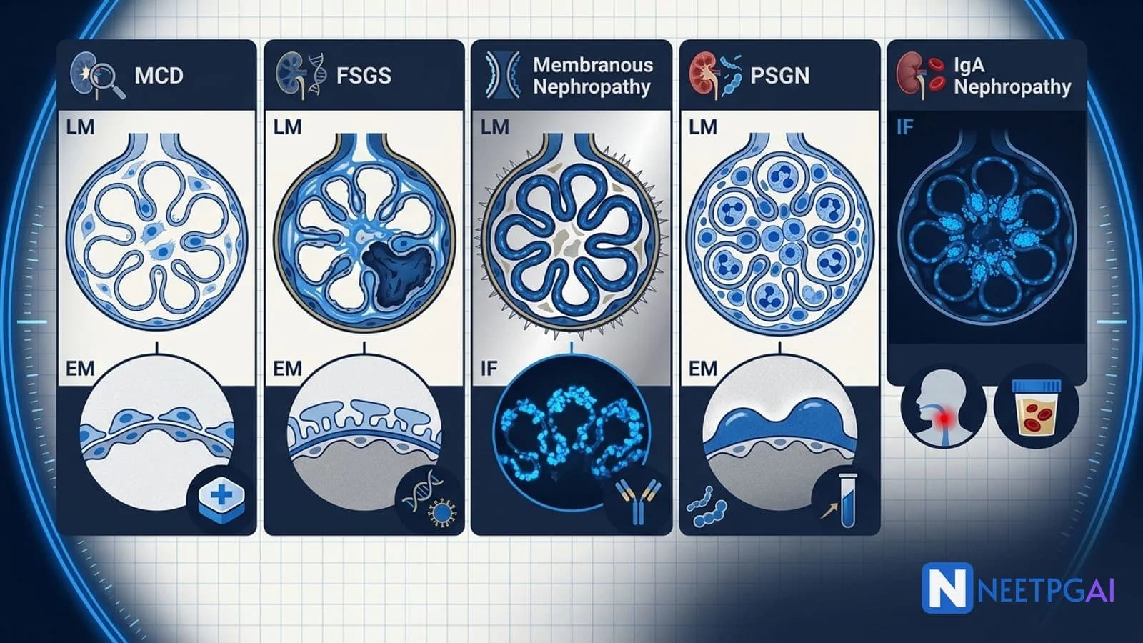
Renal biopsy image MCQs contribute 3-5 questions per NEET PG paper across pathology, nephrology, and internal medicine. Five high-yield glomerular patterns recur reliably year after year:
- Minimal change disease (MCD) — normal LM, no IF deposits, foot process effacement on EM; children with steroid-responsive nephrotic syndrome
- Focal segmental glomerulosclerosis (FSGS) — segmental sclerosis on LM, no or trivial IF, foot process effacement on EM; adult nephrotic syndrome; less steroid-responsive
- Membranous nephropathy — thickened GBM with silver-stain spikes on LM, granular subepithelial IgG plus C3 on IF, subepithelial deposits with spikes on EM; anti-PLA2R positive in 70 percent primary; adult nephrotic
- Post-streptococcal glomerulonephritis (PSGN) — endocapillary hypercellularity plus neutrophils on LM, granular IgG plus C3 on IF, subepithelial humps on EM; 1-3 weeks post-streptococcal infection; nephritic; low C3 normalising in 6-8 weeks
- IgA nephropathy — mesangial hypercellularity on LM, IgA-dominant mesangial deposits on IF, mesangial deposits on EM; commonest primary GN worldwide; synpharyngitic haematuria; MEST-C Oxford classification
Locking these 5 patterns plus 4-5 additional biopsy PYQ images (crescentic RPGN, lupus nephritis WHO classes, MPGN, amyloidosis, diabetic nephropathy) over 1-2 weeks moves biopsy-MCQ accuracy from 40 to 85 percent.
Why renal biopsy image MCQs are high-yield for NEET PG
Renal pathology is one of the highest-density MCQ topics in the pathology and internal-medicine sections of NEET PG. The reason is that the LM-IF-EM triad for each glomerular lesion is stereotyped, photogenic, and diagnostic. Nephrotic and nephritic presentations map cleanly onto biopsy findings, and the treatment approach (steroids, cytotoxics, rituximab, RAAS blockade, transplant) is different for each. Building a mental atlas of the 5 core patterns plus the systemic overlays (SLE, diabetes, amyloid) pays back reliably.
Drilling these 5 patterns plus a small library of crescentic GN, SLE nephritis, MPGN, and amyloid over 1-2 weeks moves biopsy-MCQ accuracy from 40 to 85 percent.
Foundational approach — the systematic biopsy read
Read every biopsy on 3 modalities
| Modality | What it shows | Preparation |
|---|
| Light microscopy (LM) | Architecture — glomerular cellularity, sclerosis, crescents, tubular atrophy, interstitial fibrosis, vessels | H&E, PAS (highlights basement membranes), silver stain (spikes, splitting), trichrome (fibrosis) |
| Immunofluorescence (IF) | Immune deposit pattern — location (mesangial, subendothelial, subepithelial), pattern (granular, linear), intensity, and immunoglobulin/complement class (IgG, IgA, IgM, C3, C1q, kappa, lambda) | Frozen tissue with fluoresceinated antisera |
| Electron microscopy (EM) | Podocyte foot processes, GBM structure, precise deposit location | Fixed tissue, ultrathin sections |
Systematic checklist for every glomerulus
- Cellularity — normal, mesangial hypercellular, endocapillary hypercellular, extracapillary (crescents)
- Sclerosis — global or segmental; focal (some glomeruli) vs diffuse (most)
- GBM — normal thickness, thickened, spikes, double contours (tram-tracking)
- Crescents — cellular (recent), fibrocellular, fibrous
- Deposits on IF — mesangial, subendothelial, subepithelial, granular vs linear, immunoglobulin class, complement
- EM — foot process effacement, deposit location and shape (subepithelial humps, subepithelial spikes, subendothelial deposits, mesangial deposits, dense deposits)
- Tubulointerstitium — atrophy, fibrosis, infiltrate
- Vessels — arteriolar hyalinosis, arteriolosclerosis, vasculitis
Aunt Minnie triggers for glomerular disease
| Finding | First-line diagnosis |
|---|
| Normal LM plus foot process effacement | Minimal change disease (MCD) |
| Segmental sclerosis in some glomeruli plus foot process effacement | Focal segmental glomerulosclerosis (FSGS) |
| Diffuse GBM thickening plus silver-stain spikes plus subepithelial granular IgG | Membranous nephropathy |
| Endocapillary hypercellularity plus neutrophils plus subepithelial humps | Post-streptococcal (post-infectious) GN |
| Mesangial hypercellularity plus IgA-dominant mesangial deposits | IgA nephropathy |
| Diffuse global crescents (over 50 percent) plus linear IgG on IF | Anti-GBM disease (Goodpasture) |
| Diffuse global crescents plus pauci-immune IF | ANCA-associated vasculitis (GPA, MPA, EGPA) |
| Full-house IF (IgG, IgA, IgM, C3, C1q) plus subendothelial and mesangial deposits | SLE lupus nephritis |
| Tram-track GBM double contours plus subendothelial deposits | Membranoproliferative GN (MPGN) |
| Apple-green birefringence on Congo red plus polarised light | Amyloidosis |
| Kimmelstiel-Wilson nodules plus GBM thickening | Diabetic nephropathy |
MCQ 1: 4-year-old boy with 2 weeks of periorbital and pedal oedema — biopsy shows normal-appearing glomeruli but electron microscopy reveals diffuse podocyte foot process effacement
Image description: [Renal biopsy panels of a 4-year-old boy. Panel 1 — Light microscopy (H&E and PAS) — glomeruli appear entirely normal, of normal cellularity, with no mesangial or endocapillary hypercellularity, no segmental sclerosis, no crescents, no thickened GBM, no double contours. Tubulointerstitium and vessels are normal. Panel 2 — Immunofluorescence — completely negative for IgG, IgA, IgM, C3, C1q; no immune deposits identified (or trivial IgM/C3 which is non-specific). Panel 3 — Electron microscopy — striking diffuse effacement of podocyte foot processes across the entire glomerular capillary surface (the foot processes are fused into a continuous cytoplasmic sheet along the outer aspect of the GBM). No electron-dense deposits identified in any location — subepithelial, subendothelial, or mesangial. GBM appears normal in thickness and structure. Podocyte microvillous transformation is visible.]
Clinical vignette: A 4-year-old boy is brought to the paediatric outpatient clinic in Chennai with 2 weeks of progressive periorbital puffiness worse in the mornings and pedal oedema. His parents noticed his abdomen has become slightly distended and his weight has increased by 2 kg over the last 10 days. He had a mild upper respiratory infection 3 weeks ago that resolved spontaneously. On examination — periorbital and pedal pitting oedema, mild ascites, BP 96/62 (normal for age), no rash, no arthritis. Urine dipstick shows 4+ proteinuria, no haematuria. Labs — 24-hour urine protein 4.8 g, serum albumin 2.4 g/dL, cholesterol 380 mg/dL, creatinine 0.4 mg/dL (normal), C3 and C4 normal, ANA negative, HBsAg and anti-HCV negative.
Options:
- (a) Minimal change disease (MCD)
- (b) Focal segmental glomerulosclerosis (FSGS)
- (c) Membranous nephropathy
- (d) Post-streptococcal glomerulonephritis
Correct answer: (a) Minimal change disease (MCD)
Reasoning: The triad of normal light microscopy, negative immunofluorescence (no immune deposits), and diffuse podocyte foot process effacement on electron microscopy is diagnostic of minimal change disease. MCD is the commonest cause of primary nephrotic syndrome in children (over 90 percent of primary nephrotic syndrome in children under 10 years); the clinical presentation of periorbital and pedal oedema, heavy proteinuria (over 3.5 g/24 h), hypoalbuminaemia (under 3.0 g/dL), and hyperlipidaemia fulfils classic nephrotic syndrome criteria.
FSGS would show segmental sclerosis in at least some glomeruli on LM (with matching foot process effacement on EM). Membranous nephropathy would show thickened GBM with spikes on silver stain plus granular subepithelial IgG on IF. Post-streptococcal GN would show endocapillary hypercellularity with neutrophils on LM plus granular IgG/C3 on IF plus subepithelial humps on EM, and would present as nephritic (haematuria, hypertension, low C3, oliguria) rather than pure nephrotic.
Teaching pearl — MCD:
| Aspect | Detail |
|---|
| Age and epidemiology | 90 percent of primary nephrotic syndrome in children under 10; less common in adults (10-15 percent of adult primary nephrotic) |
| Primary | Idiopathic — most cases |
| Secondary | NSAIDs, lithium, gold, Hodgkin lymphoma, thymoma, bee stings |
| First-line treatment | Corticosteroids — prednisolone 60 mg/m2/day or 2 mg/kg/day for 4-6 weeks (max 60 mg/day), then taper over 4-6 months; 80-90 percent complete remission |
| Steroid-dependent or frequently relapsing | Add second-line agent — cyclophosphamide, cyclosporine or tacrolimus, mycophenolate mofetil, rituximab |
| Complications | VTE (nephrotic syndrome is prothrombotic), infection (loss of Ig and complement in urine — pneumococcal, gram-negative peritonitis), hypovolaemia (over-diuresis risk), AKI, dyslipidaemia |
| Prognosis | Excellent in children (over 90 percent long-term renal survival); relapses common; adult prognosis slightly less favourable |
- NEET PG tests the normal-LM + effacement-on-EM triad and the steroid-responsiveness of MCD
MCQ 2: 34-year-old African-descent male with resistant nephrotic syndrome — biopsy shows segmental sclerosis of some glomeruli plus foot process effacement
Image description: [Renal biopsy panels of a 34-year-old male. Panel 1 — Light microscopy (PAS) — segmental sclerosis (pink hyaline sclerotic material with obliteration of the capillary lumen) is present in the perihilar segment of one of the sampled glomeruli; adhesion of the sclerotic tuft to Bowman's capsule is visible. Other glomeruli in the same biopsy appear normal — the lesion is focal (only some glomeruli affected) and segmental (only part of the affected glomerulus). Two glomeruli show global sclerosis (advanced disease). Mild tubular atrophy and interstitial fibrosis affect approximately 15 percent of the cortex. Panel 2 — Immunofluorescence — negative for IgG, IgA, C3, C1q; only trivial non-specific IgM and C3 staining in areas of segmental sclerosis. Panel 3 — Electron microscopy — foot process effacement over approximately 80 percent of the glomerular capillary surface (diffuse), no electron-dense deposits identified. Podocyte microvillous transformation and vacuolation are visible.]
Clinical vignette: A 34-year-old male from Kerala (originally of African descent through Siddi ancestry — an APOL1-risk-allele population) presents with 3 months of progressive generalised anasarca and hypertension. He is a truck driver, no chronic illness previously, no drug use. He was started on empirical prednisolone 60 mg/day 8 weeks ago by an outside physician with a presumptive diagnosis of MCD, but has failed to remit — urine protein remains 6.2 g/24 hours. Labs — serum albumin 2.1 g/dL, cholesterol 420 mg/dL, creatinine 1.6 mg/dL (mildly raised), BP 156/98, C3 and C4 normal, ANA negative, HIV negative, HBsAg and anti-HCV negative, syphilis serology negative, urine microscopy — occasional dysmorphic red cells, no red cell casts. A renal biopsy is performed given steroid resistance.
Options:
- (a) Minimal change disease
- (b) Focal segmental glomerulosclerosis (primary or APOL1-associated)
- (c) Membranous nephropathy
- (d) IgA nephropathy
Correct answer: (b) Focal segmental glomerulosclerosis (FSGS)
Reasoning: The finding of segmental sclerosis of some (focal) glomeruli on light microscopy, no significant immune deposits on IF, and foot process effacement on EM defines FSGS. The perihilar variant seen here is one of the five Columbia variants (perihilar, cellular, collapsing, tip, and NOS — not otherwise specified). Segmental sclerosis is the key distinguishing feature from MCD (which has normal LM). The presence of some globally sclerosed glomeruli reflects more advanced disease.
MCD would have normal LM in all sampled glomeruli. Membranous nephropathy would show thickened GBM with silver-stain spikes and granular subepithelial IgG on IF. IgA nephropathy would show mesangial hypercellularity on LM and IgA-dominant mesangial deposits on IF.
Teaching pearl — FSGS:
| Aspect | Detail |
|---|
| Epidemiology | Commonest cause of primary nephrotic syndrome in adults in Western populations; rising in India as a proportion of primary nephrotic |
| Primary (idiopathic) | Circulating permeability factor (suPAR proposed but not confirmed); variable steroid response |
| Secondary — adaptive/hyperfiltration | Obesity, single kidney (unilateral renal agenesis or nephrectomy), reflux nephropathy, low birth weight, sickle cell disease |
| Secondary — viral | HIV (collapsing variant), parvovirus B19, EBV, CMV |
| Secondary — drug | Heroin (heroin-associated nephropathy), pamidronate, interferon, lithium, anabolic steroids |
| Genetic | Podocin (NPHS2), nephrin (NPHS1), alpha-actinin-4, TRPC6, INF2, formin, laminin-beta2 (Pierson syndrome); APOL1 risk alleles in African-descent populations (G1, G2) — up to 10 times increased FSGS risk |
| Columbia variants | Perihilar (adaptive, secondary), cellular (young, aggressive), collapsing (HIV, worst prognosis), tip (best prognosis, steroid-responsive), NOS (commonest, variable) |
| First-line treatment (primary FSGS) | Corticosteroids — prednisolone 1 mg/kg/day (max 80 mg) for 4-6 months (longer course than MCD); only 30-50 percent complete remission |
| Steroid-resistant or dependent | Calcineurin inhibitors (cyclosporine, tacrolimus), mycophenolate mofetil, rituximab (variable evidence) |
| Secondary FSGS treatment | Treat underlying cause (weight loss, HIV therapy, drug withdrawal); RAAS blockade for renal protection; immunosuppression not helpful |
| Prognosis | 40-60 percent progress to end-stage kidney disease over 10-15 years without treatment; collapsing variant worst |
- NEET PG tests the segmental sclerosis definition, the primary vs secondary distinction, the APOL1 association, and the collapsing variant (HIV-associated, poor prognosis)
MCQ 3: 62-year-old man with 3 months of oedema and heavy proteinuria — biopsy shows diffusely thickened GBM with silver-stain spikes plus granular subepithelial IgG on IF
Image description: [Renal biopsy panels of a 62-year-old male. Panel 1 — Light microscopy (H&E and silver stain) — glomeruli show diffusely thickened glomerular basement membranes without hypercellularity, without crescents, without segmental sclerosis. On silver stain, characteristic "spikes" project from the outer (subepithelial) aspect of the GBM at regular intervals, giving the GBM a "comb-tooth" appearance. Tubulointerstitium and vessels normal. Panel 2 — Immunofluorescence — granular subepithelial IgG-dominant deposits in a "starry-sky" pattern along the entire GBM; granular C3 also present in the same distribution. IgA and IgM negative. IgG subclass staining shows IgG4 predominance (a feature of primary membranous). Panel 3 — Electron microscopy — subepithelial electron-dense deposits located between the podocyte foot processes and the GBM, with GBM spikes projecting between them (Ehrenreich-Churg stage II). Foot process effacement is diffuse. Panel 4 — Additional stain — anti-PLA2R immunohistochemistry positive at the glomerular basement membrane (in situ antigen demonstration). Serum anti-PLA2R by ELISA is positive at a titre of 320 RU/mL.]
Clinical vignette: A 62-year-old male retired accountant presents with 3 months of progressive generalised oedema — periorbital in the morning, pedal by evening, and now abdominal ascites. Urine is frothy. He has no chronic illness but had a 3-month history of dyspepsia last year (H. pylori positive, treated with triple therapy). No recent drug use. Labs — urine protein 8.4 g/24 hours, serum albumin 1.8 g/dL, cholesterol 460 mg/dL, creatinine 1.1 mg/dL (normal), C3 and C4 normal, ANA negative, HBsAg negative, anti-HCV negative, syphilis negative, complete blood count normal. He is enrolled for renal biopsy and simultaneous malignancy screen (CT chest-abdomen-pelvis, colonoscopy, urine cytology, PSA) given his age.
Options:
- (a) Minimal change disease
- (b) Focal segmental glomerulosclerosis
- (c) Membranous nephropathy (anti-PLA2R positive, likely primary)
- (d) Post-streptococcal glomerulonephritis
Correct answer: (c) Membranous nephropathy (anti-PLA2R positive, likely primary)
Reasoning: The triad of diffusely thickened GBM with silver-stain spikes on LM, granular subepithelial IgG plus C3 on IF (starry-sky pattern, IgG4 predominance), and subepithelial electron-dense deposits with intervening GBM spikes on EM defines membranous nephropathy. Positive anti-PLA2R serology (specificity over 95 percent) and glomerular anti-PLA2R staining strongly suggest primary membranous nephropathy, though malignancy screening is still mandatory in a patient over 65 years given the 20-30 percent paraneoplastic membranous rate in that age group.
MCD would have normal LM. FSGS would have segmental sclerosis. Post-streptococcal GN would have endocapillary hypercellularity and subepithelial humps on EM (not spikes) plus low C3.
Teaching pearl — membranous nephropathy:
| Aspect | Detail |
|---|
| Epidemiology | Common in adults, particularly middle-aged and older; commonest cause of primary nephrotic syndrome in older adults in Western populations |
| Primary (70 percent) | Autoantibody-mediated — anti-PLA2R positive in 70-80 percent; anti-THSD7A in 3-5 percent; other rare antigens (NELL-1, EXT1/EXT2, semaphorin 3B) |
| Secondary (30 percent) | Malignancy (solid tumours — lung, colon, breast, prostate, GI, kidney; screen aggressively in over-65s), infection (hepatitis B, syphilis, malaria, schistosomiasis), autoimmune (SLE class V, rheumatoid arthritis, autoimmune thyroiditis), drugs (captopril, penicillamine, gold, NSAIDs, TNF-alpha inhibitors) |
| Ehrenreich-Churg staging (EM) | Stage I — subepithelial deposits without spikes; Stage II — deposits with spikes (as our patient); Stage III — deposits incorporated into GBM with intervening basement membrane; Stage IV — resorbed deposits with irregular thickened GBM |
| First-line treatment (primary) | Rituximab — 1 g on days 1 and 15 (MENTOR trial 2019); replaced modified Ponticelli (alternating monthly cyclophosphamide and steroid for 6 months) as first-line based on non-inferior efficacy and better tolerability |
| Alternatives | Modified Ponticelli, calcineurin inhibitors (cyclosporine, tacrolimus), obinutuzumab, belimumab; expectant management for low-risk (proteinuria under 4 g/day, normal creatinine, stable) — 30 percent spontaneous remission |
| Secondary treatment | Address the underlying cause (treat malignancy, treat infection, withdraw drug) |
| Anti-PLA2R monitoring | Falling titre predicts remission by 3-6 months; rising titre predicts relapse |
| Complications | Nephrotic syndrome complications plus particularly high renal vein thrombosis risk (30 percent — screen with Doppler or CT if suspicion) — anticoagulate if albumin under 2.0 g/dL or if VTE risk factors |
| Malignancy screen | Age-appropriate — CT chest-abdomen-pelvis, colonoscopy, mammography, urine cytology, PSA; especially critical in patients over 65 |
- NEET PG tests the LM spikes, IF granular subepithelial IgG, EM subepithelial deposits, anti-PLA2R signal, MENTOR-rituximab first-line, and mandatory malignancy screen in older adults
MCQ 4: 8-year-old boy with sudden-onset haematuria, hypertension, and periorbital oedema 2 weeks after streptococcal pharyngitis — biopsy shows endocapillary hypercellularity plus subepithelial humps
Image description: [Renal biopsy panels of an 8-year-old boy. Panel 1 — Light microscopy (H&E) — glomeruli are enlarged and hypercellular — the hypercellularity is endocapillary (within the glomerular capillary lumina, distinct from mesangial hypercellularity) and includes infiltrating neutrophils among the endothelial and mesangial cells. Occasional glomeruli show early cellular crescents (small, cellular, restricted). No segmental sclerosis. Mild tubulointerstitial inflammation. Panel 2 — Immunofluorescence — granular subepithelial and mesangial IgG and C3 deposits in a "starry-sky" pattern along the GBM and in the mesangium; C3 typically dominant over IgG. IgA and IgM negative or trivial. Panel 3 — Electron microscopy — large, dome-shaped subepithelial electron-dense deposits ("humps") located on the outer aspect of the GBM between podocyte foot processes; the humps are much larger and more discrete than the subepithelial deposits of membranous. Mesangial deposits also present. Some foot process effacement.]
Clinical vignette: An 8-year-old boy presents with 2 days of tea-coloured urine, periorbital puffiness, and reduced urine output. Two weeks ago, he had a sore throat with fever that was treated with a course of amoxicillin. On examination — BP 138/92 (well above 95th centile for age), periorbital and pedal oedema, no rash, no arthritis. Labs — urine dipstick 3+ blood, 2+ protein; urine microscopy — dysmorphic red cells and red cell casts; 24-hour urine protein 1.4 g (sub-nephrotic); creatinine 0.9 mg/dL (mildly raised for age); C3 markedly reduced at 42 mg/dL (normal 90-180), C4 normal at 22 mg/dL; ASO titre 800 IU/mL (raised), throat swab culture negative (organism cleared after antibiotics). ANA negative, ANCA negative, anti-GBM negative.
Options:
- (a) Post-streptococcal (post-infectious) glomerulonephritis
- (b) IgA nephropathy
- (c) Membranous nephropathy
- (d) Lupus nephritis (SLE class IV)
Correct answer: (a) Post-streptococcal (post-infectious) glomerulonephritis
Reasoning: The classic triad of endocapillary hypercellularity with neutrophil infiltrate on LM, granular subepithelial and mesangial IgG plus C3 (starry-sky pattern) on IF, and large subepithelial "humps" on EM defines post-streptococcal (post-infectious) glomerulonephritis (PSGN). The clinical presentation of nephritic syndrome (haematuria, hypertension, oedema, oliguria, active urinary sediment with dysmorphic red cells and red cell casts) 1-3 weeks after a streptococcal pharyngitis (or 3-6 weeks after streptococcal skin infection like pyoderma) in a child is classical; raised ASO titre and low C3 (with normal C4 — the alternative pathway is activated) support the diagnosis.
IgA nephropathy would have IgA-dominant mesangial deposits on IF, normal C3, and often synpharyngitic haematuria (within days, not 2 weeks). Membranous would have thickened GBM with spikes and no hypercellularity. Lupus nephritis would have "full-house" IF (IgG, IgA, IgM, C3, C1q, kappa and lambda), positive ANA, and often extra-renal SLE features.
Teaching pearl — PSGN:
| Aspect | Detail |
|---|
| Trigger | Group A Streptococcus — M12 strain after pharyngitis (1-3 weeks incubation), M49 strain after skin infection (3-6 weeks incubation) |
| Age | Commonest in children 4-12 years; can occur at any age |
| Presentation | Nephritic syndrome — haematuria (often macroscopic, "cola-coloured"), hypertension, oedema, oliguria, sub-nephrotic proteinuria, red cell casts |
| Diagnostic labs | Raised ASO (post-pharyngitic) or anti-DNase B (post-skin), low C3, normal C4 (alternative pathway activation), positive throat or skin culture (if not treated), rising streptozyme antibodies |
| C3 dynamics | Low at presentation, normalises by 6-8 weeks — persistent low C3 beyond this should prompt reconsideration (MPGN, lupus, C3 glomerulopathy) |
| Treatment | Supportive — antihypertensives (loop diuretics for volume-driven HT, calcium channel blockers if needed), salt and fluid restriction, dialysis if AKI severe; antibiotics (penicillin) if streptococcal infection still present |
| Prognosis | Excellent in children (over 95 percent complete recovery); less favourable in adults (residual proteinuria and hypertension in 30-50 percent); rare progression to CKD |
| Complications | Hypertensive encephalopathy, acute pulmonary oedema, AKI needing short-term dialysis, RPGN (rare); long-term — small proportion progress to CKD |
| Prevention | Prompt antibiotic treatment of streptococcal pharyngitis (though evidence for PSGN prevention is weaker than for rheumatic fever prevention) |
- NEET PG tests the LM endocapillary + neutrophils, IF starry-sky IgG + C3, EM humps, low C3 with normal C4, C3 normalisation at 6-8 weeks, and the supportive-treatment nature of PSGN
MCQ 5: 26-year-old man with recurrent macroscopic haematuria within 2 days of upper respiratory infections — biopsy shows mesangial hypercellularity plus IgA-dominant mesangial deposits
Image description: [Renal biopsy panels of a 26-year-old male. Panel 1 — Light microscopy (H&E and PAS) — glomeruli show mesangial hypercellularity and matrix expansion — over 50 percent of glomeruli show mesangial cellularity above the normal 3-cells-per-mesangial-area threshold (M1 by Oxford). Occasional glomeruli show endocapillary hypercellularity (E1) and one small cellular crescent (C1 — 1-25 percent of glomeruli). No significant segmental sclerosis (S0). Tubular atrophy and interstitial fibrosis affect approximately 20 percent of the cortex (T0). Panel 2 — Immunofluorescence — strong IgA-dominant mesangial deposits in a granular pattern; C3 co-deposits in the mesangium; IgG and IgM present but less intense than IgA; kappa and lambda light chains balanced. Panel 3 — Electron microscopy — mesangial electron-dense deposits clearly visible in the mesangial matrix, with paramesangial extension in some areas; some subepithelial and subendothelial deposits (aggressive form). Foot process effacement is only partial (focal), consistent with the sub-nephrotic proteinuria in this patient.]
Clinical vignette: A 26-year-old male engineering graduate presents with 3 recurrent episodes of macroscopic haematuria over 18 months, each episode following an upper respiratory tract infection within 1-2 days (synpharyngitic pattern). Between episodes he has persistent microscopic haematuria with mild proteinuria (0.8 g/24 hours) on urine dipstick. He is otherwise well, works out at the gym, no chronic medications, no drug or alcohol use. Family history — his 55-year-old father was recently found to have creatinine 2.1 mg/dL with proteinuria on a health check but has not been biopsied. BP 132/86 (borderline hypertension), creatinine 1.1 mg/dL (normal for young adult), C3 and C4 normal, ANA negative, HBsAg and anti-HCV negative, ANCA negative, anti-GBM negative, ASO titre normal, serum IgA levels raised (500 mg/dL; normal 70-400).
Options:
- (a) Post-streptococcal glomerulonephritis
- (b) IgA nephropathy (primary)
- (c) Thin basement membrane disease
- (d) Alport syndrome
Correct answer: (b) IgA nephropathy (primary)
Reasoning: The triad of mesangial hypercellularity on LM, IgA-dominant mesangial deposits on IF, and mesangial electron-dense deposits on EM defines IgA nephropathy. The clinical picture of synpharyngitic macroscopic haematuria (within 1-2 days of a URI, not 2-3 weeks as in PSGN), persistent microscopic haematuria with mild proteinuria between episodes, borderline hypertension in a young adult of Asian ancestry, and raised serum IgA is classical. Normal C3 and C4 distinguish it from PSGN and lupus. IgA nephropathy is the commonest primary glomerulonephritis worldwide, particularly in Asian populations, and is a leading cause of CKD in young Indian adults.
PSGN would have a 2-3 week latent period from infection, low C3, raised ASO, endocapillary hypercellularity with neutrophils on LM, and subepithelial humps on EM. Thin basement membrane disease would have a uniformly thinned GBM on EM without immune deposits. Alport syndrome would have basket-weave (lamellated) GBM changes on EM plus family history of renal failure and sensorineural deafness or eye findings (anterior lenticonus).
Teaching pearl — IgA nephropathy MEST-C Oxford classification:
| Parameter | Score |
|---|
| M — Mesangial hypercellularity | 0 (under 50 percent of glomeruli) or 1 (50 percent or more) |
| E — Endocapillary hypercellularity | 0 (absent) or 1 (present) |
| S — Segmental sclerosis | 0 (absent) or 1 (present) |
| T — Tubular atrophy/interstitial fibrosis | 0 (under 25 percent), 1 (26-50 percent), 2 (over 50 percent) |
| C — Crescents (added 2016) | 0 (absent), 1 (1-25 percent of glomeruli), 2 (over 25 percent) |
-
Primary IgA nephropathy — commonest primary GN worldwide; Asian and Indian populations particularly affected; autoantibodies against galactose-deficient IgA1 (Gd-IgA1)
-
Secondary IgA nephropathy — Henoch-Schonlein purpura (IgA vasculitis with skin, joint, GI, kidney involvement — usually children), cirrhosis, celiac disease, HIV, IBD, dermatitis herpetiformis, ankylosing spondylitis
-
Treatment (KDIGO 2021):
- Aggressive RAAS blockade (ACE inhibitor or ARB titrated to maximum tolerated) — target proteinuria under 0.5-1 g/day, BP under 130/80
- If proteinuria remains over 0.75-1 g/day after 3-6 months of optimal supportive care AND T0-T1 AND preserved GFR — consider corticosteroids (TESTING trial — reduced-dose methylprednisolone; risk of infection)
- C1 or C2 crescents (crescentic IgA — a form of RPGN) — corticosteroids plus cyclophosphamide or rituximab (though evidence is weaker for IgA than for ANCA-vasculitis)
- Newer targeted therapies — budesonide (Nefecon, targeted-release oral corticosteroid to Peyer's patches in the ileum, reduces Gd-IgA1 production) — approved in 2021 based on NefIgArd trial; sparsentan (dual endothelin-angiotensin receptor blocker) — approved 2023 based on PROTECT trial
- Advanced disease (T2, extensive C, GFR under 30) — limited immunosuppression benefit; prepare for RRT
-
Prognosis — approximately 30 percent progress to end-stage kidney disease over 20 years; predictors include proteinuria over 1 g/day, hypertension, reduced GFR, higher MEST-C scores
-
Post-transplant recurrence — up to 30 percent, but graft loss uncommon in first decade
-
NEET PG tests the mesangial IgA deposits on IF, the synpharyngitic haematuria clinical pattern, the MEST-C classification, and the RAAS-first supportive care approach
Common pitfalls in glomerular biopsy image MCQs
Pitfall 1: Confusing MCD with FSGS
Both show foot process effacement on EM. The discriminator is LM — MCD has normal LM, FSGS has segmental sclerosis in some glomeruli. Sampling error is a real concern in FSGS because the lesion is focal — a biopsy that samples only unaffected glomeruli can misdiagnose FSGS as MCD. Repeat biopsy or careful review of all sampled glomeruli is important; steroid resistance in a presumed MCD case should always prompt reconsideration of FSGS.
Pitfall 2: Mixing up spikes (membranous) and humps (PSGN) on EM
Spikes are GBM projections between subepithelial deposits in membranous nephropathy — small and repetitive. Humps are large dome-shaped subepithelial deposits in PSGN — bigger and less repetitive. The clinical picture is opposite — membranous is nephrotic in adults, PSGN is nephritic in children. C3 is normal in membranous but low in PSGN.
Pitfall 3: Missing the "starry-sky" pattern
Both PSGN and membranous nephropathy show granular subepithelial IgG (with C3) — the "starry-sky" pattern. The discriminator is LM (endocapillary hypercellularity in PSGN, thickened GBM in membranous), EM (humps vs spikes), and clinical (nephritic vs nephrotic, child vs adult, low C3 vs normal C3).
Pitfall 4: Overlooking IgA-dominant deposits
Every biopsy of a young Asian adult with recurrent haematuria should have IF looked at carefully for IgA dominance. The mesangial IgA pattern is diagnostic. Serum IgA is raised in only 50 percent — its absence does not exclude IgA nephropathy.
Pitfall 5: Forgetting to screen for malignancy in older membranous
New-onset membranous in a patient over 65 has a 20-30 percent paraneoplastic rate — anti-PLA2R positivity does not fully exclude secondary membranous with a coincident tumour. Do CT chest-abdomen-pelvis, colonoscopy, mammography, urine cytology, and PSA before starting immunosuppression.
How to study glomerular pathology for NEET PG
- Memorise the LM-IF-EM triad for each of the 5 patterns — MCD, FSGS, membranous, PSGN, IgA
- Learn the nephrotic vs nephritic mapping — MCD, FSGS, membranous, diabetic → nephrotic; PSGN, IgA, MPGN, RPGN, lupus (III/IV) → nephritic (with overlap)
- Learn primary vs secondary distinctions for each — because treatment is different (immunosuppression vs treat-the-cause)
- Learn the classifications — Columbia variants for FSGS, Ehrenreich-Churg for membranous, MEST-C Oxford for IgA
- Learn the treatments — MCD (steroids), FSGS (steroids longer plus CNI), membranous (rituximab MENTOR), PSGN (supportive), IgA (RAAS plus steroids for high-risk)
- Learn the anti-PLA2R serology — specificity over 95 percent for primary membranous
- Learn crescentic RPGN — anti-GBM (Goodpasture, linear IgG), immune-complex (lupus, PSGN, IgA — granular deposits), pauci-immune (ANCA vasculitis — GPA, MPA, EGPA)
- Practice 10-15 biopsy MCQs per day for 2-3 weeks; NEETPGAI pathology bank has a tagged renal set
- Pair biopsies with clinical vignettes — age, ancestry, infection history, drug history, systemic features
- Learn the SLE lupus nephritis WHO/ISN-RPS classes I-VI — commonly tested alongside these 5 core patterns
Key takeaways
- Renal biopsy image MCQs contribute 3-5 questions per NEET PG paper
- MCD — normal LM, no IF, foot process effacement on EM; children with steroid-responsive nephrotic
- FSGS — segmental sclerosis on LM, no IF, foot process effacement on EM; adult nephrotic; less steroid-responsive; APOL1 risk in African-descent populations
- Membranous — thickened GBM with silver-stain spikes on LM, granular subepithelial IgG plus C3 on IF, subepithelial deposits with spikes on EM; anti-PLA2R positive in 70 percent primary; MENTOR-rituximab first-line
- PSGN — endocapillary hypercellularity plus neutrophils on LM, granular subepithelial IgG plus C3 (starry-sky) on IF, subepithelial "humps" on EM; low C3 normalising in 6-8 weeks; supportive treatment
- IgA nephropathy — mesangial hypercellularity on LM, IgA-dominant mesangial deposits on IF, mesangial deposits on EM; synpharyngitic haematuria; MEST-C Oxford classification; RAAS-first supportive care
- Anti-PLA2R specificity over 95 percent for primary membranous — falling titre predicts remission
- Always screen for malignancy in new-onset membranous over 65 (20-30 percent paraneoplastic)
- Aggressive RAAS blockade is the backbone for every non-nephrotic proteinuric disease
- IgA nephropathy is the commonest primary GN worldwide, particularly in Asian populations
- Diabetic nephropathy is the commonest cause of nephrotic syndrome in India overall (driven by T2DM epidemic)
Frequently Asked Questions
What are the key light microscopy, immunofluorescence, and electron microscopy findings for each of the five common glomerular lesions?
The five commonest glomerular lesions tested on NEET PG have stereotyped LM, IF, and EM findings. Minimal change disease (MCD) shows normal glomeruli on light microscopy (no hypercellularity, no sclerosis, no crescents), no immune deposits on immunofluorescence (or trivial IgM/C3), and diffuse foot process effacement on electron microscopy — this triad is diagnostic. Focal segmental glomerulosclerosis (FSGS) shows segmental sclerosis of some (focal) glomeruli on LM, no significant immune deposits (or non-specific IgM/C3 in areas of sclerosis) on IF, and foot process effacement on EM; the segmental nature of the lesion and the sclerosis distinguish it from MCD. Membranous nephropathy shows diffusely thickened glomerular basement membrane (GBM) with characteristic 'spikes' on silver stain on LM, granular subepithelial IgG (typically IgG4) and C3 on IF (the 'starry-sky' pattern), and subepithelial electron-dense deposits with intervening GBM spikes on EM (Ehrenreich-Churg staging I-IV). Post-streptococcal (post-infectious) glomerulonephritis shows diffuse endocapillary hypercellularity with neutrophil infiltrate on LM, granular IgG and C3 subepithelial and mesangial deposits on IF (the 'starry-sky' pattern), and subepithelial 'humps' (large dome-shaped electron-dense deposits) on EM. IgA nephropathy shows mesangial hypercellularity and expansion on LM (with variable crescents in aggressive disease), IgA-dominant mesangial deposits on IF (plus C3, sometimes IgG or IgM), and mesangial electron-dense deposits on EM. The MEST-C Oxford classification quantitatively scores mesangial hypercellularity (M), endocapillary proliferation (E), segmental sclerosis (S), tubular atrophy/interstitial fibrosis (T), and crescents (C) to guide prognosis and immunosuppression. NEET PG tests the LM-IF-EM triad for each and the pathognomonic EM findings (foot process effacement in MCD, spikes in membranous, humps in PSGN, mesangial deposits in IgA).
How do the clinical presentations of nephrotic and nephritic syndromes differ and which pathologies present as each?
Nephrotic syndrome is defined by heavy proteinuria greater than 3.5 g per 24 hours (or urine protein-creatinine ratio greater than 3.5 g/g), hypoalbuminaemia (serum albumin under 3.0 g/dL), oedema (typically periorbital, dependent, or generalised anasarca), and hyperlipidaemia; complications include VTE risk (particularly renal vein thrombosis in membranous), infection risk (loss of immunoglobulins and complement in urine), and accelerated atherosclerosis. Common pathologies that present as nephrotic syndrome are minimal change disease (commonest cause in children, over 90 percent of primary nephrotic syndrome in children under 10; steroid-responsive), focal segmental glomerulosclerosis (commonest cause of primary nephrotic syndrome in adults in Western populations; less steroid-responsive), membranous nephropathy (common in adults, especially older; anti-PLA2R positive in 70 percent primary), diabetic nephropathy (the commonest cause overall in India, driven by the T2DM epidemic), and secondary causes like SLE (class V lupus nephritis), amyloidosis, and myeloma. Nephritic syndrome is defined by haematuria (often dysmorphic red cells and red cell casts on urine microscopy), variable proteinuria (usually sub-nephrotic under 3 g/day but can overlap), oliguria, hypertension, and acute kidney injury; often with active urinary sediment on microscopy. Common pathologies presenting as nephritic syndrome are post-infectious glomerulonephritis (classical after group A Streptococcus pharyngitis or skin infection, especially in children), IgA nephropathy (the commonest primary GN worldwide, particularly in Asian populations; recurrent macroscopic haematuria within days of URI or exercise — synpharyngitic), rapidly progressive glomerulonephritis (RPGN — crescentic GN — three types: anti-GBM/Goodpasture, immune-complex like SLE and PSGN and IgA, pauci-immune ANCA-associated), membranoproliferative GN (MPGN), Alport syndrome, and thin basement membrane disease. Some pathologies present as mixed nephrotic-nephritic (SLE class III/IV, MPGN, severe IgA, crescentic overlays). NEET PG tests both syndromic overlap and the pathology-per-syndrome mapping.
What is the primary vs secondary distinction in each glomerular lesion and why does it matter for treatment?
Every glomerular lesion has both primary (idiopathic, autoimmune) and secondary (drug, infection, systemic disease, malignancy) causes, and the distinction changes management fundamentally. In minimal change disease, primary MCD (over 90 percent of cases in children) is idiopathic and treated with corticosteroids (prednisolone 60 mg/m2/day or 2 mg/kg/day for 4-6 weeks then taper — 80-90 percent complete remission); secondary MCD can be triggered by NSAIDs, lithium, gold, Hodgkin lymphoma, thymoma, and bee stings — treatment is withdrawal of the offending agent and treatment of the underlying condition. In FSGS, primary FSGS is idiopathic (with variable steroid response); secondary FSGS results from adaptive/hyperfiltration causes (obesity, single kidney, reflux nephropathy, sickle cell), viral (HIV — collapsing variant, parvovirus B19), drug (heroin, pamidronate, interferon), and genetic (podocin NPHS2, alpha-actinin-4, TRPC6, APOL1 in African-descent populations); the treatment focus is on the underlying cause, not immunosuppression. In membranous nephropathy, primary membranous (70 percent) is driven by autoantibodies against PLA2R (phospholipase A2 receptor on podocytes; anti-PLA2R positive in serum in 70 percent) — treatment is immunosuppression (rituximab is now first-line based on MENTOR, replacing modified Ponticelli of alternating cyclophosphamide and steroid); secondary membranous (30 percent) is triggered by hepatitis B, SLE (class V), malignancy (solid tumours, particularly in older patients — screen for malignancy in over-65s), and drugs (captopril, penicillamine, gold, NSAIDs) — treatment addresses the underlying cause. In IgA nephropathy, primary IgA is idiopathic (autoantibodies against galactose-deficient IgA1 — the current pathogenic model); secondary IgA occurs in Henoch-Schonlein purpura (IgA vasculitis with skin, joint, GI involvement in children), cirrhosis, celiac disease, HIV, IBD, and dermatitis herpetiformis — again treatment addresses the underlying cause plus RAAS blockade for renal protection. In post-streptococcal GN, the disease is entirely post-infectious (usually 1-3 weeks after streptococcal pharyngitis with the M12 strain or streptococcal skin infection with the M49 strain) and treatment is supportive because the disease is self-limited in most children; adults may have residual proteinuria. NEET PG tests the primary-secondary distinction because treatment differs radically.
What is the MEST-C Oxford classification of IgA nephropathy and how does it guide treatment?
The Oxford MEST-C classification of IgA nephropathy is a validated histopathological scoring system that predicts renal outcome and increasingly guides immunosuppression decisions. Each biopsy is scored across five parameters. M (mesangial hypercellularity) — 0 if under 50 percent of glomeruli show mesangial hypercellularity, 1 if 50 percent or more. E (endocapillary hypercellularity) — 0 if absent, 1 if present. S (segmental sclerosis) — 0 if absent, 1 if present. T (tubular atrophy and interstitial fibrosis) — 0 if under 25 percent, 1 if 26-50 percent, 2 if over 50 percent. C (crescents, added in the 2016 revision) — 0 if absent, 1 if 1-25 percent, 2 if over 25 percent. Higher scores predict worse renal outcome. Standard treatment is aggressive RAAS blockade (ACE inhibitor or ARB titrated to maximum tolerated dose) with a target proteinuria under 0.5-1 g/day; if proteinuria remains over 0.75-1 g/day despite 3-6 months of optimal supportive care AND the patient has T0-T1 histology and preserved GFR, immunosuppression may be added (corticosteroids — the TESTING trial supports methylprednisolone with a reduced-dose regimen to limit infection risk). C1 or C2 crescents raise consideration for combined corticosteroids plus cyclophosphamide or rituximab (crescentic IgA is a form of rapidly progressive glomerulonephritis). Newer targeted therapies include budesonide (Nefecon — a targeted-release oral corticosteroid to reduce galactose-deficient IgA1 production in the gut) and sparsentan (dual endothelin-angiotensin receptor blocker). Advanced disease with T2, extensive C, or GFR under 30 has limited immunosuppression benefit and needs preparation for renal replacement therapy. NEET PG tests the MEST-C parameters and the RAAS-first supportive-care approach.
How is anti-PLA2R serology used to distinguish primary from secondary membranous nephropathy and what does it mean for treatment?
Anti-phospholipase A2 receptor (anti-PLA2R) antibodies are highly specific for primary (idiopathic) membranous nephropathy — the M-type PLA2 receptor is expressed on podocytes and is the target antigen in approximately 70-80 percent of primary membranous cases. Anti-PLA2R is measured by ELISA (or indirect immunofluorescence on transfected cells) in serum, and the antigen can also be demonstrated in the glomerular basement membrane on kidney biopsy by immunohistochemistry. A positive serum anti-PLA2R has a specificity of over 95 percent for primary membranous, allowing a non-invasive presumptive diagnosis in many cases (though biopsy remains standard for confirmation, especially in atypical presentations). A negative anti-PLA2R does not exclude primary membranous — a second antigen, thrombospondin type-1 domain-containing 7A (THSD7A), accounts for 3-5 percent of primary cases; other rarer antigens include exostosin 1/2 (EXT1/EXT2, seen in lupus membranous), NELL-1 (associated with malignancy-related and native), and semaphorin 3B. For treatment, anti-PLA2R titre correlates with disease activity — high or rising titre suggests active disease and impending nephrotic flare; falling titre precedes clinical remission by 3-6 months and can guide immunosuppression tapering. The MENTOR trial (2019) established rituximab (two 1 g infusions 2 weeks apart) as first-line therapy for primary membranous with anti-PLA2R positivity — comparable to modified Ponticelli (alternating monthly cyclophosphamide and steroid for 6 months) in efficacy but with better tolerability. For secondary membranous (30 percent — hepatitis B, SLE, malignancy, drugs), the treatment focus is on the underlying cause. In older patients (age over 65) with new-onset membranous, active malignancy screening (CT chest-abdomen-pelvis, colonoscopy, mammography, urine cytology) is critical because 20-30 percent of new-onset membranous in the elderly is paraneoplastic. NEET PG tests the anti-PLA2R specificity, the MENTOR trial rituximab-first indication, and the malignancy-screening advice in older patients.
This content is for educational purposes for NEET PG exam preparation. It is not a substitute for professional medical advice, diagnosis, or treatment. Clinical information has been reviewed by qualified medical professionals.
Written by: NEETPGAI Editorial Team
Reviewed by: Pending SME Review
Last reviewed: August 2026