Version 1.0 — Published September 2026
Quick Answer
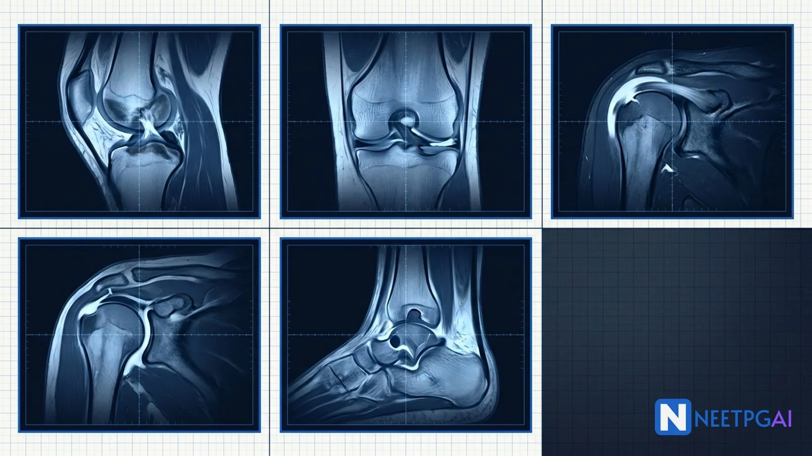
Orthopedic MRI image MCQs contribute 2-3 questions per NEET PG paper, mostly in Surgery and Orthopedics. Five high-yield patterns recur reliably:
- ACL tear — discontinuity or wavy ACL, kissing bone bruise on lateral femoral condyle and posterior lateral tibia, PCL buckling, anterior tibial translation; young active — arthroscopic reconstruction with hamstring/BPTB graft
- Bucket-handle meniscal tear — double-PCL sign, absent bow-tie, locked knee; arthroscopic partial meniscectomy for irreparable, repair for peripheral red-red zone
- Rotator cuff supraspinatus tear — coronal oblique T2 shows tendon discontinuity, retraction, subacromial-subdeltoid bursal fluid; arthroscopic repair for symptomatic full-thickness; reverse total shoulder arthroplasty for massive irreparable with cuff arthropathy
- SLAP tear — MR arthrogram shows contrast extension into superior labrum-biceps anchor; O'Brien test positive; arthroscopic SLAP repair or biceps tenodesis
- Talar osteochondral defect — postero-medial (or antero-lateral) talar dome subchondral defect with oedema and fluid under fragment; microfracture, OATS, or ACI by size
Locking these 5 patterns plus MRI vs USG choice, timing of MRI after injury, and grade-of-tear classifications moves orthopedic-MRI-MCQ accuracy from 45 to 85 percent.
Why orthopedic MRI image MCQs are high-yield for NEET PG
Orthopedic imaging cuts across sports medicine, general surgery, radiology, and emergency medicine. MRI is the gold standard for soft-tissue and cartilage assessment, and NEET PG tests the pattern-recognition-plus-management logic heavily. India's rising sports injury burden, growing PMJAY coverage of arthroscopy, and the widespread availability of MRI at tertiary and many secondary centres make these clinical scenarios high-relevance.
Foundational approach — the systematic MRI read
Read the joint MRI in this order
| Structure | What to look for |
|---|
| Alignment and effusion | Joint congruity, joint effusion volume and location, loose bodies |
| Bones | Subchondral bone oedema (T2/STIR high signal), fractures, contusions, bone bruise patterns |
| Cartilage | Chondral defect, thinning, delamination |
| Ligaments | Continuity, thickness, signal intensity, wavy or buckled appearance |
| Tendons | Continuity, retraction, tendinosis vs partial vs full-thickness tear |
| Menisci (knee) / labrum (shoulder, hip) | Linear high signal extending to articular surface, displacement, discoid morphology |
| Muscles | Volume, fatty atrophy (Goutallier grade), strain |
| Bursae | Fluid collections indicating adjacent tendon pathology |
MCQ 1: 22-year-old footballer with knee giving way after a non-contact pivot injury
Image description: [Sagittal proton density fat-saturated MRI of the right knee of a 22-year-old male. The image shows: discontinuity and wavy fibres of the ACL at its mid-portion with increased T2 signal (oedema and haemorrhage within the ligament substance). PCL is buckled with an increased PCL angle greater than 115 degrees. Anterior tibial translation of greater than 7 mm relative to the femur. Kissing bone bruise pattern — high T2 marrow signal in the mid-portion of the LATERAL femoral condyle and the POSTERIOR lateral tibial plateau. Joint effusion — moderate. Coronal PDFS image shows an intact medial collateral ligament and a small peripheral tear of the lateral meniscus posterior horn. No Segond fracture visible.]
Clinical vignette: A 22-year-old male semi-professional footballer sustained a non-contact pivot injury to the right knee while landing from a jump 3 days ago. He heard a "pop" and immediately fell, unable to continue. On examination, positive Lachman test (grade 2), positive anterior drawer, positive pivot shift; moderate effusion; joint-line tenderness. He is otherwise fit, wants to return to competitive sport.
Options:
- (a) Complete ACL tear with lateral meniscus tear and bone bruise pattern
- (b) PCL tear
- (c) MCL tear
- (d) Patellar dislocation
Correct answer: (a) Complete ACL tear with lateral meniscus tear and bone bruise pattern
Reasoning: Classical MRI findings of an acute ACL tear — discontinuity of the ligament fibres, increased T2 signal within the ligament substance, a wavy or bowed appearance, secondary signs of anterior tibial translation greater than 5 mm and PCL buckling, and the pathognomonic kissing bone bruise pattern on the lateral femoral condyle and posterior lateral tibia produced by the pivot-shift mechanism. This patient has all of these plus a lateral meniscus tear (common in acute ACL rupture). His clinical picture (positive Lachman, positive pivot shift, non-contact pivot with pop) is the classical acute ACL rupture.
PCL tear has an intact ACL, disrupted PCL with high T2 signal, and posterior tibial sag on sagittal imaging. MCL tear shows disruption or oedema at the medial collateral ligament on coronal imaging. Patellar dislocation shows a medial patellar contusion, lateral femoral condyle contusion in a different pattern, and disrupted medial patellofemoral ligament.
Teaching pearl — ACL tear management:
| Aspect | Recommendation |
|---|
| Acute (first 2 weeks) | RICE, brace, physiotherapy for range of motion and quadriceps activation |
| Definitive treatment for young active or high-demand | Arthroscopic ACL reconstruction with hamstring tendon (semitendinosus/gracilis) autograft OR bone-patellar-tendon-bone (BPTB) autograft OR quadriceps tendon autograft OR allograft |
| Timing of surgery | Typically 4-6 weeks after injury (once inflammation settled) to reduce risk of arthrofibrosis |
| Conservative | Elderly, sedentary, low-demand patients; some may function well with intensive quadriceps rehabilitation alone |
| Rehabilitation timeline | Return to jogging 3-4 months; sport-specific drills 4-6 months; return to competitive sport 6-9 months (functional testing gates return) |
| Associated injuries | Address concurrent meniscal tears (repair vs partial meniscectomy), MCL (usually heals with brace), and chondral defects |
- O'Donoghue unhappy triad — ACL plus MCL plus medial meniscus (classical); some modern authors specify lateral meniscus for acute injury (medial in chronic ACL insufficiency)
- Segond fracture — avulsion of the lateral tibial plateau at the anterolateral ligament insertion — pathognomonic for ACL tear
- NEET PG tests the bone bruise pattern, the PCL buckling sign, the Segond fracture, and the graft options
MCQ 2: 28-year-old with a locked knee after twisting on a flexed weight-bearing leg
Image description: [Sagittal proton density fat-saturated MRI of the left knee of a 28-year-old male. The image shows: a linear low-signal fragment lying parallel and inferior to the intact PCL in the intercondylar notch — the double-PCL sign pathognomonic for a displaced bucket-handle meniscal tear. Absent bow-tie appearance of the medial meniscus on the peripheral sagittal slices (loss of the normal bow-tie shape on two consecutive 4-5 mm slices) indicating the meniscus body has displaced. Coronal PDFS shows a fragment in the intercondylar notch and a truncated medial meniscus in its native position. ACL appears intact. Small joint effusion. No bone bruise pattern.]
Clinical vignette: A 28-year-old male construction worker sustained a twisting injury to the left knee while stepping down from a scaffold 2 weeks ago. Since then, he has been unable to fully extend the knee (mechanical block, "locked knee"), with pain along the medial joint line. On examination, positive McMurray test (medial), positive Apley grinding, tenderness at the medial joint line, and a springy block to full extension. No ligamentous laxity.
Options:
- (a) Bucket-handle tear of the medial meniscus
- (b) Discoid lateral meniscus
- (c) Complete ACL tear
- (d) Loose body from osteochondritis dissecans
Correct answer: (a) Bucket-handle tear of the medial meniscus
Reasoning: A bucket-handle meniscal tear is a longitudinal vertical tear in which the inner fragment displaces centrally into the intercondylar notch — resembling the handle of a bucket flipped inward. The double-PCL sign (displaced meniscal fragment lying parallel and inferior to the intact PCL) is pathognomonic on sagittal MRI. This patient has the sign plus the absent bow-tie sign (indicating the meniscus body has displaced) plus the classical clinical presentation of a locked knee with mechanical block to extension after a twisting injury on a flexed weight-bearing leg. Most bucket-handle tears involve the medial meniscus (three times more common than lateral).
Discoid lateral meniscus is a congenital enlarged lateral meniscus with a thickened peripheral edge; MRI shows a bow-tie shape on more than three consecutive sagittal slices. Complete ACL tear does not cause a locked knee mechanically; it causes instability. Loose body from OCD would show a chondral or osteochondral fragment elsewhere in the joint with a corresponding donor site.
Teaching pearl — meniscal tear management:
| Aspect | Recommendation |
|---|
| Reducible bucket-handle | Urgent arthroscopic reduction and repair if peripheral (red-red or red-white zone) in a young patient — preserves meniscal function |
| Non-reducible or degenerate bucket-handle | Arthroscopic partial meniscectomy — removes the displaced fragment, leaves peripheral rim intact |
| Peripheral longitudinal tear in vascular zone (red-red) | Arthroscopic repair (inside-out, outside-in, or all-inside sutures) |
| Radial tear | Repair increasingly attempted (biomechanically important for hoop stress); partial meniscectomy for larger tears |
| Horizontal cleavage tear | Usually degenerative in older patients; partial meniscectomy if symptomatic |
| Concomitant ACL tear | Both addressed at same operation |
| Discoid meniscus | Saucerisation (partial resection) if symptomatic |
- Meniscal vascular zones — red-red (peripheral 3 mm, vascularised — best repair candidate); red-white (middle third, variable); white-white (inner third, avascular, does not heal — remove)
- NEET PG tests the double-PCL sign, the absent bow-tie sign, the mechanical locked knee presentation, and the repair-vs-remove decision
MCQ 3: 62-year-old with 6-month shoulder pain and inability to lift the arm above the head
Image description: [Coronal oblique T2 fat-saturated MRI of the right shoulder of a 62-year-old female. The image shows: full-thickness discontinuity of the supraspinatus tendon at its insertion on the greater tuberosity with retraction of the muscle-tendon unit to the level of the glenoid (measured retraction 3.5 cm — large tear). Fluid in the subacromial-subdeltoid bursa with communication to the glenohumeral joint (indicating full-thickness tear). Superior migration of the humeral head with an acromio-humeral distance of 5 mm (normal greater than 7 mm — indicates cuff arthropathy). Fatty atrophy of the supraspinatus muscle belly on axial T1 (Goutallier grade 3). Subscapularis intact.]
Clinical vignette: A 62-year-old female homemaker presents with 6 months of progressive right shoulder pain and inability to raise the arm above the head. No specific injury. She reports night pain that wakes her up when lying on the affected side. On examination, positive drop arm test, weak external rotation, positive Neer impingement sign, positive Hawkins-Kennedy. Range of motion — active abduction limited to 60 degrees, passive full. No sensory deficit.
Options:
- (a) Adhesive capsulitis (frozen shoulder)
- (b) Large full-thickness supraspinatus tear with cuff arthropathy
- (c) Anterior shoulder dislocation
- (d) Rotator cuff tendinopathy without tear
Correct answer: (b) Large full-thickness supraspinatus tear with cuff arthropathy
Reasoning: Classical MRI findings of a full-thickness supraspinatus tear — increased T2 signal and fluid-filled gap at the insertion, retraction of the muscle-tendon unit, fluid in the subacromial-subdeltoid bursa, fatty atrophy of the muscle belly (Goutallier grades 3-4 predict poor surgical outcome), and superior humeral head migration with acromio-humeral distance less than 7 mm (indicates massive irreparable tear with cuff arthropathy). This patient has all the features. Supraspinatus is the most commonly torn cuff tendon (95 percent of cuff tears), classically at the critical zone (a hypovascular area 1-2 cm proximal to the greater tuberosity insertion).
Adhesive capsulitis has restricted passive range of motion (this patient has full passive), not tendon discontinuity. Anterior shoulder dislocation shows humeral head displacement anteriorly and inferiorly with a Hill-Sachs lesion. Rotator cuff tendinopathy without tear shows increased T2 signal in the tendon substance without discontinuity.
Teaching pearl — rotator cuff tear management:
| Aspect | Recommendation |
|---|
| Partial-thickness tear (Ellman grade 1-2) | Physical therapy 6-12 weeks (scapular stabilisers, cuff strengthening) plus NSAIDs plus subacromial corticosteroid injection; arthroscopic debridement or repair if failed conservative |
| Full-thickness tear less than 5 cm (Cofield small-medium-large) | Arthroscopic repair (single-row or double-row suture-anchor) in active symptomatic patients or failed conservative |
| Massive irreparable tear (greater than 5 cm retraction with Goutallier 3-4 fatty atrophy) | Options — superior capsular reconstruction, lower trapezius transfer, latissimus dorsi transfer, reverse total shoulder arthroplasty (if cuff arthropathy present) |
| Cuff arthropathy (acromio-humeral distance less than 7 mm, glenohumeral arthritis) | Reverse total shoulder arthroplasty is the definitive treatment |
| Elderly, low-demand, non-repairable | Physical therapy for compensatory muscle recruitment; corticosteroid injections; accept functional limitations |
- Cofield full-thickness classification — small less than 1 cm, medium 1-3 cm, large 3-5 cm, massive greater than 5 cm
- Goutallier fatty atrophy grades 0-4 — grades 3-4 predict poor surgical outcome (fat has replaced muscle, no functional recovery)
- NEET PG tests the supraspinatus predominance, Cofield size classification, Goutallier grade, and reverse-shoulder-arthroplasty indication for cuff arthropathy
MCQ 4: 28-year-old cricket bowler with deep shoulder pain, positive O'Brien test, and MR arthrogram findings
Image description: [Coronal oblique T1 fat-saturated MR arthrogram (intra-articular gadolinium) of the right shoulder of a 28-year-old male. The image shows: contrast tracking into the superior labrum at the interface with the biceps anchor, with a detachment of the superior labrum-biceps anchor complex from the underlying glenoid rim. The contrast column extends anteriorly and posteriorly across the superior labrum consistent with a Type II SLAP tear. Middle glenohumeral ligament is normal (no Buford complex). The rest of the labrum (anterior, posterior, inferior) is intact. Rotator cuff is intact.]
Clinical vignette: A 28-year-old right-handed cricket fast bowler presents with 4 months of deep-seated right shoulder pain during the delivery stride, worse with the follow-through. He denies dislocation. On examination, positive O'Brien active compression test (pain with resisted forward flexion at 90 degrees with elbow extended and thumb down, relieved by thumb up), positive Speed test, tender bicipital groove, full range of motion, negative apprehension test, negative sulcus sign.
Options:
- (a) Bankart lesion
- (b) Hill-Sachs lesion
- (c) Type II SLAP tear
- (d) Rotator cuff tear
Correct answer: (c) Type II SLAP tear
Reasoning: A SLAP tear is a superior labrum anterior-to-posterior tear — a lesion of the superior glenoid labrum involving the anchor of the long head of biceps tendon. Type II (Snyder classification) — detachment of the labrum-biceps anchor from the underlying glenoid — is the commonest surgical SLAP. This patient has the classical presentation — overhead athlete with deep shoulder pain worse with the follow-through and positive O'Brien and Speed tests. MR arthrogram shows contrast tracking into the labrum-biceps anchor interface — the direct SLAP finding.
Bankart lesion is a tear of the anteroinferior labrum from a prior anterior shoulder dislocation; MR arthrogram shows contrast tracking into an anteroinferior labral defect. Hill-Sachs lesion is a compression fracture of the posterolateral humeral head from anterior dislocation, seen on axial imaging. Rotator cuff tear shows tendon discontinuity, not labral pathology.
Teaching pearl — SLAP tear management:
- Snyder classification — Type I fraying/degeneration of the free edge; Type II detachment of the labrum-biceps anchor (commonest, surgical); Type III bucket-handle tear of the labrum with intact biceps anchor; Type IV bucket-handle tear extending into the biceps tendon
- Physical exam tests — O'Brien active compression, Speed test, Yergason test — sensitivity around 60-80 percent, specificity around 60-70 percent individually; combinations improve accuracy
- Diagnostic imaging — MR arthrogram is gold standard; standard MRI misses 30 percent of SLAP tears
- Non-operative — physical therapy 6-12 weeks (scapular stabilisation, biceps stretching) is first-line for the general population
- Surgical — arthroscopic SLAP repair (suture anchor fixation of the labrum-biceps anchor) in young athletes; biceps tenodesis (releasing the biceps from the labrum and re-anchoring it to the humerus) in patients over 40 or when SLAP repair fails; the modern trend is towards tenodesis over repair in many patient groups
- Return to overhead sport — variable; inferior for baseball pitchers compared to other athletes
- Normal variants to know — sublabral recess (superior), sublabral foramen (anterosuperior), Buford complex (absent anterosuperior labrum with a thick middle glenohumeral ligament — do not confuse with a tear)
MCQ 5: 35-year-old with chronic ankle pain after a remote inversion sprain that never fully resolved
Image description: [Coronal T2 fat-saturated MRI of the left ankle of a 35-year-old female. The image shows: a subchondral defect of the postero-medial talar dome measuring 8 mm in diameter and 6 mm in depth, with high T2 signal representing bone oedema in the underlying subchondral bone, a rim of high T2 signal separating the fragment from the parent bone (fluid under fragment indicating instability), and a small subchondral cyst adjacent to the defect. The overlying cartilage is disrupted. No loose body visible. Sagittal PDFS confirms the location. No malleolar fracture.]
Clinical vignette: A 35-year-old female recreational runner presents with 8 months of chronic deep left ankle pain and occasional mechanical catching, following a remote inversion ankle sprain 2 years ago that she treated conservatively at home. The initial sprain seemed to heal but pain gradually returned and has never fully resolved. On examination, tenderness deep to the medial malleolus, small effusion, restricted plantar flexion, positive anterior drawer.
Options:
- (a) Anterior talofibular ligament tear (chronic)
- (b) Osteochondral lesion of the talus (postero-medial dome)
- (c) Achilles tendinopathy
- (d) Tarsal tunnel syndrome
Correct answer: (b) Osteochondral lesion of the talus (postero-medial dome)
Reasoning: An osteochondral lesion of the talus (OLT) is a defect involving the articular cartilage and subchondral bone of the talar dome. Classical location — postero-medial talar dome (55-60 percent) or antero-lateral talar dome (40-45 percent); postero-medial lesions are typically deeper and more cystic; antero-lateral lesions are shallower and more clearly post-traumatic. Symptoms — chronic deep ankle pain, mechanical catching or locking, effusion after a remote ankle injury that never fully resolved. MRI is the definitive modality — coronal T2 fat-saturated shows a subchondral bone defect with high signal (oedema) and, when unstable, fluid extending under the fragment. This patient has the classical postero-medial dome presentation with a Hepple grade IV (fluid rim indicating instability).
ATFL tear (chronic) shows attenuation or thickening of the ligament without a subchondral bone defect. Achilles tendinopathy shows fusiform thickening and increased T2 signal within the tendon. Tarsal tunnel syndrome is a compression neuropathy of the tibial nerve behind the medial malleolus; MRI may show a mass or oedema but not a talar dome defect.
Teaching pearl — talar OCD management:
| Lesion size / stability | Treatment |
|---|
| Stable Berndt-Harty I-II, small, minimally symptomatic | Non-operative — immobilisation, non-weight-bearing 6-12 weeks, physiotherapy |
| Unstable lesion, less than 1.5 cm2 | Arthroscopic microfracture — creates channels to underlying marrow, allowing fibrocartilage repair |
| 1-2 cm2 defect | Osteochondral autograft transfer (OATS) — plug harvested from ipsilateral knee |
| Greater than 2 cm2 defect or failed microfracture | Autologous chondrocyte implantation (ACI) — two-stage cell-based repair; or matrix-induced ACI (MACI) — single-stage on collagen scaffold |
| Large defect, salvage | Osteochondral allograft |
| End-stage ankle arthritis from failed OCD | Ankle arthrodesis or arthroplasty |
- Berndt-Harty (radiographic) stages I-IV and Hepple (MRI) grades I-V — used interchangeably in Indian textbooks
- Postero-medial vs antero-lateral split — postero-medial commoner, deeper, more cystic; antero-lateral more clearly post-traumatic, shallower
- NEET PG tests the location split, the ankle sprain mechanism, and the microfracture-OATS-ACI ladder based on lesion size
Common pitfalls in orthopedic MRI image MCQs
Pitfall 1: MRI vs USG for rotator cuff
Both are valid. MRI is more sensitive for partial tears, fatty atrophy grading, and associated pathology; USG is dynamic, quick, and can compare with the contralateral shoulder in real time. For the exam, MRI is the standard answer for definitive rotator cuff assessment; USG is acceptable for screening in expert hands.
Pitfall 2: Missing the difference between complete and high-grade partial tears
A high-grade partial tear (over 50 percent of tendon thickness) may retract and mimic a full-thickness tear on MRI. The discriminator is fluid tracking from the joint into the bursa (indicates full-thickness communication). NEET PG can be pedantic — read the description carefully.
Pitfall 3: Timing of MRI after acute injury
An MRI too soon after acute injury may show diffuse oedema and haemorrhage that obscures anatomy. Best timing is 4-6 weeks after acute injury (allows effusion to clear) unless emergent (locked knee, suspected occult fracture, tumour). Chronic injuries can be imaged anytime.
Pitfall 4: Interpreting normal variants as pathology
Sublabral recess (superior labrum), sublabral foramen (anterosuperior), and Buford complex (absent anterosuperior labrum with thick middle glenohumeral ligament) are normal variants — do not confuse with SLAP or Bankart tear. Discoid lateral meniscus is a congenital enlarged meniscus with a bow-tie on more than three consecutive sagittal slices.
Pitfall 5: When arthroscopy is needed vs when MRI is sufficient
MRI is diagnostic in most cases; arthroscopy is needed when MRI is equivocal and clinical suspicion is high, when a mechanical block is present (locked knee — go straight to arthroscopy), when definitive treatment is planned in the same session, or when soft-tissue pathology cannot be visualised (small chondral defects, subtle labral pathology).
How to study orthopedic MRI for NEET PG
- Learn the 5 core patterns — ACL tear, bucket-handle meniscus, rotator cuff, SLAP, talar OCD
- Memorise the pathognomonic signs — kissing bone bruise (ACL), PCL buckling (ACL), double-PCL sign (bucket-handle), absent bow-tie (bucket-handle), contrast into labrum-biceps anchor (SLAP), fluid under fragment (unstable OCD)
- Learn the size classifications — Cofield for cuff (small, medium, large, massive), Goutallier for fatty atrophy, Berndt-Harty and Hepple for OCD
- Learn the treatment ladders — arthroscopic repair vs partial meniscectomy, arthroscopic cuff repair vs reverse total shoulder arthroplasty, microfracture vs OATS vs ACI
- Learn the return-to-sport timelines — 6-9 months for ACL reconstruction, variable for SLAP
- Practice 10-15 orthopedic MRI MCQs per day for 2 weeks
- Pair with plain radiograph reads — Segond fracture (ACL), tibial spine avulsion, sub-luxation patterns
- India specifics — rising sports injuries, PMJAY orthopedic coverage, arthroscopy access mostly tertiary
Key takeaways
- Orthopedic MRI image MCQs contribute 2-3 questions per NEET PG paper
- ACL tear — kissing bone bruise (lateral femoral condyle and posterior lateral tibia), PCL buckling, anterior tibial translation, wavy ligament
- Bucket-handle meniscus — double-PCL sign, absent bow-tie, locked knee
- Rotator cuff supraspinatus — commonest (95 percent), Cofield size, Goutallier grade, reverse total shoulder for cuff arthropathy
- SLAP tear — Snyder Type II commonest, O'Brien test positive, MR arthrogram gold standard
- Talar OCD — postero-medial (deeper, cystic) or antero-lateral (shallower, post-traumatic); microfracture, OATS, ACI ladder by size
- Segond fracture pathognomonic for ACL tear
- O'Donoghue triad — ACL plus MCL plus meniscus
- MRI 4-6 weeks after acute injury for best clarity
- India — PMJAY orthopedic coverage, arthroscopy access mostly tertiary, rising sports injury burden
Frequently Asked Questions
What are the classical MRI findings of an anterior cruciate ligament (ACL) tear and the associated injuries to look for?
The normal ACL is a taut, straight, low-signal band on MRI. An acute ACL tear shows discontinuity of the ligament fibres, increased T2 signal within the ligament substance, a wavy or bowed appearance, non-visualisation of the ligament, and horizontal orientation of the residual fibres. Secondary signs — anterior tibial translation greater than 5 mm relative to the femur; increased PCL angle (PCL buckling); deep lateral femoral condyle sulcus greater than 1.5 mm; uncovering of the posterior horn of the lateral meniscus. Classical bone bruise (kissing contusion) pattern — bone marrow oedema in the LATERAL femoral condyle and the POSTERIOR lateral tibial plateau, produced by the pivot-shift mechanism. Associated injuries — medial meniscal tear (more common in chronic ACL insufficiency), lateral meniscal tear (more common at time of acute rupture), MCL sprain (O'Donoghue triad), Segond fracture (avulsion of the lateral tibial plateau at the anterolateral ligament — pathognomonic for ACL tear), and posterolateral corner injury.
What is a bucket-handle meniscal tear and what are the pathognomonic MRI signs?
A bucket-handle tear is a longitudinal vertical meniscal tear in which the inner fragment displaces centrally into the intercondylar notch. Most involve the medial meniscus (three times more common than lateral) and are often associated with ACL insufficiency. Patients typically present with a mechanical block to full knee extension (locked knee) and pain along the joint line. Classical MRI signs. Double-PCL sign — the displaced meniscal fragment lies parallel and inferior to the intact PCL. Absent bow-tie sign — the normal peripheral portion of the meniscus normally appears as a bow-tie on at least two consecutive 4-5 mm sagittal slices; loss of this on all slices indicates a displaced fragment. Double-anterior-horn sign, coronal fragment-in-notch sign, and flipped fragment sign. Treatment — arthroscopic partial meniscectomy for irreparable tears; arthroscopic repair for peripheral tears in the vascularised red-red or red-white zone in a young active patient; concomitant ACL reconstruction if the ACL is torn.
How is a rotator cuff tear evaluated on MRI and what are the treatment principles?
Supraspinatus is the most commonly torn (95 percent), at the critical zone. MRI shoulder coronal oblique T2 fat-saturated is the workhorse sequence. Classical findings — increased T2 signal within the tendon; fluid-filled gap or full-thickness discontinuity; retraction of the muscle-tendon unit (small less than 1 cm, medium 1-3 cm, large 3-5 cm, massive greater than 5 cm); fatty atrophy of the muscle belly (Goutallier grades 0-4; grades 3-4 predict poor surgical outcome); fluid in the subacromial-subdeltoid bursa; superior migration of the humeral head with acromio-humeral distance less than 7 mm (indicates massive irreparable tear with cuff arthropathy). Treatment — physical therapy plus NSAIDs and subacromial corticosteroid injection for degenerative tears in older patients; arthroscopic repair for symptomatic full-thickness tears in active patients or failed conservative; reverse total shoulder arthroplasty for massive irreparable tears with cuff arthropathy.
What is a SLAP tear and how is it diagnosed and treated?
A SLAP tear is a superior labrum anterior-to-posterior tear. Snyder classification — Type I fraying; Type II detachment of the labrum-biceps anchor (commonest, surgical); Type III bucket-handle tear of the labrum with intact biceps anchor; Type IV bucket-handle tear extending into the biceps tendon. Mechanisms — repetitive overhead activity, forceful biceps contraction, fall on outstretched arm, or shoulder dislocation. Symptoms — deep-seated shoulder pain, pain with overhead activity, clicking. Physical examination tests — O'Brien active compression test, Speed test, Yergason test. MR arthrogram is the gold standard because contrast fills the labral defect; distinguishes SLAP from normal variants (sublabral recess, sublabral foramen, Buford complex). Treatment — physical therapy first line in the general population; arthroscopic SLAP repair or biceps tenodesis (favoured in patients over 40 or when SLAP repair fails) in surgical candidates.
What is a talar osteochondral defect and how is it treated?
An osteochondral lesion of the talus (OLT) is a defect involving the articular cartilage and subchondral bone. Aetiology is traumatic in most cases (post-inversion or eversion ankle sprain). Classical location — postero-medial talar dome (55-60 percent) or antero-lateral talar dome (40-45 percent); lateral lesions are more commonly post-traumatic and shallow, medial are deeper and more cystic. Symptoms — chronic deep ankle pain, mechanical catching, effusion. MRI is the definitive modality — coronal T2 fat-saturated shows a subchondral bone defect with high signal (oedema) and, when unstable, fluid extending under the fragment. Berndt and Harty (radiographic) and Hepple (MRI) classifications stage lesions from I through V. Treatment — non-operative for stable I-II; arthroscopic microfracture (defects under 1.5 cm2); osteochondral autograft transfer (OATS) for 1-2 cm2 defects; autologous chondrocyte implantation (ACI) or MACI for defects over 2 cm2 or failed microfracture; osteochondral allograft for salvage. Ankle arthrodesis or arthroplasty for end-stage arthritis.
This content is for educational purposes for NEET PG exam preparation. It is not a substitute for professional medical advice, diagnosis, or treatment. Clinical information has been reviewed by qualified medical professionals.
Written by: NEETPGAI Editorial Team
Reviewed by: Pending SME Review
Last reviewed: September 2026