Quick Answer
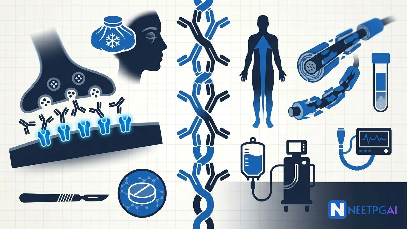
Neuromuscular disorders are a NEET PG favourite because localisation is so testable.
- Myasthenia gravis (MG) — post-synaptic AChR (or MuSK) antibody disease; fatigable weakness worst at end of day; ocular then bulbar then generalised.
- Diagnosis — ice-pack test, repetitive nerve stimulation (decremental), single-fiber EMG (most sensitive), AChR/MuSK antibodies, CT thorax for thymoma.
- Treatment — pyridostigmine first-line; steroids plus azathioprine/mycophenolate; IVIG or plasma exchange for crisis; thymectomy for AChR+ generalised MG.
- Guillain-Barré (GBS) — post-infectious (Campylobacter) ascending flaccid paralysis with areflexia; albuminocytologic dissociation in CSF.
- GBS treatment — IVIG or plasma exchange (equivalent); no steroids.
- LEMS — pre-synaptic anti-VGCC antibodies, paraneoplastic with SCLC, incremental RNS.
Neuromuscular junction and peripheral nerve disorders are among the most heavily tested topics in NEET PG neurology because they pivot on precise localisation, antibody-driven mechanisms and life-saving decisions in the emergency department. Myasthenic crisis and Guillain-Barré syndrome are two of the classic causes of acute neuromuscular respiratory failure that every intern will manage.
This NEETPGAI deep dive walks through myasthenia gravis, Guillain-Barré syndrome and Lambert-Eaton myasthenic syndrome — mechanism, presentation, diagnostic pearls, treatment ladders, India-specific epidemiology and the exam traps AIIMS and INI-CET stems love. Pair this with the multiple sclerosis and demyelinating disease guide for the broader neuro-immunology framework.
Myasthenia gravis — pathophysiology
Myasthenia gravis is the archetypal autoimmune disease of the neuromuscular junction. Most patients have IgG antibodies against the nicotinic acetylcholine receptor (AChR) on the post-synaptic muscle membrane. The antibodies cross-link and internalise receptors, activate complement, and simplify the post-synaptic folds — the safety margin of neuromuscular transmission collapses.
- AChR antibodies — positive in around 85 percent of generalised MG and 50 percent of ocular MG.
- MuSK (muscle-specific kinase) antibodies — 5 to 10 percent of generalised MG, more common in women, prominent bulbar and neck weakness, poor response to acetylcholinesterase inhibitors.
- LRP4 antibodies — a small minority of double-seronegative patients.
- Thymic pathology — thymic hyperplasia in 65 percent of AChR-positive patients under 40 years; thymoma in about 10 to 15 percent, more often in older patients.
Association with other autoimmune disease is common — autoimmune thyroid disease, SLE, rheumatoid arthritis, pernicious anaemia.
Clinical presentation of MG
MG classically presents with fluctuating, fatigable weakness that worsens through the day and improves with rest. The distribution progresses in a fairly stereotyped pattern.
| Stage | Features |
|---|
| Ocular MG | Ptosis (often asymmetric), diplopia; extraocular muscles affected without pupillary involvement — pupil sparing is a key differentiator from third-nerve palsy |
| Bulbar | Dysphagia, nasal regurgitation, dysarthria, jaw fatigue while chewing, snarling smile (myasthenic sneer) |
| Generalised | Proximal limb weakness, neck flexion weakness, respiratory muscle involvement |
| Myasthenic crisis | Respiratory failure requiring mechanical ventilation — a neurological emergency |
Around 15 percent of patients remain purely ocular; the rest progress to generalised disease, most within the first two years.
Diagnosis of MG
A systematic bedside plus laboratory work-up is expected in NEET PG stems.
| Test | Utility |
|---|
| Ice-pack test | Apply ice over ptotic lid for 2 minutes; improvement supports MG (cold slows acetylcholinesterase) |
| Edrophonium (Tensilon) test | Short-acting AChE inhibitor causes transient improvement; rarely used now due to muscarinic side effects |
| Repetitive nerve stimulation (RNS) | Decremental response greater than 10 percent at low-frequency (2 to 3 Hz) stimulation |
| Single-fiber EMG | Increased jitter — most sensitive test (95 percent), used when antibody and RNS are inconclusive |
| AChR antibodies | Highly specific; positive in most generalised MG |
| MuSK antibodies | Order if AChR-negative and clinically MG |
| CT/MRI thorax | Mandatory in every MG patient to look for thymoma |
| TFT, ANA, B12 | Screen for associated autoimmune disease |
Treatment of MG
Management is a stepwise ladder from symptomatic to disease-modifying to rescue therapies.
- Symptomatic — pyridostigmine — oral acetylcholinesterase inhibitor (longer-acting than neostigmine or edrophonium); typical dose 30 to 60 mg every 4 to 6 hours titrated to symptoms. Muscarinic side effects (abdominal cramps, diarrhoea, hypersalivation) limit dose. Poor response in MuSK MG.
- Chronic immunosuppression — oral prednisolone is the mainstay; start low and up-titrate to avoid initial worsening. Steroid-sparing agents include azathioprine (check TPMT), mycophenolate mofetil, methotrexate, ciclosporin and tacrolimus.
- Thymectomy — indicated for every patient with thymoma. For non-thymomatous generalised AChR-positive MG in patients under 65 years, the MGTX trial (2016) confirmed clinical benefit and reduced steroid burden.
- Rescue for crisis or refractory disease — IVIG (2 g/kg over 2 to 5 days) or plasma exchange (5 exchanges over 7 to 10 days) work within days.
- Biologics for refractory disease — eculizumab (C5 complement inhibitor) for refractory AChR-positive generalised MG; rituximab is particularly effective in MuSK MG; efgartigimod (FcRn inhibitor) approved 2021; ravulizumab and rozanolixizumab are the newer additions.
Drugs that unmask or worsen MG must be flagged in every prescription — aminoglycosides, fluoroquinolones, macrolides, telithromycin, magnesium sulfate, beta-blockers, procainamide, quinidine, chloroquine, penicillamine, statins and iodinated contrast can all destabilise MG.
Myasthenic crisis versus cholinergic crisis
A clinical trap is distinguishing myasthenic crisis (undertreatment or precipitated worsening) from cholinergic crisis (pyridostigmine excess with muscarinic overload). Both present with generalised weakness, but cholinergic crisis has SLUDGE — salivation, lacrimation, urination, defecation, GI cramps, emesis — plus miosis and bradycardia. In practice, most crises in modern care are myasthenic; low-dose edrophonium was historically used to differentiate but is now rarely available.
Manage crisis in an ICU with bedside forced vital capacity (FVC) and negative inspiratory force (NIF); electively intubate if FVC falls below 15 mL/kg or NIF is weaker (less negative) than −20 cmH2O — do not wait for hypoxia. IVIG or plasma exchange are equivalent first-line rescues; treat the trigger (infection, aspiration, offending drug); optimise steroids and long-term immunosuppression. Pyridostigmine is often held during ventilation to reduce airway secretions.
Lambert-Eaton myasthenic syndrome (LEMS)
LEMS is the pre-synaptic mirror image of MG.
- Mechanism — IgG antibodies against pre-synaptic voltage-gated calcium channels (VGCC) reduce acetylcholine release.
- Association — around 60 percent are paraneoplastic, almost always with small cell lung cancer; the rest are primary autoimmune.
- Clinical — proximal lower-limb weakness, hyporeflexia (which briefly returns after sustained contraction — post-tetanic facilitation), autonomic features (dry mouth, erectile dysfunction, orthostatic hypotension). Ocular and bulbar involvement is mild compared to MG.
- Key differentiator — brief improvement of strength with sustained effort ("facilitation"); RNS shows an incremental response of over 60 percent (classically described as over 100 percent) at high frequency (20 to 50 Hz).
- Treatment — treat the underlying malignancy; symptomatic 3,4-diaminopyridine (amifampridine) enhances ACh release; add pyridostigmine, immunosuppression or IVIG for refractory cases.
Guillain-Barré syndrome — pathophysiology
Guillain-Barré syndrome is an acute inflammatory polyradiculoneuropathy triggered in most cases by molecular mimicry after an infection. The commonest antecedent is Campylobacter jejuni gastroenteritis, followed by CMV, EBV, Mycoplasma pneumoniae, influenza, hepatitis E, Zika, HIV seroconversion and COVID-19. The Pune GBS outbreak (January 2025), linked to contaminated water and Campylobacter jejuni, has kept this topic high-yield.
The main subtypes are:
| Subtype | Features |
|---|
| AIDP (acute inflammatory demyelinating polyradiculoneuropathy) | Commonest form globally including India; demyelinating on nerve conduction |
| AMAN (acute motor axonal neuropathy) | Axonal; more severe; often post-Campylobacter with GM1 antibodies |
| AMSAN | Motor and sensory axonal — poor prognosis |
| Miller Fisher syndrome | Ophthalmoplegia, ataxia, areflexia; anti-GQ1b antibodies |
| Pharyngeal-cervical-brachial | Bulbar weakness with descending pattern |
Clinical presentation of GBS
- Progressive, symmetric, ascending flaccid weakness reaching nadir within 4 weeks (most by 2 weeks, over 90 percent by 4 weeks).
- Areflexia or hyporeflexia — a defining feature.
- Minimal sensory symptoms — usually paraesthesiae in fingers and toes; back and radicular pain are common and often overlooked.
- Autonomic dysfunction in about 65 percent — labile blood pressure, arrhythmias, ileus, urinary retention, SIADH.
- Respiratory failure in about 25 percent, requiring mechanical ventilation.
- Cranial nerve involvement — facial diplegia is the most common.
Diagnosis of GBS
| Test | Finding |
|---|
| CSF analysis | Albuminocytologic dissociation — high protein with fewer than 10 cells/mm3; may be normal in week 1 |
| Nerve conduction studies | Prolonged distal motor latency, conduction block, absent F waves, demyelinating pattern in AIDP; low CMAP amplitudes in AMAN |
| Antibody panels | Anti-GM1 (AMAN), anti-GQ1b (Miller Fisher) |
| MRI spine (gadolinium) | Nerve root enhancement — useful when diagnosis unclear |
| Bedside FVC | Essential every 4 to 6 hours to detect respiratory failure |
| ECG monitoring | For autonomic arrhythmia |
Brighton criteria are used for research and case ascertainment. Look for the classic distinguishers from spinal cord pathology — GBS is areflexic without a sensory level or sphincter disturbance.
Treatment of GBS
Immunotherapy accelerates recovery when started within four weeks of onset.
- IVIG — 2 g/kg over 5 days; convenient, widely available.
- Plasma exchange — 5 exchanges over 1 to 2 weeks; equivalent to IVIG in adults.
- Combining them offers no benefit and adds cost.
- Steroids alone are ineffective and can worsen outcome — do not use as monotherapy.
- Supportive care — DVT prophylaxis, neuropathic pain control (gabapentin, pregabalin), bladder care, physiotherapy, nutrition, meticulous ICU care.
- Ventilatory support — intubate electively if FVC less than 20 mL/kg, maximum inspiratory pressure less than 30 cmH2O or expiratory less than 40 cmH2O (the 20/30/40 rule).
- Autonomic monitoring — treat sustained tachyarrhythmias but avoid over-treating labile blood pressure.
Around 80 percent recover fully or with mild residual deficit; 3 to 7 percent die from respiratory failure, autonomic collapse or pulmonary embolism; the rest have significant long-term disability. Early ICU care and prompt ventilation are the biggest determinants of survival.
NEET PG MCQ traps
- Ptosis with pupil sparing plus fatigability — think MG, not third-nerve palsy.
- Single-fiber EMG — most sensitive test in MG.
- AChR-negative MG with prominent bulbar weakness — order MuSK antibodies.
- Every MG patient — CT thorax to rule out thymoma.
- Drugs that worsen MG — aminoglycosides, fluoroquinolones, macrolides, magnesium sulfate, beta-blockers, procainamide.
- Cholinergic crisis — SLUDGE plus miosis and bradycardia; myasthenic crisis is weakness with normal or dilated pupils.
- LEMS — proximal weakness improving briefly with effort; incremental RNS; screen for SCLC.
- Albuminocytologic dissociation — hallmark of GBS.
- Miller Fisher triad — ophthalmoplegia, ataxia, areflexia with anti-GQ1b.
- Post-Campylobacter GBS — AMAN subtype, GM1 antibodies.
- Steroids in GBS — ineffective and potentially harmful as monotherapy.
- IVIG versus plasma exchange in GBS — equivalent; do not combine.
- 20/30/40 rule — intubation thresholds in GBS.
- Facial diplegia — the classic cranial-nerve sign in GBS.
- Autonomic dysfunction — commoner in GBS than MG; drives ICU-level arrhythmia and BP swings.
Recent updates and India context
- Myasthenia gravis biologics — eculizumab, ravulizumab, efgartigimod, rozanolixizumab and zilucoplan now expand refractory-MG options; India has limited access outside select tertiary centres due to cost.
- MGTX trial (2016) — established thymectomy benefit in AChR-positive non-thymomatous generalised MG under 65 years; forms the basis of current AAN and ICMR guidance.
- International MG consensus 2020 update — introduced treatment-target definitions ("minimal manifestations" as the therapeutic goal) and steroid-tapering guidance.
- Pune GBS outbreak January 2025 — over 200 suspected cases linked to contaminated water and Campylobacter jejuni; kept GBS in the recent-news bank for NEET PG.
- Post-COVID GBS — reported as a rare vaccine-associated event and more commonly as a post-infectious complication of SARS-CoV-2.
- India protocols — most tertiary centres use IVIG-first for GBS given plasma exchange logistics; MG immunotherapy centres around low-dose prednisolone plus azathioprine or mycophenolate.
- Digital ICU tools — bedside spirometry apps and NIF meters now standard in Level-3 ICUs across India for GBS and MG-crisis monitoring.
Frequently asked questions
What is the difference between myasthenia gravis and Lambert-Eaton myasthenic syndrome?
Myasthenia gravis is a post-synaptic disorder caused by antibodies against acetylcholine receptors, presenting with fatigable weakness that worsens with activity and improves with rest. LEMS is a pre-synaptic disorder driven by antibodies against voltage-gated calcium channels, most commonly paraneoplastic with small cell lung cancer. LEMS weakness paradoxically improves briefly with sustained effort (facilitation), and repetitive nerve stimulation shows an incremental response, opposite to the decremental pattern seen in MG.
When is thymectomy indicated in myasthenia gravis?
Thymectomy is indicated in all patients with a thymoma regardless of MG severity, because thymoma is a neoplasm needing removal. In non-thymomatous generalised MG, the MGTX trial confirmed thymectomy improves clinical outcomes in AChR-antibody-positive patients under 65 years, reducing steroid requirement and hospitalisations. It is not routinely done in ocular-only MG, in seronegative or MuSK-antibody-positive MG, or in older patients where surgical risk outweighs the modest benefit.
What is the classic CSF finding in Guillain-Barré syndrome?
GBS shows albuminocytologic dissociation — a raised CSF protein (often over 100 mg/dL by the second week) with a normal or only mildly elevated cell count (usually fewer than 10 cells/mm3). Elevated CSF pleocytosis should raise suspicion of an alternative diagnosis such as HIV seroconversion, Lyme disease, sarcoidosis or leptomeningeal malignancy. CSF protein may be normal in the first week and typically peaks around week 2 to 3, so an early lumbar puncture does not exclude GBS.
Is IVIG or plasma exchange preferred in Guillain-Barré syndrome?
IVIG and plasma exchange are equally effective in GBS when started within four weeks of symptom onset — no head-to-head trial shows superiority for either. Combining them offers no additional benefit and adds cost. Choice depends on availability, contraindications and patient factors. IVIG is generally more accessible and simpler to administer, while plasma exchange is preferred if IVIG is contraindicated (IgA deficiency with anti-IgA antibodies, hyperviscosity, thromboembolic disease). Steroids are not effective in GBS and are contraindicated as monotherapy.
What triggers a myasthenic crisis and how is it managed?
Myasthenic crisis is respiratory failure requiring mechanical ventilation, precipitated by infection, surgery, aspiration, pregnancy, tapering of immunosuppression or offending drugs (aminoglycosides, fluoroquinolones, macrolides, magnesium sulfate, beta-blockers, procainamide). Management is ICU admission with bedside FVC and NIF monitoring, elective intubation before frank respiratory failure, IVIG or plasma exchange as first-line rescue, continuation or introduction of steroids and long-term immunosuppression, and treatment of the precipitant. Pyridostigmine is often held during crisis to reduce secretions.
This content is for educational purposes for NEET PG exam preparation. It is not a substitute for professional medical advice, diagnosis, or treatment. Clinical information has been reviewed by qualified medical professionals.
Written by: NEETPGAI Editorial Team
Reviewed by: NEETPGAI Medical Advisory Board
Last reviewed: September 2026