Quick Answer
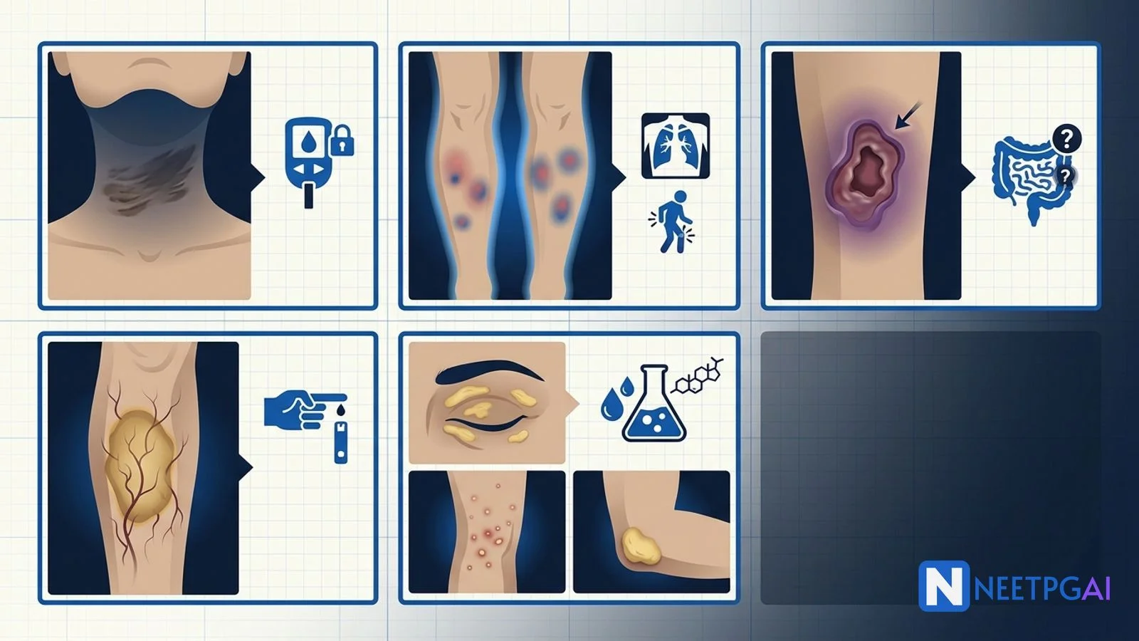
Cutaneous manifestations of systemic disease contribute 2-4 questions per NEET PG paper across dermatology, medicine, and pathology sections. Five patterns recur reliably:
- Acanthosis nigricans — velvety hyperpigmented plaques in flexural areas; insulin resistance (obesity, T2DM, PCOS, Cushing); sudden extensive onset in non-obese adult → screen for gastric adenocarcinoma; tripe palms = paraneoplastic
- Erythema nodosum — tender bilateral pretibial nodules; Löfgren syndrome (sarcoidosis + BHL + arthritis); IBD activity; streptococcal; TB (important in India); OCPs; pregnancy
- Pyoderma gangrenosum — rapidly progressing painful ulcer with violaceous undermined edge + pathergy; IBD, RA, haematological malignancy; aggressive surgical debridement is CONTRAINDICATED
- Necrobiosis lipoidica — yellow-brown atrophic pretibial plaques with telangiectasias; diabetes (60 percent overt, 20 percent IGT); may precede diabetes diagnosis; treatment often unsatisfactory
- Xanthomas — cholesterol deposits by pattern → eruptive (severe hypertriglyceridaemia), tuberous (familial hypercholesterolaemia, dysbetalipoproteinaemia), tendinous (homozygous FH), palmar (dysbetalipoproteinaemia), xanthelasma (50 percent normolipidemic)
Locking these 5 patterns plus a small library of genodermatoses (NF1, tuberous sclerosis, Sturge-Weber) and vasculitic patterns over 1-2 weeks moves cutaneous-systemic MCQ accuracy from 40 to 80 percent.
Why cutaneous manifestations of systemic disease are high-yield for NEET PG
The skin is a window to systemic disease and NEET PG tests this connection heavily because it forces integration across dermatology, endocrinology, gastroenterology, rheumatology, oncology, and haematology. Each pattern has a stereotyped morphology and location; pair the image with the clinical vignette (age, systemic symptoms, drug history, family history) and the answer typically falls out.
Drilling these 5 patterns plus a set of vasculitic patterns, genodermatoses, and paraneoplastic skin signs over 1-2 weeks moves the cutaneous-systemic MCQ accuracy from 40 to 80 percent.
Foundational approach — the systematic skin lesion read
Read every skin image on 5 axes
- Distribution — localised vs generalised, flexural vs extensor, photodistributed, symmetric vs asymmetric
- Morphology — macule, papule, nodule, plaque, vesicle, bulla, ulcer; primary vs secondary lesions
- Colour — erythematous, violaceous, yellow-orange, hyperpigmented, hypopigmented, purpuric
- Surface — smooth, verrucous, scaly, atrophic, crusted, ulcerated
- Border — well-demarcated, ill-defined, undermined, raised
Cutaneous marker of systemic disease — quick reference
| Skin finding | First-line systemic association |
|---|
| Velvety hyperpigmented flexural plaques | Insulin resistance (T2DM, PCOS, obesity); malignancy if sudden and extensive |
| Tender pretibial red nodules | Sarcoidosis (Löfgren), IBD, streptococcal, TB, OCPs |
| Rapidly progressing ulcer with undermined violaceous edge | Pyoderma gangrenosum (IBD, RA, haematological malignancy) |
| Yellow-brown atrophic pretibial plaques with telangiectasias | Necrobiosis lipoidica (diabetes) |
| Yellow-orange papules with halo (crops) | Eruptive xanthomas (severe hypertriglyceridaemia) |
| Café-au-lait spots + axillary freckling + neurofibromas | NF1 |
| Facial angiofibromas + ash-leaf spots + shagreen patch | Tuberous sclerosis |
| Port-wine stain in V1 distribution | Sturge-Weber |
| Butterfly malar rash | SLE |
| Heliotrope rash + Gottron papules | Dermatomyositis |
| Sausage digits + nail pitting | Psoriatic arthritis |
| Purpuric palpable rash on lower legs | Small vessel vasculitis (IgA vasculitis, cryoglobulinaemia) |
MCQ 1: 14-year-old obese girl with velvety hyperpigmented plaques on the posterior neck and axillae
Image description: [Clinical photograph of a 14-year-old girl. Panel 1 — Posterior neck — a well-demarcated, velvety, hyperpigmented (brown-black) plaque with a slightly thickened, papillomatous surface covers the posterior and lateral neck symmetrically. The lesion has a smooth, soft, velvety texture on palpation. Panel 2 — Bilateral axillae — similar velvety hyperpigmented plaques in both axillae, with a few overlying skin tags (acrochordons) — a common associated finding. Panel 3 — Groin and inframammary areas — subtle involvement. Panel 4 — Body habitus — the patient is obese (BMI 34), with abdominal striae and a fatty neck-shoulder pad. Investigations — fasting glucose 118 mg/dL (impaired), HbA1c 6.4 percent, fasting insulin 42 mIU/mL (elevated), HOMA-IR 12 (marked insulin resistance), lipid panel — triglycerides 240 mg/dL, HDL 32 mg/dL (metabolic syndrome). LH:FSH ratio 3:1 with elevated total testosterone on subsequent workup.]
Clinical vignette: A 14-year-old obese Indian girl is brought by her mother for evaluation of a "dark neck". Her mother has noticed a gradually darkening of her daughter's posterior neck over the last 2 years, initially attributed to poor hygiene. She has irregular menstrual cycles (menarche at 12, cycles every 45-90 days) and mild facial acne with early hirsutism (fine dark hair on upper lip and chin). She reports increased appetite, especially for sweets, and has gained 8 kilograms in the last 18 months. Family history — her father has type 2 diabetes and her mother has PCOS. Examination — BMI 34, waist circumference 88 centimetres, mild central obesity, no Cushingoid features, no galactorrhoea.
Options:
- (a) Post-inflammatory hyperpigmentation
- (b) Acanthosis nigricans (obesity-associated, insulin resistance)
- (c) Addison disease
- (d) Confluent and reticulated papillomatosis of Gougerot-Carteaud
Correct answer: (b) Acanthosis nigricans (obesity-associated, insulin resistance)
Reasoning: Velvety, hyperpigmented plaques with a papillomatous surface in flexural areas (posterior neck, axillae, groin) with associated obesity, insulin resistance (elevated HOMA-IR), and features of PCOS defines obesity-associated acanthosis nigricans (AN). The pathophysiology is hyperinsulinaemia acting on IGF-1 receptors on keratinocytes and fibroblasts, producing epidermal hyperplasia and hyperpigmentation.
Post-inflammatory hyperpigmentation follows a preceding inflammatory event and is not typically velvety. Addison disease causes generalised hyperpigmentation including palmar creases, mucous membranes, and scars, with associated hypotension and hyponatraemia. Confluent and reticulated papillomatosis has a distinct reticulate pattern on the trunk in young adults, is not typically flexural, and does not have the metabolic association.
Teaching pearl — acanthosis nigricans:
| Type | Associations |
|---|
| Benign (obesity-associated) | Commonest form; insulin resistance (T2DM, PCOS, obesity, metabolic syndrome) |
| Endocrine | Acromegaly, Cushing syndrome, Addison disease, hypothyroidism |
| Drug-induced | Systemic glucocorticoids, oral contraceptives, niacin, insulin, testosterone |
| Malignant (paraneoplastic) | Sudden onset in non-obese adult, extensive, mucous membranes and palmar involvement (tripe palms), pruritus; commonest primary is gastric adenocarcinoma; also lung, colon, breast, ovary |
| Familial | Autosomal dominant, non-obese, no metabolic disease |
Red-flag features for malignancy workup:
- Sudden onset in non-obese adult
- Rapid progression over weeks-months
- Extensive body surface involvement
- Mucous membrane involvement
- Palmar/plantar involvement (tripe palms)
- Pruritus
- Weight loss
Workup — upper GI endoscopy, chest CT, colonoscopy, mammography, pelvic ultrasound, tumour markers.
Treatment:
-
Benign — weight loss, metformin, lifestyle modification, topical retinoids and keratolytics (limited cosmetic improvement)
-
Paraneoplastic — treat the underlying malignancy (skin lesions often resolve)
-
NEET PG tests the insulin-resistance mechanism, the obesity/T2DM/PCOS association, the tripe-palms sign of paraneoplastic AN, and the gastric adenocarcinoma link
MCQ 2: 28-year-old woman with fever, ankle arthritis, and bilateral tender pretibial nodules — chest X-ray shows bilateral hilar lymphadenopathy
Image description: [Clinical photograph and imaging of a 28-year-old woman. Panel 1 — Anterior shins bilateral — tender, red-purple, poorly demarcated nodules (1-5 centimetres) on the pretibial area of both legs; lesions do NOT ulcerate; some are turning yellow-brown (mimicking bruises — erythema contusiforme). Panel 2 — Both ankles — swelling and erythema of both ankle joints with restricted range of motion. Panel 3 — Chest X-ray posteroanterior — symmetric bilateral hilar lymphadenopathy (BHL, egg-shell calcification NOT present) without parenchymal opacities or pleural effusion. Panel 4 — Investigations — ESR 68, CRP raised, serum ACE 92 IU/L (elevated), calcium mildly elevated, ASO titre normal, Mantoux test negative, HIV negative. Skin biopsy of a nodule shows septal panniculitis with lymphocytic infiltrate — consistent with erythema nodosum.]
Clinical vignette: A 28-year-old female schoolteacher presents with 10 days of fever, painful ankles bilaterally, and painful red bumps on her shins. She reports 2 weeks of low-grade fever, mild dry cough, and easy fatigue. No history of sore throat, no GI symptoms, no drug use, no recent travel, no OCP use. She is nulliparous. On examination — bilateral pretibial tender red-purple nodules, symmetric ankle synovitis with restricted range of motion, no other joint involvement, no rash elsewhere, no lymphadenopathy. Vitals — temperature 38.4 degrees Celsius, otherwise stable. Chest examination normal.
Options:
- (a) Löfgren syndrome (acute sarcoidosis)
- (b) Post-streptococcal erythema nodosum
- (c) Rheumatoid arthritis with rheumatoid nodules
- (d) Nodular vasculitis (erythema induratum)
Correct answer: (a) Löfgren syndrome (acute sarcoidosis)
Reasoning: The triad of erythema nodosum (tender bilateral pretibial nodules), bilateral hilar lymphadenopathy on chest X-ray, and migratory arthritis (usually ankles or knees), often with fever, is Löfgren syndrome — an acute presentation of sarcoidosis with over 80 percent spontaneous remission within 2 years. The elevated serum ACE and mildly elevated calcium support sarcoidosis. It is more common in Scandinavian and North Indian populations; young women in the third to fourth decades.
Post-streptococcal erythema nodosum would have a raised ASO titre and typically presents 2-3 weeks after pharyngitis. Rheumatoid arthritis with rheumatoid nodules would have a chronic symmetric polyarthritis of small joints (MCP, PIP, wrists), positive rheumatoid factor and anti-CCP, and firm subcutaneous nodules at pressure points — different pattern. Erythema induratum is nodular vasculitis on the calves posteriorly, often associated with tuberculosis in India.
Teaching pearl — erythema nodosum systemic associations:
| Association | Clinical clues |
|---|
| Sarcoidosis (Löfgren syndrome) | EN + BHL on CXR + migratory ankle arthritis + fever; over 80 percent spontaneous remission |
| Inflammatory bowel disease | Crohn or UC; often correlates with disease activity; abdominal pain, diarrhoea, blood in stool |
| Streptococcal infection | Post-pharyngitis, raised ASO, 2-3 weeks after infection |
| Tuberculosis | Primary or reactivation; important in India; positive Mantoux; treat with ATT |
| Medications | Oral contraceptives, sulfonamides, penicillins, HRT, halides |
| Pregnancy | Hormonally driven; often in first or second trimester |
| Other infections | Yersinia enterocolitica, coccidioidomycosis, histoplasmosis, hepatitis B/C, EBV |
| Malignancy | Lymphoma, leukaemia |
| Autoimmune | Behçet, IgA vasculitis, SLE (rare) |
Investigation of new erythema nodosum:
- Chest X-ray (BHL of sarcoidosis, TB findings)
- ASO titre and throat swab
- Mantoux test (TB — India)
- CBC, ESR/CRP, calcium, LFT
- Stool for GI evaluation if bowel symptoms
- Serum ACE (sarcoidosis, not specific)
- Biopsy rarely needed unless atypical
Treatment:
-
Supportive — NSAIDs, leg elevation, compression stockings
-
Treat underlying — antibiotics for strep, ATT for TB, IBD-directed therapy
-
Potassium iodide — second-line
-
Systemic steroids — reserved for severe or recurrent disease after excluding infection
-
NEET PG tests Löfgren syndrome (the triad), the IBD activity link, and the TB association in Indian populations
MCQ 3: 34-year-old woman with Crohn disease presents with a rapidly enlarging painful ulcer on the lower leg with a violaceous undermined edge
Image description: [Clinical photograph of a 34-year-old woman. Panel 1 — Left lower leg pretibial area — a large (approximately 8 x 6 centimetres) ulcer with a violaceous, dusky, undermined edge and a necrotic yellow-black base with purulent discharge. The ulcer border is elevated and the surrounding skin is erythematous. The centre of the ulcer is deep with visible subcutaneous tissue. Panel 2 — Progression over 2 weeks — the ulcer has doubled in size after a well-intentioned surgical debridement attempt at another hospital (pathergy). Panel 3 — Small satellite pustule at a needle-stick site from a recent blood draw (pathergy). Panel 4 — Colonoscopy — active Crohn disease with deep skip lesions, cobblestoning, and rectal sparing in the terminal ileum and ascending colon. Investigations — CBC — mild anaemia, elevated CRP and ESR, wound swab grew mixed skin flora (not pathogenic), biopsy of the ulcer edge shows dense neutrophil-rich infiltrate without vasculitis (non-specific).]
Clinical vignette: A 34-year-old female software engineer with known Crohn disease (diagnosed 3 years ago, currently on azathioprine and mesalazine) presents with a painful ulcer on her left lower leg that started as a small pustule 3 weeks ago after a minor abrasion from gardening. Over 2 weeks the ulcer rapidly enlarged and became extremely painful. A surgical team at another hospital attempted debridement 1 week ago, after which the ulcer dramatically worsened and a new pustule developed at a nearby venipuncture site. Examination — as above; abdominal examination shows mild right-lower-quadrant tenderness (active Crohn). She has taken diclofenac for pain with no relief.
Options:
- (a) Necrotising fasciitis
- (b) Pyoderma gangrenosum (Crohn-associated)
- (c) Cutaneous vasculitis (leukocytoclastic)
- (d) Chronic venous ulcer with secondary infection
Correct answer: (b) Pyoderma gangrenosum (Crohn-associated)
Reasoning: A rapidly progressing painful ulcer with a violaceous undermined edge, in a patient with active inflammatory bowel disease, that dramatically worsened after surgical debridement (pathergy phenomenon) and developed new lesions at a venipuncture site (further pathergy), defines pyoderma gangrenosum. The wound swab growing mixed flora without a single pathogen and biopsy showing non-specific neutrophil-rich inflammation supports the diagnosis. Aggressive surgical debridement is CONTRAINDICATED — it triggered the dramatic worsening.
Necrotising fasciitis would show rapidly spreading crepitus, systemic toxicity, high fever, and hypotension. Cutaneous vasculitis would show palpable purpura on the lower legs, positive vasculitis on biopsy, and often systemic features (fever, arthralgia, renal). Chronic venous ulcer would be shallow, over the medial malleolus, with surrounding lipodermatosclerosis and haemosiderin staining.
Teaching pearl — pyoderma gangrenosum:
| Aspect | Detail |
|---|
| Definition | Rare, painful, ulcerating neutrophilic dermatosis; violaceous undermined edge; necrotic base |
| Location | Lower extremities commonest; can occur anywhere; peristomal (colostomy, ileostomy) common in IBD |
| Pathergy | Trauma (surgical debridement, biopsy, needle stick, minor abrasion) worsens the ulcer and triggers new lesions; aggressive surgical debridement is CONTRAINDICATED |
| Systemic associations (50-70 percent) | Inflammatory bowel disease (Crohn, UC — up to 30 percent), rheumatoid arthritis, seronegative arthropathies, haematological (MDS, AML, IgA monoclonal gammopathy — paraneoplastic), other autoimmune |
| Diagnosis | Clinical; biopsy non-specific (neutrophil-rich infiltrate); biopsy often done only to exclude other causes (infection, vasculitis, malignancy) |
| Topical treatment | Potent corticosteroids, tacrolimus for small lesions |
| Intralesional | Corticosteroids for larger lesions |
| Systemic — first-line | Oral corticosteroids (often high-dose 1 mg/kg/day), cyclosporine |
| Systemic — second-line | Mycophenolate mofetil, methotrexate, dapsone, colchicine |
| Biologics (especially with IBD) | TNF-alpha inhibitors — infliximab, adalimumab (proven benefit) |
| Wound care | Non-adherent dressings, atraumatic, meticulous management of underlying systemic disease |
| Analgesia | Ulcers extremely painful; adequate opioid analgesia often needed |
| Skin grafting | Only after immunosuppression has controlled active disease and pathergy phase passed |
- NEET PG tests the pathergy phenomenon, IBD and RA associations, biopsy non-specific nature, and the surgical debridement contraindication
MCQ 4: 52-year-old woman with 20-year history of type 1 diabetes presents with yellow-brown atrophic plaques on the shins
Image description: [Clinical photograph of a 52-year-old woman. Panel 1 — Anterior shins bilateral — well-demarcated, yellow-brown, atrophic plaques with a raised violaceous border, measuring 3-6 centimetres each, on the pretibial area. The plaque surfaces are shiny and waxy with visible telangiectasias coursing through the thinned epidermis. One of the plaques on the right shin has a small superficial ulceration in the centre. Panel 2 — Close-up — the border of the plaque is slightly raised and inflammatory, while the central atrophic area appears yellow-orange due to underlying lipid deposition and thinned epidermis. Panel 3 — Histopathology (biopsy done for one atypical plaque) — palisading granulomas surrounding areas of collagen degeneration and lipid deposition in the dermis, consistent with necrobiosis lipoidica. Panel 4 — HbA1c 8.4 percent (poorly controlled diabetes), fasting glucose 178 mg/dL, urine microalbumin positive (early diabetic nephropathy), fundoscopy shows moderate non-proliferative diabetic retinopathy.]
Clinical vignette: A 52-year-old female with type 1 diabetes since age 32 (poorly controlled — most recent HbA1c 8.4 percent, on basal-bolus insulin) presents with slowly progressive yellow-brown plaques on her shins noted over the last 3-4 years. She reports that some plaques ulcerated with minor bumps and healed slowly. She has diabetic nephropathy (microalbuminuria) and moderate non-proliferative diabetic retinopathy. She has NO history of thyroid disease, vitiligo, or coeliac disease.
Options:
- (a) Diabetic dermopathy (shin spots)
- (b) Necrobiosis lipoidica
- (c) Granuloma annulare
- (d) Erythema nodosum
Correct answer: (b) Necrobiosis lipoidica
Reasoning: Yellow-brown atrophic plaques with raised violaceous border, waxy shiny surface with visible telangiectasias, on the pretibial area in a patient with long-standing diabetes, plus histopathology showing palisading granulomas with collagen degeneration and lipid deposition, defines necrobiosis lipoidica. The tendency to ulcerate with minor trauma and slow healing is characteristic. Approximately 60 percent of patients with necrobiosis lipoidica have overt diabetes and another 20 percent have impaired glucose tolerance; it may precede the diagnosis of diabetes by months to years.
Diabetic dermopathy (shin spots) are small brown atrophic macules, commonest cutaneous marker of diabetes, but they are smaller and less inflammatory. Granuloma annulare appears as ring-shaped skin-coloured to erythematous papules with a raised border, typically on the dorsa of hands or feet, and the association with diabetes is debated (localised granuloma annulare not strongly associated; disseminated granuloma annulare has some association). Erythema nodosum has tender, ill-defined, red-purple nodules that do not ulcerate or scar.
Teaching pearl — cutaneous manifestations of diabetes:
| Condition | Features |
|---|
| Necrobiosis lipoidica | Yellow-brown atrophic pretibial plaques with telangiectasias; may precede diabetes diagnosis; treatment often unsatisfactory; glycaemic control alone does NOT reliably improve lesions |
| Diabetic dermopathy (shin spots) | Small brown atrophic macules on shins; commonest cutaneous marker of diabetes; often asymptomatic; correlates with microvascular disease |
| Diabetic bullae (bullosis diabeticorum) | Tense clear blisters on extremities in poorly controlled diabetes; heal without scarring; supportive treatment |
| Acanthosis nigricans | Insulin resistance marker (T2DM, PCOS, obesity); see MCQ 1 |
| Granuloma annulare | Ring-shaped skin-coloured to erythematous papules with raised border on dorsa of hands/feet; localised or generalised; association with diabetes debated |
| Scleredema diabeticorum | Indurated skin on upper back and posterior neck in long-standing T2DM; treatment difficult |
| Diabetic foot ulcers | Neuropathic (painless, plantar surface pressure points) vs ischaemic (painful, distal digits, dry gangrene); multidisciplinary care |
| Skin infections | Recurrent bacterial (staphylococcal folliculitis, cellulitis, foot infections), fungal (candidal intertrigo, tinea pedis), rhinocerebral mucormycosis (uncontrolled diabetes with ketoacidosis — emergency) |
Treatment of necrobiosis lipoidica:
-
First-line — potent topical corticosteroids (active border), intralesional corticosteroids, topical or intralesional calcineurin inhibitors, glycaemic control (though glycaemic control alone does not reliably improve lesions)
-
Second-line — pentoxifylline, aspirin, dipyridamole, systemic corticosteroids, hyperbaric oxygen
-
Emerging — TNF inhibitors, JAK inhibitors for refractory disease
-
Laser — cosmetic for atrophic scars
-
Ulcerated lesions — wound care, infection surveillance
-
NEET PG tests the pretibial location, association with diabetes, the fact that it may precede diabetes diagnosis, and the treatment-refractory nature
MCQ 5: 22-year-old man with sudden appearance of hundreds of small yellow papules with erythematous halos on the buttocks, elbows, and knees
Image description: [Clinical photograph and lab of a 22-year-old male. Panel 1 — Buttocks, elbows, and knees — hundreds of 1-4 millimetre yellow-orange papules with erythematous halos, appearing in crops, symmetric distribution over extensor surfaces. Lesions are non-tender and non-pruritic. Panel 2 — Chest and abdomen — sparser but similar lesions. Panel 3 — Fundoscopy — lipaemia retinalis (creamy-white appearance of retinal vessels from elevated triglycerides), no diabetic changes. Panel 4 — Investigations — fasting lipid panel — total cholesterol 340 mg/dL, LDL 90 mg/dL, HDL 22 mg/dL, triglycerides 3200 mg/dL (grossly elevated), serum appears milky (lipaemic), amylase 480 U/L (raised — pancreatitis risk), fasting glucose 180 mg/dL, HbA1c 9.6 percent (undiagnosed poorly controlled diabetes), family history — brother has diabetes with hypertriglyceridaemia.]
Clinical vignette: A 22-year-old male university student presents with sudden appearance of hundreds of small yellow bumps on his buttocks, elbows, and knees over the last 3 weeks. He reports mild epigastric discomfort but no vomiting. He has been drinking 2-3 cans of sweetened cola per day and eating a diet high in refined carbohydrates and fried foods. Recent 5-kilogram weight loss over 2 months (with polyuria and polydipsia — undiagnosed diabetes). Family history — father died of premature coronary artery disease at age 45, brother has type 2 diabetes with high triglycerides.
Options:
- (a) Molluscum contagiosum
- (b) Eruptive xanthomas (severe hypertriglyceridaemia)
- (c) Tuberous xanthomas (familial hypercholesterolaemia)
- (d) Sarcoidosis (papular)
Correct answer: (b) Eruptive xanthomas (severe hypertriglyceridaemia)
Reasoning: Sudden crops of 1-4 millimetre yellow-orange papules with erythematous halos on extensor surfaces, in a patient with grossly elevated triglycerides (over 1000 mg/dL — chylomicronaemia range), lipaemia retinalis, and lipaemic serum, defines eruptive xanthomas. The concurrent finding of poorly controlled diabetes (HbA1c 9.6 percent) is a common trigger — uncontrolled diabetes causes secondary hypertriglyceridaemia through insulin deficiency, increased VLDL, and reduced lipoprotein lipase activity. There is also a suggestion of familial hypertriglyceridaemia given the family history.
Molluscum contagiosum lesions have a central umbilication and are typically flesh-coloured or pearly, not yellow-orange, and appear in the pubic area or elsewhere with sexual contact. Tuberous xanthomas are larger firm nodules on extensor surfaces, associated with familial hypercholesterolaemia. Sarcoidosis presents with a variety of papular lesions but rarely mimics eruptive xanthomas in this crop pattern.
Teaching pearl — xanthomas and lipid disorders:
| Xanthoma type | Morphology | Associated lipid disorder |
|---|
| Eruptive xanthomas | 1-4 mm yellow-orange papules with erythematous halos, in crops on buttocks/elbows/knees | Severe hypertriglyceridaemia (over 1000 mg/dL) — chylomicronaemia syndrome, familial LPL deficiency, uncontrolled diabetes; resolves rapidly with triglyceride lowering |
| Tuberous xanthomas | Firm yellow-red nodules on extensor surfaces (elbows, knees, knuckles) | Familial dysbetalipoproteinaemia (Type III, ApoE2 homozygosity), familial hypercholesterolaemia |
| Tendinous xanthomas | Firm painless subcutaneous nodules along tendons (Achilles, extensor tendons of fingers, plantar fascia) | Homozygous familial hypercholesterolaemia (LDL receptor mutations), heterozygous FH |
| Palmar xanthomas (xanthoma striatum palmare) | Yellow-orange plaques or linear streaks in palmar creases | Pathognomonic of familial dysbetalipoproteinaemia (Type III) |
| Plane xanthomas | Flat yellow-orange patches on any body surface | Various dyslipidaemias, primary biliary cholangitis |
| Xanthelasma palpebrarum | Yellow soft plaques on eyelids, medial canthus area | Approximately 50 percent normolipidemic — NOT a reliable marker; treatment cosmetic |
Workup of any patient with xanthomas:
- Fasting lipid panel (total cholesterol, LDL, HDL, triglycerides)
- Apolipoprotein E genotype for suspected Type III
- Family screening (first-degree relatives)
- Cardiovascular risk assessment (ASCVD score, imaging where indicated)
- Fasting glucose and HbA1c (secondary hypertriglyceridaemia)
- Thyroid function (hypothyroidism causes secondary hyperlipidaemia)
- LFT (secondary causes)
Treatment:
- Eruptive xanthomas — fibrates (fenofibrate, gemfibrozil), omega-3 fatty acids, diet (low fat, low simple carbohydrate), insulin/glycaemic control for diabetic patients; resolves in weeks
- Familial hypercholesterolaemia — high-intensity statin, ezetimibe, PCSK9 inhibitors (evolocumab, alirocumab) for refractory cases, LDL apheresis for homozygous FH
- Xanthelasma — cosmetic treatment (topical trichloroacetic acid, laser, surgical excision); lipid panel to identify occult hyperlipidaemia
Bonus — cutaneous markers of hereditary systemic disease
Not primary MCQs above but frequently paired distractors:
Neurofibromatosis type 1 (NF1)
- Diagnostic criteria (need 2 or more) — 6 or more café-au-lait spots larger than 5 mm prepubertal (larger than 15 mm postpubertal), axillary or inguinal freckling (Crowe sign), 2 or more neurofibromas or 1 plexiform neurofibroma, optic pathway glioma, 2 or more Lisch nodules (iris hamartomas), sphenoid dysplasia or tibial pseudarthrosis, first-degree relative with NF1
- Autosomal dominant, NF1 gene on 17q11 encoding neurofibromin (a tumour suppressor)
Tuberous sclerosis complex
- Facial angiofibromas (adenoma sebaceum), ash-leaf spots (hypopigmented macules), shagreen patch (thickened leathery skin, lumbosacral), subungual and periungual fibromas (Koenen tumours)
- Systemic — mental retardation, epilepsy (infantile spasms, Vogt triad), cortical tubers, subependymal nodules, giant cell astrocytoma, cardiac rhabdomyoma, renal angiomyolipoma, lymphangioleiomyomatosis
- Autosomal dominant, TSC1 (hamartin) on 9q, TSC2 (tuberin) on 16p — regulate mTOR pathway; treatment includes mTOR inhibitors (everolimus)
Sturge-Weber syndrome
- Port-wine stain (naevus flammeus) in trigeminal V1 distribution (forehead and upper eyelid)
- Leptomeningeal angioma (tram-track calcifications on CT), seizures, hemiparesis, mental retardation, glaucoma (ipsilateral)
Common pitfalls in cutaneous-systemic MCQs
Pitfall 1 — Ignoring the paraneoplastic red flags
Sudden onset extensive acanthosis nigricans in a non-obese adult with tripe palms is not simple obesity-associated AN — screen for gastric adenocarcinoma. Similarly, sudden crops of xanthelasma or dermatomyositis in an older adult should prompt malignancy workup.
Pitfall 2 — Debriding pyoderma gangrenosum
Aggressive surgical debridement is CONTRAINDICATED — pathergy worsens the ulcer. Immunosuppression first, careful atraumatic wound care, grafting only after inflammation controls.
Pitfall 3 — Assuming diabetic dermopathy explains yellow-brown pretibial plaques
Diabetic dermopathy is small brown atrophic macules. Yellow-brown atrophic plaques with telangiectasias in a diabetic patient are necrobiosis lipoidica — a different entity with different treatment implications.
Pitfall 4 — Missing Löfgren syndrome
The triad of erythema nodosum + bilateral hilar lymphadenopathy + migratory arthritis (usually ankles) is Löfgren syndrome — acute sarcoidosis with over 80 percent spontaneous remission. Do not miss this benign self-limited entity by starting aggressive immunosuppression.
Pitfall 5 — Treating xanthelasma without checking lipids
Approximately 50 percent of xanthelasma patients have normal lipid levels. Xanthelasma is NOT a reliable marker of hyperlipidaemia — but you should still get a lipid panel to identify the 50 percent who do have occult dyslipidaemia and need treatment.
How to study cutaneous manifestations of systemic disease for NEET PG
- Memorise the 5 core patterns — acanthosis nigricans, erythema nodosum, pyoderma gangrenosum, necrobiosis lipoidica, xanthomas — plus the tripe-palms and Löfgren-triad rules
- Learn the xanthoma-lipid mapping — pattern to disorder (eruptive to hypertriglyceridaemia, tendinous to homozygous FH, palmar to dysbetalipoproteinaemia)
- Learn the genodermatoses — NF1 diagnostic criteria, tuberous sclerosis features, Sturge-Weber
- Learn the diabetes cutaneous manifestations — dermopathy, necrobiosis, bullae, acanthosis, granuloma annulare, scleredema, infections
- Learn the paraneoplastic skin signs — acanthosis nigricans + tripe palms (gastric), dermatomyositis (ovarian, lung), erythema gyratum repens (lung), necrolytic migratory erythema (glucagonoma)
- Learn the vasculitic patterns — palpable purpura on lower legs (IgA vasculitis), livedo reticularis (antiphospholipid, cholesterol emboli), splinter haemorrhages (endocarditis)
- Practice 10-15 cutaneous-systemic MCQs per day for 2-3 weeks using NEETPGAI dermatology bank
- Pair every image with a systemic vignette — age, associated symptoms, drug history, family history
- Learn the ATT drug rash patterns — INH-induced pellagra, rifampin-induced red discolouration of secretions
- Learn the cutaneous LGV, syphilis, and HIV stigmata — high-yield for STI/HIV MCQs
Key takeaways
- Cutaneous manifestations of systemic disease contribute 2-4 questions per NEET PG paper
- Acanthosis nigricans — velvety flexural hyperpigmentation; insulin resistance; malignancy screen if sudden extensive in non-obese adult (tripe palms)
- Erythema nodosum — tender bilateral pretibial nodules; Löfgren syndrome triad; IBD activity; streptococcal; TB (important in India)
- Pyoderma gangrenosum — rapidly progressing painful ulcer with violaceous undermined edge; pathergy; IBD, RA, haematological malignancy; aggressive debridement CONTRAINDICATED
- Necrobiosis lipoidica — yellow-brown atrophic pretibial plaques with telangiectasias; diabetes; may precede diabetes diagnosis
- Xanthomas — cholesterol deposits by pattern → eruptive (severe hypertriglyceridaemia), tuberous and tendinous (familial hypercholesterolaemia), palmar (dysbetalipoproteinaemia), xanthelasma (50 percent normolipidemic)
- The xanthoma-morphology to lipid-disorder mapping is the single most tested element in the xanthoma family
- Genodermatoses and paraneoplastic skin signs are common distractor content
Frequently asked questions
What are the systemic associations of acanthosis nigricans and how do you distinguish benign from paraneoplastic disease?
Acanthosis nigricans is a velvety, hyperpigmented, hyperkeratotic thickening of the skin most commonly seen in intertriginous areas — the posterior and lateral neck, axillae, groin, umbilicus, and antecubital and popliteal fossae. It represents a marker of insulin resistance and is one of the commonest cutaneous signs of systemic disease. Benign (obesity-associated) acanthosis nigricans is the commonest form, driven by hyperinsulinaemia acting on IGF-1 receptors on keratinocytes and fibroblasts producing epidermal hyperplasia — it is strongly associated with obesity, type 2 diabetes mellitus, polycystic ovary syndrome (PCOS), metabolic syndrome, and Cushing syndrome, and is seen commonly in Indian adolescents and young adults with the growing metabolic-syndrome epidemic. Drug-induced acanthosis nigricans is triggered by systemic glucocorticoids, oral contraceptives, niacin, insulin, and testosterone therapy. Endocrine forms are seen in acromegaly, Addison disease, and hypothyroidism. The critical distinction is malignant (paraneoplastic) acanthosis nigricans — sudden-onset extensive acanthosis nigricans in a non-obese adult, with rapid progression, involvement of mucous membranes and palms/soles (tripe palms — velvety palmar thickening), pruritus, and associated skin tags is a red-flag for underlying malignancy, most commonly gastric adenocarcinoma (over 60 percent of cases) but also lung, colon, breast, ovary, and pancreatic tumours. Workup should include upper GI endoscopy, chest CT, and tumour-specific evaluation. Treatment of benign acanthosis nigricans is aimed at the underlying insulin resistance — weight loss, metformin, and lifestyle modification; topical retinoids and keratolytics offer limited cosmetic improvement. Paraneoplastic acanthosis nigricans resolves with treatment of the underlying malignancy. NEET PG tests the insulin-resistance mechanism, the association with obesity and T2DM, the malignancy screen indications, and the tripe-palms sign.
What are the systemic associations of erythema nodosum and what is Löfgren syndrome?
Erythema nodosum is a septal panniculitis presenting as tender, red-to-purple nodules bilaterally on the shins, less commonly on the thighs, arms, or trunk. It is a delayed hypersensitivity reaction to a triggering antigen and is one of the commonest reactive dermatoses. The lesions are painful but do not ulcerate or scar; they resolve over 3-6 weeks passing through colour changes (red to purple to yellow-brown, mimicking bruises — erythema contusiforme). Fever, arthralgia (especially of ankles), and malaise commonly accompany the skin findings. The main systemic associations tested on NEET PG are sarcoidosis (Löfgren syndrome — the triad of erythema nodosum, bilateral hilar lymphadenopathy on chest X-ray, and migratory arthritis of ankles or knees, with fever — an acute presentation of sarcoidosis with over 80 percent spontaneous remission rate within 2 years, more common in Scandinavian and North Indian populations), inflammatory bowel disease (both Crohn disease and ulcerative colitis — often correlates with disease activity), streptococcal infection (post-streptococcal erythema nodosum in children and young adults, 2-3 weeks after pharyngitis, low ASO titre helpful), tuberculosis (primary TB infection or reactivation — important in India; tuberculin skin test positive; treat with ATT), medications (oral contraceptive pills, sulfonamides, penicillins, hormone replacement therapy), pregnancy (hormonally driven), and other infections (Yersinia enterocolitica, coccidioidomycosis, histoplasmosis, hepatitis B and C, EBV). Malignancy (lymphoma, leukaemia) and other autoimmune diseases (Behçet, IgA vasculitis) are less common triggers. Investigation of a new patient with erythema nodosum includes a targeted history for GI symptoms and recent infections, chest X-ray (for hilar lymphadenopathy of sarcoidosis or TB findings), ASO titre and throat swab, Mantoux test, complete blood count, and inflammatory markers; further testing (colonoscopy, biopsy of the erythema nodosum lesion is rarely needed) is guided by clinical suspicion. Treatment is largely supportive — NSAIDs, leg elevation, compression stockings, and treatment of the underlying cause; potassium iodide is a second-line agent; systemic steroids reserved for severe or recurrent disease after excluding infection. NEET PG heavily tests Löfgren syndrome (the triad), the association with IBD activity, and the TB association in Indian populations.
What is pyoderma gangrenosum and why is aggressive surgical debridement contraindicated?
Pyoderma gangrenosum is a rare, painful, ulcerating neutrophilic dermatosis characterised by rapidly progressing ulcers with a violaceous undermined border and a necrotic base, most commonly on the lower extremities but can occur anywhere including peristomal skin in patients with an ileostomy or colostomy. The pathogenesis involves dysregulated neutrophil function and is strongly associated with systemic disease — approximately 50-70 percent of cases have an identifiable systemic association. The key associations are inflammatory bowel disease (both Crohn disease and ulcerative colitis — up to 30 percent of pyoderma gangrenosum cases; skin activity often correlates with bowel activity), rheumatoid arthritis and other seronegative arthropathies, haematological disorders (myelodysplastic syndrome, acute myeloid leukaemia, monoclonal gammopathies especially IgA — the paraneoplastic form), and less commonly other autoimmune diseases. The pathergy phenomenon is critical — trauma to the skin (including surgical debridement, biopsy, needle sticks, or trauma) worsens the ulcer and causes new lesions to develop at the trauma site. This is why aggressive surgical debridement is CONTRAINDICATED in classic pyoderma gangrenosum — attempting to remove the necrotic base surgically triggers further pathergy-driven ulceration and typically dramatically worsens the ulcer. The diagnosis is clinical (there is no specific test or biopsy finding — biopsy shows neutrophil-rich inflammation but is not diagnostic; biopsy is often done only to exclude other causes like infection, vasculitis, or malignancy). Treatment focuses on immunosuppression — topical potent corticosteroids and calcineurin inhibitors for small lesions, intralesional corticosteroids for larger, and systemic therapy (oral corticosteroids often high-dose, cyclosporine, mycophenolate mofetil, methotrexate, dapsone, colchicine, and increasingly TNF-alpha inhibitors like infliximab and adalimumab, especially where IBD coexists — infliximab has been shown effective in multiple case series and small trials). Wound care with non-adherent dressings, meticulous management of the underlying systemic disease, and adequate analgesia (the ulcers are extremely painful) are foundations. Skin grafting, if needed for large residual ulcers, should be attempted only after immunosuppression has controlled active disease and the pathergy phase has passed. NEET PG tests the pathergy phenomenon, the IBD and RA associations, the biopsy-non-specific nature, and the contraindication of aggressive surgical debridement.
What is necrobiosis lipoidica and how does it differ from other cutaneous manifestations of diabetes?
Necrobiosis lipoidica (formerly necrobiosis lipoidica diabeticorum) is a rare granulomatous dermatosis that classically presents as well-demarcated, yellow-brown, atrophic plaques with a raised violaceous border, most commonly on the shins (pretibial area), less commonly on the arms, face, or scalp. Telangiectasias are visible through the thinned skin of the plaques, giving them a shiny, waxy appearance. The plaques often ulcerate with minor trauma and heal slowly. It is strongly associated with diabetes mellitus — approximately 60 percent of patients have overt diabetes and another 20 percent have impaired glucose tolerance, while 15-20 percent have neither at presentation but many go on to develop diabetes years later. Necrobiosis lipoidica may precede the diagnosis of diabetes by months to years, so a new patient without known diabetes should have fasting glucose, HbA1c, and OGTT. It is more common in women and typically presents in the third to fifth decades. Pathologically it shows collagen degeneration with palisading granulomas and lipid deposition. The differential diagnosis includes other diabetic cutaneous manifestations tested on NEET PG — diabetic dermopathy (small brown atrophic macules on the shins, the commonest cutaneous marker of diabetes and often asymptomatic), diabetic bullae (bullosis diabeticorum — tense clear blisters on the extremities that heal without scarring), acanthosis nigricans (marker of insulin resistance, see earlier), granuloma annulare (localised, generalised, or perforating variants; association with diabetes is debated but described), scleredema diabeticorum (indurated skin on the upper back and posterior neck), and diabetic foot ulcers and infections. Treatment of necrobiosis lipoidica is difficult and often unsatisfactory. First-line therapies include potent topical corticosteroids to the active border, intralesional corticosteroids, topical or intralesional calcineurin inhibitors, and glycaemic control (though glycaemic control alone does not consistently improve the lesions — an important NEET PG point). Second-line therapies include pentoxifylline, aspirin, dipyridamole, systemic corticosteroids, and hyperbaric oxygen. Emerging therapies include TNF inhibitors and JAK inhibitors for refractory disease. Laser therapy is used cosmetically for the atrophic scars. Ulcerated lesions require wound care and infection surveillance. NEET PG tests the pretibial location, the association with diabetes, the fact that it may precede diabetes diagnosis, and the treatment-refractory nature.
What are the different types of xanthomas and what lipid disorders do they indicate?
Xanthomas are focal accumulations of cholesterol and cholesterol esters within tissue macrophages (foam cells), presenting as yellow to yellow-orange papules, nodules, or plaques. They are cutaneous markers of specific dyslipidaemias and are heavily tested on NEET PG because each morphological type points to a specific lipid disorder. Eruptive xanthomas are 1-4 mm yellow-orange papules with erythematous halos, appearing in crops on the buttocks, elbows, knees, and extensor surfaces; they are strongly associated with severe hypertriglyceridaemia (typically serum triglycerides above 1000 mg/dL — chylomicronaemia syndrome, familial lipoprotein lipase deficiency, or uncontrolled diabetes with secondary hypertriglyceridaemia); they resolve rapidly (weeks) with triglyceride-lowering therapy (fibrates, omega-3 fatty acids, diet, insulin for diabetic patients). Tuberous xanthomas are firm, non-tender, yellow to red nodules on extensor surfaces especially elbows, knees, and knuckles; they are associated with familial dysbetalipoproteinaemia (Type III hyperlipoproteinaemia, ApoE2 homozygosity) and familial hypercholesterolaemia. Tendinous xanthomas are firm, painless, subcutaneous nodules along tendons — classically the Achilles tendon, extensor tendons of the fingers, and the plantar fascia; they are pathognomonic of severe hypercholesterolaemia, most notably homozygous familial hypercholesterolaemia (LDL receptor mutations, extremely high LDL from birth) and to a lesser extent heterozygous familial hypercholesterolaemia; a young patient with Achilles tendon xanthoma and premature coronary artery disease is a classic vignette. Palmar xanthomas (xanthoma striatum palmare) are yellow-orange plaques or linear streaks in the palmar creases and are pathognomonic of familial dysbetalipoproteinaemia. Plane xanthomas are flat, yellow-orange patches on any body surface. Xanthelasma palpebrarum is the most common xanthoma — yellow soft plaques on the eyelids, medial canthus area, more common in women; approximately 50 percent of xanthelasma patients have normal lipid levels, so xanthelasma is NOT a reliable marker of hyperlipidaemia and requires a lipid panel for confirmation; treatment is cosmetic (topical trichloroacetic acid, laser, surgical excision). Verruciform xanthomas are rare oral or genital plaques and are not related to lipid disorders. Workup of any patient with xanthomas includes fasting lipid panel, apolipoprotein E genotype (for suspected type III), family screening, and cardiovascular risk assessment. Treatment includes lipid-lowering therapy (statins for hypercholesterolaemia, fibrates and omega-3 for hypertriglyceridaemia, ezetimibe and PCSK9 inhibitors for refractory cases), diet, and treatment of secondary causes. NEET PG heavily tests each xanthoma morphology with its lipid disorder — the pattern-lipid mapping is the highest-yield element.
This content is for educational purposes for NEET PG exam preparation. It is not a substitute for professional medical advice, diagnosis, or treatment. Clinical information has been reviewed by qualified medical professionals.
Written by: NEETPGAI Editorial Team
Reviewed by: Pending SME Review
Last reviewed: September 2026