Version 1.0 - Published October 2026
Quick Answer
SICS uses a 6.5-7 mm sutureless self-sealing scleral tunnel to deliver the entire nucleus manually and is the Indian high-volume workhorse (Rs 2000-5000 total cost, 85-90 percent 6/9 outcomes). Phacoemulsification uses a 2.2-2.8 mm clear corneal incision with ultrasonic nucleus fragmentation (divide-and-conquer or phaco-chop) and foldable IOL - faster recovery and less astigmatism but 10-20x costlier. Posterior capsule rupture (1-5 percent) and endophthalmitis (0.07 percent with intracameral cefuroxime, 0.35 percent without) are the headline complications you must know for NEET PG and INI-CET.
Why This Matters for NEET PG and INI-CET
Cataract remains the leading cause of reversible blindness in India - the National Blindness and Visual Impairment Survey 2015-19 reported 66.2 percent of all blindness in Indians over 50 is from cataract. India performs approximately 6.5 million cataract surgeries per year, among the highest absolute volumes globally. For NEET PG and INI-CET, this translates to 4-7 ophthalmology questions per paper, with cataract surgery as the single most tested ophthalmology topic alongside glaucoma and retinal detachment.
High-yield exam territory covers four domains: (1) SICS technique steps and the Indian context, (2) phacoemulsification techniques (divide-and-conquer vs phaco-chop vs stop-and-chop), (3) IOL selection (monofocal, multifocal, toric, EDOF), and (4) complication recognition - particularly posterior capsule rupture, dropped nucleus, endophthalmitis (acute vs chronic), TASS, and PCO with Nd:YAG capsulotomy. Image-based MCQs often show pupillary flare, hypopyon, or an Elschnig pearl-laden posterior capsule and ask you to match the complication to its organism, timing or management.
INI-CET and AIIMS papers additionally test the ESCRS 2007 endophthalmitis trial, the EVS 1995 management algorithm, and the Nishi square-edge IOL effect on PCO. A confident clinical reasoner will outperform a rote memoriser - the examiner wants you to pick SICS for a rock-hard nucleus in a 68 year old village patient with pseudoexfoliation and phaco for a clear cornea 55 year old urban professional with moderate NC 2 cataract.
Step 1 - Preoperative Evaluation and Biometry
Every patient destined for cataract surgery needs a structured preoperative work-up because surgical planning depends on it:
- Visual acuity (uncorrected and best-corrected) and glare testing (Brightness Acuity Tester).
- Slit lamp - LOCS III grading (NC = nuclear colour 1-6, NO = nuclear opalescence 1-6, C = cortical 1-5, P = posterior subcapsular 1-5). A black (NC6) cataract is the SICS indication par excellence.
- Intraocular pressure - Goldmann applanation. Rule out glaucoma; adjust surgical plan if cup-disc ratio is high.
- Fundus examination after dilation - rule out age-related macular degeneration, diabetic retinopathy, high myopic degeneration, retinal detachment. A dense cataract blocking fundus view mandates B-scan ultrasonography.
- Pupil dilation - poor dilation (less than 5 mm) predicts difficult surgery; plan for iris hooks, Malyugin ring or pupil-expanding viscoelastic.
- Corneal endothelial cell count - specular microscopy; a count below 1500 cells/mm2 raises risk of pseudophakic bullous keratopathy; SICS or femtosecond-assisted phaco preferred.
- Biometry - axial length by IOLMaster 700 or Lenstar (optical, 0.02 mm precision) or A-scan immersion ultrasound. Keratometry by Pentacam, IOLMaster or manual K-reader. IOL power calculation uses SRK/T (axial length 22-26 mm), Hoffer Q (short eyes less than 22 mm), Haigis or Barrett Universal II (modern gold standard).
Step 2 - Small Incision Cataract Surgery (SICS) Technique
SICS is the Indian high-volume cataract procedure of choice. The steps:
- Peribulbar or sub-Tenon anaesthesia - 5 mL lignocaine 2 percent plus bupivacaine 0.5 percent plus hyaluronidase.
- Superior rectus bridle suture (optional) and conjunctival peritomy.
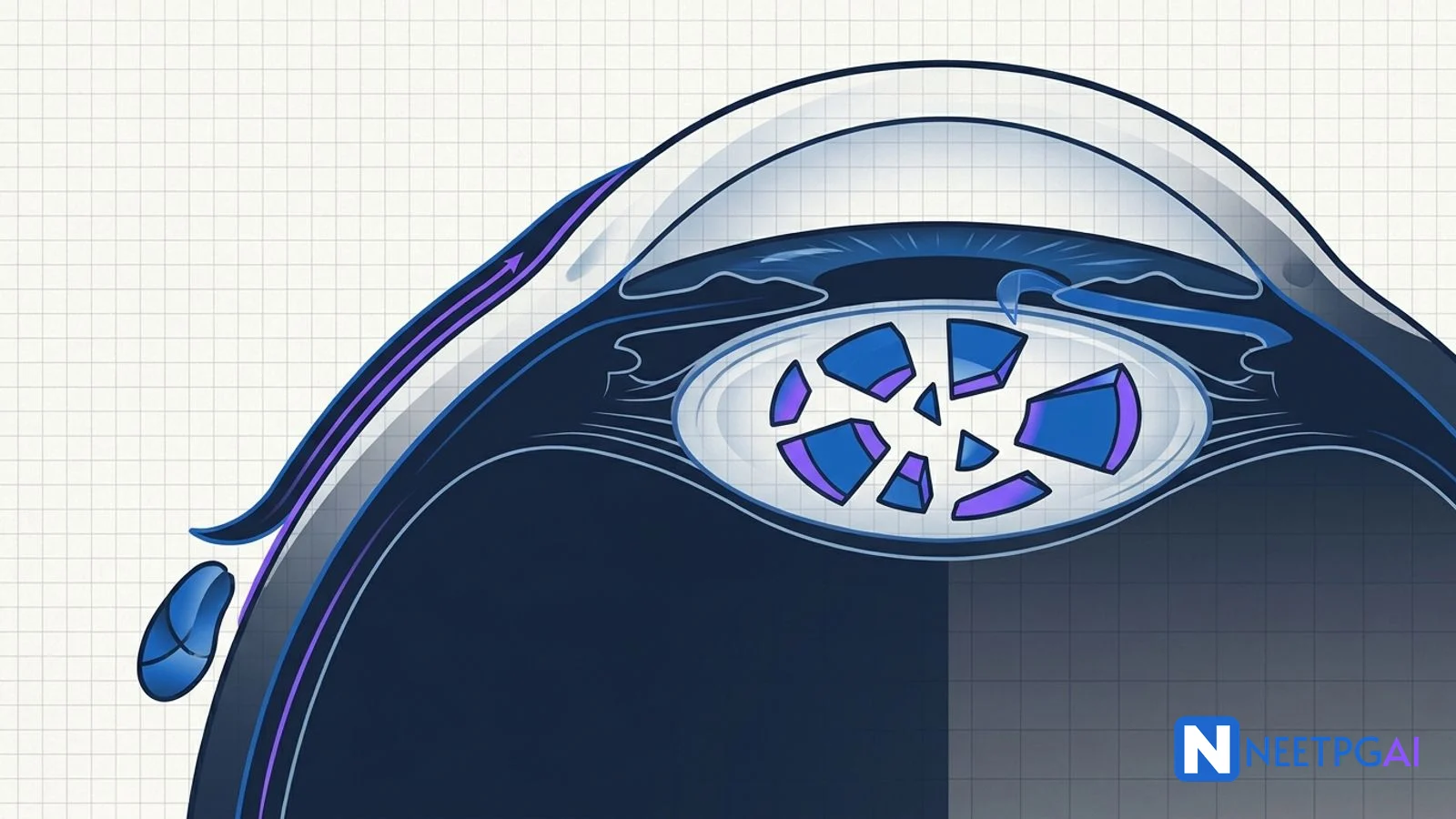
- Scleral tunnel - a 6.5-7 mm chord length frown or straight incision 1.5-2 mm behind the limbus, dissected forward into clear cornea as a self-sealing valve.
- Side-port paracentesis at the limbus, 2 clock hours from the main.
- Capsulorhexis - 5-6 mm continuous curvilinear capsulorhexis (CCC) using Utrata forceps or cystotome. Trypan blue 0.06 percent stains the capsule in white cataract.
- Hydrodissection - BSS cannula injection beneath the capsule to free the nucleus.
- Nucleus prolapse into the anterior chamber - gentle pressure or hydroprolapse. Viscoelastic (methylcellulose or sodium hyaluronate) placed above and below the nucleus to protect endothelium and posterior capsule.
- Nucleus delivery - four classical techniques:
- Viscoexpression: viscoelastic injected behind the nucleus pushes it out through the tunnel.
- Fishhook (Blumenthal): a Sinskey hook engages the nucleus and pulls it out.
- Phacosandwich (Fry technique): nucleus held between a Sinskey hook above and a lens loop below, delivered as a sandwich.
- Irrigating vectis: nucleus floated out on a vectis using BSS pressure.
- Cortical cleanup - two-way irrigation-aspiration cannula or Simcoe cannula.
- IOL implantation - rigid PMMA single-piece 6.0 mm optic in the bag.
- Wound closure - the self-sealing tunnel usually needs no suture; hydrate the paracentesis to seal.
Mean surgically induced astigmatism with a 6.5 mm temporal tunnel is 1.0-1.5 D, functionally equivalent to a well-done phaco.
Step 3 - Phacoemulsification Technique
Phacoemulsification is the global gold standard where resources and corneal health permit:
- Topical anaesthesia (proparacaine or lignocaine 2 percent jelly) +/- intracameral preservative-free lignocaine 1 percent 0.1 mL.
- Clear corneal incision - 2.2-2.8 mm trapezoidal keratome at the temporal limbus. Smaller incisions (micro-coaxial 1.8 mm) are also used.
- Side-port paracentesis at 90 degrees from the main.
- Capsulorhexis - 5.0-5.5 mm CCC, slightly smaller than the IOL optic for 360 degree overlap.
- Hydrodissection and hydrodelineation (separates nucleus from epinucleus).
- Nucleus removal - choose a technique by cataract hardness:
- Divide and conquer (Gimbel) - trainee-friendly; sculpt a central trench, crack the nucleus into quadrants, emulsify each. Best for soft to moderate (NC 2-3) cataracts.
- Phaco chop (Nagahara, horizontal; Koch, vertical) - chop the nucleus with a chopper against the phaco tip; faster, lower ultrasound energy. Best for moderate-to-hard (NC 3-5) cataracts and endothelial preservation.
- Stop and chop (Koch) - sculpt a single central groove, crack the nucleus into two halves, then chop each half; hybrid of the above two.
- Cortex removal - bimanual irrigation-aspiration.
- IOL implantation - foldable acrylic single-piece or 3-piece in the bag through the 2.8 mm incision using an injector.
- Wound closure - stromal hydration; 10-0 nylon suture only if leaking.
Phacoemulsification consumables: phaco needle tip, infusion sleeve, I/A tip, viscoelastic (OVD), foldable IOL plus injector cartridge - approximately Rs 3000-15000 per case versus Rs 500-2000 for SICS.
Step 4 - IOL Types and Selection
A structured matrix:
| IOL Type | Material | Design | Best Patient | Watch For |
|---|
| Monofocal PMMA | PMMA rigid | Single-piece 6.0 mm | SICS, cost-sensitive | Needs 6.5 mm incision |
| Monofocal acrylic foldable | Hydrophobic acrylic | Square-edge single-piece | Standard phaco | Default modern choice |
| Monofocal 3-piece | Hydrophobic acrylic optic, PMMA haptics | Haptic length 13 mm | Sulcus placement, large bag | Rotational stability |
| Toric monofocal | Hydrophobic acrylic | Axis markings on IOL | Cornea >1.0-1.5 D cylinder | Axis alignment critical; 1 degree off = 3.3 percent loss |
| Multifocal diffractive | Hydrophobic acrylic | Concentric diffractive rings | Motivated patients, spectacle-free | Halos, glare, loss of contrast |
| Multifocal refractive | Hydrophobic acrylic | Zonal refractive | Similar to above | Pupil size dependent |
| Trifocal | Hydrophobic acrylic | Distance + intermediate + near | Highly motivated | Dysphotopsia 10-20 percent |
| EDOF (Symfony, Vivity) | Hydrophobic acrylic | Echelette diffractive or non-diffractive | Distance + intermediate with fewer halos | Still needs reading glasses for fine near |
Absolute contraindications to multifocal/trifocal IOLs: age-related macular degeneration, glaucomatous optic neuropathy with significant field loss, significant irregular astigmatism, previous refractive surgery with unknown K-readings, large scotopic pupils, significant posterior capsule plaque, professional night driver.
Step 5 - Intraoperative Complications
Posterior Capsule Rupture (PCR)
Incidence 1-5 percent in expert hands, up to 10-15 percent in trainees. Signs during phaco: sudden deepening of the chamber, pupil snap, iris flutter, loss of followability, vitreous presenting at the pupil. Management:
- Do not withdraw the phaco tip rapidly - keep infusion on, maintain chamber.
- Inject dispersive viscoelastic (HPMC or sodium hyaluronate) to tamponade vitreous posteriorly.
- Stop phaco; convert to manual cortex removal.
- Perform limited anterior vitrectomy - cutter rate 800-1500 cpm, vacuum 100-200 mmHg, low infusion. Dry vitrectomy preferred.
- Triamcinolone acetonide 2-4 mg/mL stains residual vitreous white and makes it visible.
- Retrieve all nucleus and cortex. If a large fragment drops into vitreous - stop, close, refer for pars plana vitrectomy within 7 days.
- IOL choice after PCR: a 3-piece IOL in the sulcus (never a single-piece acrylic in sulcus - haptic bulk causes uveitis-glaucoma-hyphema). If capsulorhexis is intact, perform optic capture (sulcus haptics, optic captured through the CCC) for superior centration. If no capsular support - anterior chamber IOL or scleral-fixated IOL (glued or sutured).
Dropped Nucleus
A piece of nucleus falls through the ruptured capsule into the vitreous. Risk factors: hard cataract, pseudoexfoliation, high myopia with lax zonules, trauma. Never attempt to retrieve from the anterior approach - fishing a dropped nucleus with instruments tears retina. Refer for pars plana vitrectomy with phacofragmentation within 7-14 days to prevent fragment-induced glaucoma and uveitis.
Suprachoroidal Haemorrhage
A catastrophic bleed in the suprachoroidal space during open-globe surgery - sudden shallowing of chamber, loss of red reflex, dark mass under conjunctiva, severe pain. Risk factors: elderly, hypertensive, atherosclerotic, high myopic, aphakic. Immediate management: close the wound as fast as possible (temporary suture), raise the head, treat IOP. Later - delayed drainage via posterior sclerotomy if the eye is salvageable.
Toxic Anterior Segment Syndrome (TASS)
A sterile toxic inflammatory reaction to intraocular contaminant (detergent residue on instruments, preservative in BSS, denatured viscoelastic, endotoxin). Presents at 12-48 hours post-op with diffuse corneal oedema, severe anterior chamber reaction, raised IOP, NO vitritis, NO pain disproportionate to signs (unlike endophthalmitis). Treatment: intensive topical steroids every hour, cycloplegic, IOP control. Distinction from endophthalmitis is critical because management differs.
Step 6 - Postoperative Complications
Acute Endophthalmitis (first 6 weeks, typically 3-7 days)
- Organisms: coagulase-negative Staphylococcus (70 percent), S. aureus, Streptococcus, Pseudomonas.
- Presentation: pain disproportionate to signs, decreased vision, hypopyon, vitritis, red eye, absent red reflex.
- Management (EVS 1995 algorithm):
- Visual acuity better than hand motions: vitreous tap + intravitreal injection of vancomycin 1 mg in 0.1 mL + ceftazidime 2.25 mg in 0.1 mL (or amikacin 0.4 mg).
- Visual acuity hand motions or worse: pars plana vitrectomy + intravitreal antibiotics.
- Oral or IV antibiotics: no proven benefit in the EVS trial. Oral moxifloxacin may be used empirically.
- Topical fortified vancomycin 50 mg/mL + ceftazidime 50 mg/mL hourly.
- Prophylaxis: povidone iodine 5 percent in sac x 3 min + intracameral cefuroxime 1 mg (ESCRS 2007 - fivefold reduction).
Chronic Endophthalmitis (after 6 weeks)
- Propionibacterium acnes: indolent, white plaque on posterior capsule, mild uveitis responsive to steroids but recurring. Treatment: capsulectomy + intravitreal vancomycin +/- IOL exchange.
- Candida albicans / Aspergillus: cottony vitreous infiltrate. Treatment: intravitreal amphotericin B 5-10 microgram or voriconazole 100 microgram + pars plana vitrectomy + systemic voriconazole.
Cystoid Macular Oedema (Irvine-Gass syndrome)
Peaks at 4-6 weeks. Decreased vision, OCT shows cystic spaces in the inner nuclear and outer plexiform layers, FFA shows petaloid leakage. Treatment: topical NSAID (nepafenac or ketorolac) + topical steroid; intravitreal triamcinolone or anti-VEGF if refractory. Prophylactic topical NSAID in diabetics and uveitic eyes.
Posterior Capsular Opacification (PCO)
Commonest late complication (10-50 percent at 2-5 years). Fibrotic (opaque) or regeneratory (Elschnig pearls). Square-edge acrylic IOLs reduce incidence to under 5 percent (Nishi effect). Treatment: Nd:YAG laser capsulotomy - cruciate or circular 3-4 mm opening, 1-2 mJ pulses, total 20-80 mJ. Complications: transient IOP spike (prophylactic brimonidine), IOL pitting, CME (1-2 percent), retinal detachment (fourfold higher, especially in myopes).
Step 7 - SICS vs Phaco Decision Tree
Dense/black cataract (NC 5-6)?           -> SICS
Hard cataract + endothelial count <1500? -> SICS (or femto phaco)
Zonular weakness/PXF/trauma?             -> SICS or phaco + CTR
Shallow AC/nanophthalmos?                -> SICS
Clear cornea + NC 2-3 + good endothelium -> Phaco (gold standard)
Need rapid visual recovery, high astigmatism tolerance low? -> Phaco + toric
Rural/high-volume/cost-constrained?      -> SICS
Trainee learning?                        -> SICS first, phaco later
High-Yield One-Liners
- ESCRS 2007 trial: intracameral cefuroxime 1 mg in 0.1 mL reduces endophthalmitis fivefold.
- Povidone iodine 5 percent in conjunctival sac for 3 minutes is the single most evidence-based prophylaxis.
- EVS 1995 trial: PPV only if VA worse than hand motions; tap-and-inject otherwise. No benefit from systemic antibiotics.
- Commonest organism in acute post-cataract endophthalmitis: coagulase-negative Staphylococcus (70 percent).
- Chronic endophthalmitis organism: Propionibacterium acnes (indolent, white plaque on capsule).
- PCR incidence: 1-5 percent in experts, 10-15 percent in trainees.
- Dropped nucleus needs pars plana vitrectomy within 7-14 days.
- Nishi effect: square-edge acrylic IOL optic prevents LEC migration, reducing PCO.
- Nd:YAG capsulotomy: 1-2 mJ pulses, 3-4 mm opening, cruciate pattern. Prophylactic brimonidine for IOP spike.
- Toric IOL: 1 degree axis misalignment loses 3.3 percent cylinder correction.
- Multifocal IOL contraindicated in AMD, glaucoma with field loss, large pupils, night drivers.
- TASS vs endophthalmitis: TASS = sterile, 12-48 hrs, no pain disproportionate, no vitritis.
- Phaco-chop uses less ultrasound energy than divide-and-conquer - endothelial friendly.
- SICS induced astigmatism: 1.0-1.5 D with 6.5 mm temporal tunnel.
- CCC size: 5-5.5 mm (slightly smaller than optic for 360 degree overlap and in-the-bag retention).
Common Mistakes
- Confusing TASS with endophthalmitis - starting intravitreal antibiotics in a sterile toxic reaction (TASS has NO vitritis, NO disproportionate pain, presents at 12-48 hrs; endophthalmitis presents at 3-7 days with vitritis and hypopyon).
- Placing a single-piece acrylic IOL in the sulcus - single-piece haptics are bulky and cause uveitis-glaucoma-hyphema syndrome. Always use a 3-piece IOL for sulcus fixation.
- Attempting to retrieve a dropped nucleus from the anterior approach - instruments passing through the capsular opening into the vitreous cause retinal tears. Close the eye and refer for pars plana vitrectomy.
- Starting systemic antibiotics in post-cataract endophthalmitis - the EVS 1995 trial showed no benefit from oral or IV antibiotics. Intravitreal antibiotics are the mainstay.
- Choosing multifocal IOL in a patient with age-related macular degeneration - contrast loss from the multifocal design worsens the already compromised macula; always rule out AMD, glaucoma, and irregular astigmatism before offering premium IOLs.
Related Topics on NEETPGAI
Practice This on NEETPGAI
Cataract surgery is a recurring INI-CET favourite - PCR management, endophthalmitis prophylaxis bundles, and IOL selection appear almost every year. Build your clinical reasoning on NEETPGAI's free 28,000+ MCQ bank, which includes real previous year questions with step-by-step explanations. Sign up free and start with the ophthalmology practice set today.