Version 1.0 - Published October 2026
Quick Answer
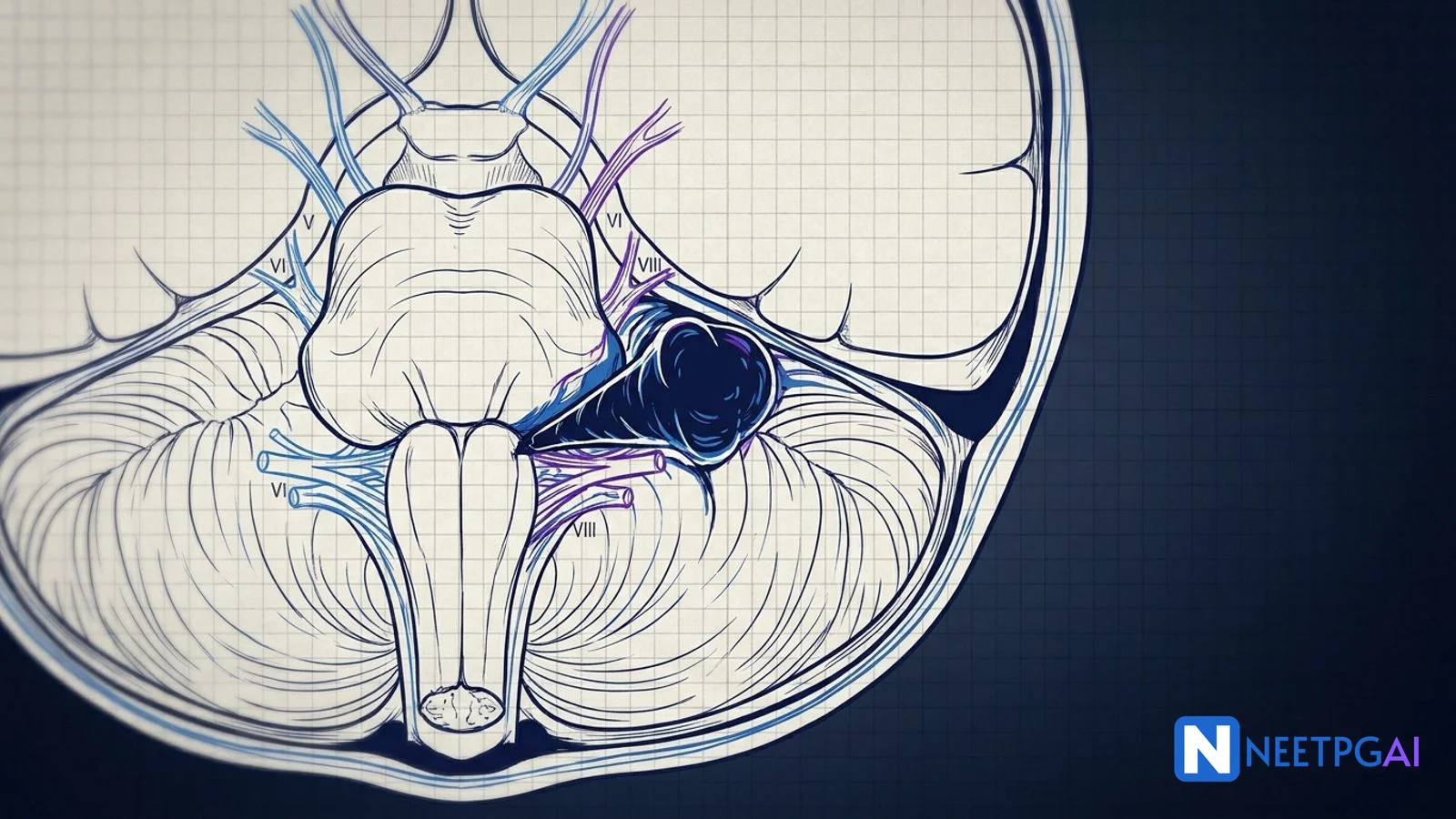
Acoustic neuroma is a benign CPA mass causing unilateral SNHL, tinnitus, and imbalance - diagnose with gadolinium MRI ("ice-cream cone" sign) and treat by observation, stereotactic radiosurgery, or microsurgery depending on size and hearing. Cholesteatoma is a destructive squamous epithelial sac in the middle ear/mastoid that classically produces an "unsafe" attic or marginal perforation with foul scanty discharge and bone erosion on CT - the definitive management is modified radical or canal-wall-down mastoidectomy. Glomus tumors (paragangliomas) are hypervascular lesions arising from middle ear (tympanicum - red mass behind TM, pulsatile tinnitus, Brown sign) or jugular foramen (jugulare - cranial nerve IX-XII palsies); staged by Fisch classification and treated by preoperative embolization plus surgical excision.
Why This Matters for NEET PG and INI-CET
ENT contributes roughly 10-12 questions per NEET PG paper, and otology dominates the subject - acoustic neuroma, cholesteatoma, and glomus tumors appear almost every year because they integrate radiology, cranial nerve examination, operative anatomy, and medicolegal nuance (hearing preservation decisions, consent for facial nerve risk). INI-CET and FMGE lean even harder on image-based identification of CPA masses on MRI and attic retraction pockets on otoscopy clinical photographs.
The three tumors in this guide also show up in the Medicine, Neurology, and Radiology components as part of CPA mass differentials and chronic suppurative otitis media (CSOM) complications. The Supreme Court mandated 1 October 2026 NBEMS answer key for the 30 August 2026 NEET PG paper confirmed at least two otology items keyed on these conditions, so recent trend data points firmly to high testing frequency.
Expect clinical vignettes that stem from pure-tone audiometry readouts, otoscopy clinical photographs, CT temporal bone cuts, MRI gadolinium sequences, and operative anatomy. The highest-yield traps are: confusing CPA masses, distinguishing safe from unsafe perforations, and getting Fisch classification wrong because candidates mix up Types B and C.
Acoustic Neuroma (Vestibular Schwannoma)
Pathology and Site of Origin
Despite the name, acoustic neuroma arises from the Schwann cells of the vestibular (not cochlear) division of CN VIII, specifically the superior vestibular nerve in roughly 90% of cases. It originates at the Obersteiner-Redlich zone - the glial-Schwann cell junction within the internal auditory canal. Growth is slow (1-2 mm per year in most, up to 10 mm per year in a small subset) and histology shows Antoni A (compact Verocay bodies) and Antoni B (loose myxoid) areas on microscopy.
Unilateral tumors are sporadic; bilateral tumors are pathognomonic of neurofibromatosis type 2 (NF2), an autosomal dominant disorder from the merlin (NF2) gene on chromosome 22q12. NF2 patients typically present younger (teens to twenties) and have associated meningiomas, ependymomas, and juvenile posterior subcapsular lens opacities (MISME syndrome - Multiple Inherited Schwannomas, Meningiomas, and Ependymomas).
Clinical Features
The classic triad is unilateral SNHL, tinnitus, and disequilibrium. Growth into the CPA produces predictable cranial nerve compression:
| Tumor Size | Clinical Feature |
|---|
| Intracanalicular (less than 10 mm) | Pure SNHL, tinnitus, maybe mild imbalance |
| Small CPA (10-15 mm) | Add trigeminal numbness, decreased corneal reflex |
| Medium (15-25 mm) | Facial numbness, headache, cerebellar signs |
| Large (25-40 mm) | Brainstem compression, hydrocephalus, long-tract signs |
| Giant (greater than 40 mm) | Lower cranial nerves IX-XII, coma |
The corneal reflex is the earliest sign of trigeminal involvement and is more sensitive than facial sensation testing. Facial nerve weakness is a very late finding - early facial palsy should prompt consideration of facial nerve schwannoma or malignancy.
Workup
Audiometry - retrocochlear pattern: asymmetric high-frequency SNHL, word recognition score (WRS) worse than expected for pure-tone average, rollover (WRS declines with louder stimuli), absent acoustic reflexes, and reflex decay.
BAERA - prolonged wave I-III interpeak latency, interaural wave V latency greater than 0.2 ms, and absent or distorted waves. BAERA sensitivity is 90-95% for tumors greater than 1 cm but misses purely intracanalicular lesions.
Gadolinium-enhanced MRI - gold standard. Shows an intensely enhancing mass with the characteristic ice-cream cone sign (intracanalicular stem plus CPA scoop). CISS or FIESTA sequences give exquisite anatomic detail of IAC contents without contrast.
Vestibular testing (caloric, VNG) - reduced response on the affected side but not required for diagnosis.
Treatment Options
Observation - serial MRI every 6-12 months. Appropriate for small tumors in elderly, asymptomatic patients, or tumors in the only hearing ear. About 50% of small lesions do not grow.
Stereotactic radiosurgery (SRS) - Gamma Knife or CyberKnife, marginal dose 12-13 Gy. Control rates greater than 90% at 10 years for tumors up to 2.5-3 cm. Hearing preservation 50-70% at 5 years. Not curative; tiny risk of radiation-induced malignancy.
Microsurgical excision - three approaches:
| Approach | Indication | Trade-off |
|---|
| Translabyrinthine | Non-serviceable hearing, any tumor size | Sacrifices hearing, best facial nerve exposure |
| Retrosigmoid (suboccipital) | Hearing preservation attempt, any size | Longer approach, cerebellar retraction |
| Middle fossa | Small intracanalicular tumors (less than 1.5 cm), good hearing | Risk of temporal lobe retraction, facial nerve injury |
House-Brackmann grading is used to score postoperative facial function (I-VI). Target preservation is grade I-II.
Cholesteatoma
Definition and Classification
Cholesteatoma is NOT a true tumor - it is an abnormal collection of keratinizing stratified squamous epithelium in the middle ear or mastoid air cell system. The epithelium relentlessly accumulates keratin debris, generates bone-resorbing enzymes and cytokines, and erodes adjacent structures. Classification:
Congenital - behind an intact tympanic membrane in a child with no prior otologic history. Typically a pearly white mass in the anterosuperior quadrant. Levenson criteria define it.
Acquired primary - from retraction of the pars flaccida (Shrapnell membrane) due to chronic Eustachian tube dysfunction. Progresses through Tos staging (I-IV).
Acquired secondary - squamous epithelium migrates through a pre-existing marginal perforation of pars tensa.
Safe vs Unsafe Perforations
| Feature | Safe (Tubotympanic, Central) | Unsafe (Atticoantral, Marginal) |
|---|
| Location | Pars tensa, central | Pars flaccida (attic) or marginal |
| Margin | Rim of TM visible all around | Extends to annulus, no rim |
| Discharge | Mucoid, non-foul, profuse | Scanty, foul, cheesy |
| Granulation | Rare | Common, aural polyp |
| Cholesteatoma | Absent | Essentially always present |
| Bone erosion | No | Yes (scutum, ossicles, LSCC) |
| Complications | Rare | Facial palsy, LSCC fistula, meningitis, brain abscess |
| Management | Conservative then tympanoplasty | Urgent CT and mastoidectomy |
The Shrapnell membrane (pars flaccida) sits above the lateral process of the malleus and lacks the middle fibrous layer, which is why it is prone to retraction.
Clinical Features and Examination
Scanty foul-smelling otorrhea plus progressive conductive hearing loss is the hallmark. Vertigo on tragal pressure (positive fistula sign - Hennebert sign) suggests a lateral semicircular canal fistula. Facial twitching or weakness means facial nerve canal erosion. Severe deep otalgia and headache suggest intracranial extension.
On otoscopy look for: pars flaccida retraction pocket, white pearly debris in the attic, posterosuperior marginal perforation, granulation tissue, aural polyp, and ossicular erosion visible through a large perforation.
Imaging - CT Temporal Bone
High-resolution non-contrast CT in axial and coronal planes is standard preoperative imaging. Classic findings:
- Soft tissue mass in Prussak's space (attic)
- Scutum erosion (the lateral attic wall)
- Ossicular erosion - most commonly the long process of the incus, then the stapes superstructure, then the malleus head
- Lateral semicircular canal fistula (bony defect in LSCC)
- Tegmen tympani or tegmen mastoideum dehiscence (risk of CSF leak and meningitis)
- Facial canal dehiscence (tympanic segment most common)
- Sigmoid sinus plate erosion (risk of thrombosis)
MRI with diffusion-weighted imaging (DWI non-EPI) is adjunctive - cholesteatoma restricts diffusion (bright on DWI) and distinguishes it from granulation or scar tissue in revision surgery.
Surgical Management
The surgical principle is to eradicate disease, obtain a safe dry ear, and preserve or reconstruct hearing. Options:
| Procedure | Features | Indication |
|---|
| Atticotomy | Removes scutum, exposes attic | Localized attic cholesteatoma |
| Cortical mastoidectomy | Opens mastoid air cells, posterior canal wall intact | Acute mastoiditis, cochlear implant access |
| Canal-wall-up tympanomastoidectomy (CWU) | Preserves posterior canal wall, better anatomy, higher recurrence | Limited disease, well-pneumatized mastoid |
| Canal-wall-down (CWD) / Modified radical mastoidectomy | Removes posterior canal wall, creates common cavity | Extensive disease, poor pneumatization, revision |
| Radical mastoidectomy | Historical, no hearing reconstruction | Rarely done now |
Second-look surgery at 9-18 months (or MRI DWI follow-up) is standard after CWU because residual disease rates reach 20-30%. Ossiculoplasty with PORP (partial) or TORP (total) ossicular replacement prosthesis restores conductive hearing.
Glomus Tumors (Paragangliomas)
Origin and Types
Glomus tumors are hypervascular, slow-growing paragangliomas arising from chemoreceptor paraganglia of the autonomic nervous system in the temporal bone and skull base. Four types based on origin:
| Type | Origin | Classic Finding |
|---|
| Glomus tympanicum | Jacobson's nerve (tympanic branch of CN IX) on cochlear promontory | Red mass behind TM, pulsatile tinnitus, positive Brown sign |
| Glomus jugulare | Jugular bulb adventitia (Arnold's nerve, branch of CN X) | CN IX-XII palsies (jugular foramen syndrome) |
| Glomus vagale | Nodose ganglion of CN X in parapharyngeal space | High vagal palsy, pulsatile neck mass |
| Carotid body tumor | Carotid bifurcation | Painless slow-growing neck mass, splays ICA from ECA (lyre sign) |
Multicentricity occurs in 10% of sporadic cases and up to 80% of familial cases. Familial paragangliomas (SDHB, SDHC, SDHD, SDHAF2 mutations) are autosomal dominant and may be functional (catecholamine-secreting). Screening includes 24-hour urinary metanephrines and genetic testing.
Clinical Features
Glomus tympanicum - unilateral pulsatile tinnitus (synchronous with heartbeat), conductive hearing loss, aural fullness. Otoscopy shows a pulsatile red-blue mass behind an intact TM. The Brown sign is pathognomonic: pneumatic otoscopy with positive pressure blanches the mass, which then reperfuses. The rising sun sign - a mass rising above the inferior annulus - supports the diagnosis.
Glomus jugulare - as above plus cranial nerve palsies. Jugular foramen syndrome (Vernet syndrome) involves CN IX, X, XI - dysphagia, hoarseness, aspiration, shoulder droop. Add CN XII (Collet-Sicard syndrome) if hypoglossal canal is involved. Horner syndrome if sympathetic chain is affected (Villaret syndrome).
Investigations
| Test | Finding |
|---|
| CT temporal bone | Enhancing mass; permeative "moth-eaten" bone erosion of jugular foramen, carotid canal, or hypotympanum |
| MRI with gadolinium | T1 hypointense, T2 hyperintense, intense enhancement; characteristic "salt-and-pepper" appearance from flow voids (greater than 2 cm) |
| MRA / CTA | Hypervascular mass; defines carotid encasement |
| Four-vessel angiography | Phoenix sign (persistent intense tumor blush); essential for preoperative embolization |
| Octreotide scintigraphy / 68Ga-DOTATATE PET | Detects multicentric and metastatic disease |
| 24-hour urinary metanephrines, catecholamines | Rules out functional (secretory) tumor |
Fisch Classification
| Type | Extent | Approach |
|---|
| A | Middle ear only (glomus tympanicum) | Transcanal or transmastoid |
| B | Tympanomastoid, no infralabyrinthine extension | Extended transmastoid |
| C | Infralabyrinthine, into carotid canal (C1-C4 subtypes) | Fisch Type A infratemporal fossa approach |
| D | Intracranial extension (D1 less than 2 cm, D2 2-4 cm, D3 unresectable) | Staged skull base approach plus SRS |
Treatment Principles
- Preoperative embolization 24-48 hours before surgery to reduce blood loss (polyvinyl alcohol particles, Onyx).
- Surgical resection via Fisch infratemporal fossa approach - may require anterior transposition of the facial nerve and ligation of the internal jugular vein and sigmoid sinus.
- Stereotactic radiosurgery for small tumors in poor surgical candidates, residual or recurrent disease. Control rates greater than 90%.
- Observation for elderly asymptomatic patients - some tumors grow very slowly.
Alpha-adrenergic blockade (phenoxybenzamine) is given 10-14 days preoperatively for functional tumors to prevent intraoperative hypertensive crisis.
CPA Mass Differential - The Must-Know Table
| Lesion | Frequency | Key Imaging Feature |
|---|
| Vestibular schwannoma | 80-90% | Ice-cream cone, enhances brightly, widens IAC |
| Meningioma | 5-10% | Dural tail sign, broad dural base, calcification, hyperostosis |
| Epidermoid cyst | 5% | DWI restricted (bright), does not enhance, insinuates around structures |
| Arachnoid cyst | 1% | Follows CSF on all sequences, no enhancement |
| Facial nerve schwannoma | Rare | Along CN VII course, labyrinthine segment widened |
| Metastasis | Rare | Multiple, enhances variably, known primary |
The epidermoid cyst is the classic trap - it mimics a schwannoma on T1/T2 but does not enhance and lights up on DWI.
High-Yield One-Liners
- Acoustic neuroma arises from the superior vestibular nerve at the Obersteiner-Redlich zone.
- Ice-cream cone sign on gadolinium MRI is pathognomonic of vestibular schwannoma.
- Bilateral acoustic neuromas equal neurofibromatosis type 2 (NF2) - chromosome 22q12, merlin gene.
- Early corneal reflex loss is the most sensitive sign of CN V involvement in CPA mass.
- Facial nerve weakness is a LATE sign in acoustic neuroma - early CN VII palsy suggests facial schwannoma or malignancy.
- Translabyrinthine approach sacrifices hearing but offers the best facial nerve exposure.
- Middle fossa approach is for small intracanalicular tumors less than 1.5 cm with serviceable hearing.
- SRS marginal dose for vestibular schwannoma is 12-13 Gy, 10-year control greater than 90%.
- Safe perforation sits in the pars tensa centrally; unsafe perforation sits in the pars flaccida (attic) or margin of pars tensa.
- Cholesteatoma erodes the long process of incus first (most common ossicular erosion).
- Positive fistula sign (Hennebert sign) in CSOM means lateral semicircular canal fistula.
- Prussak's space is the attic recess bounded by pars flaccida laterally and the neck of malleus medially.
- CWD mastoidectomy removes the posterior canal wall; CWU preserves it.
- MRI DWI (non-EPI) shows cholesteatoma as bright - distinguishes from granulation on second-look.
- Glomus tympanicum arises from Jacobson's nerve on the promontory.
- Positive Brown sign (blanching on pneumatic otoscopy) is pathognomonic of glomus tympanicum.
- Phoenix sign on angiography means persistent tumor blush despite feeder embolization.
- Fisch Type A glomus is middle-ear-only; Type D has intracranial extension.
- Alpha-blockade (phenoxybenzamine) is given preoperatively for functional glomus tumors.
- SDHB mutations in paragangliomas carry the highest malignancy risk.
Common Mistakes
-
Treating unilateral SNHL without imaging - any adult with asymmetric sensorineural hearing loss (greater than 15 dB difference at any frequency, or asymmetric word recognition) needs gadolinium MRI to rule out vestibular schwannoma, even if presbycusis seems likely.
-
Confusing safe and unsafe perforations in exam vignettes - the examiner tests location (central vs attic/marginal), not size. A large central perforation with mucoid discharge is safe; a tiny attic retraction with scanty foul discharge is unsafe.
-
Over-reading pulsatile tinnitus as benign - unilateral pulsatile tinnitus plus a red mass behind the TM is glomus tympanicum until proven otherwise. Never biopsy transcanal without imaging first - torrential bleeding has been fatal.
-
Forgetting NF2 screening in young or bilateral cases - patients less than 30 years with acoustic neuroma or any patient with bilateral tumors need NF2 genetic testing, screening MRI of the spine, and ophthalmology referral for juvenile posterior subcapsular cataracts.
-
Mis-staging glomus jugulare as glomus tympanicum - if the mass extends below the level of the inferior tympanic ring or if CN IX-XII palsies are present, it is Fisch Type C or D glomus jugulare, not Type A. The approach, embolization strategy, and prognosis differ substantially.
Related Topics on NEETPGAI
Practice This on NEETPGAI
Lock in these three otology tumors with real NEET PG and INI-CET PYQs, image-based MCQs with annotated CT and MRI stems, and spaced-repetition revision cards.
Start free on NEETPGAI - 28,000+ MCQs, authentic PYQs, AI tutor to break down any ice-cream cone, scutum erosion, or Fisch classification question until it clicks.