Version 1.0 - Published October 2026
Quick Answer
Seven high-yield pediatric dermatology image MCQs covering neonatal rashes (erythema toxicum neonatorum, miliaria rubra, mongolian spot, infantile seborrheic dermatitis/cradle cap) and school-age dermatoses (atopic dermatitis age-specific distribution, molluscum contagiosum, impetigo contagiosa, scabies burrows). Pattern-match the lesion morphology to age and distribution - that is 90 percent of the exam yield.
Why This Matters for NEET PG and INI-CET
Pediatric dermatology image MCQs appear every NEET PG cycle. The exam loves this topic because the diagnosis is visual, the differentials are tight, and the age-specific morphology creates clean right-and-wrong distinctions. INI-CET additionally probes the pathophysiology (eosinophils in erythema toxicum, Wright stain, exfoliative toxin in bullous impetigo, mite scybala in scabies).
The high-yield matrix for your mental image-bank:
- Neonatal rashes present in first 48 hours: erythema toxicum neonatorum (eosinophils), transient neonatal pustular melanosis (neutrophils, dark-skinned infants), milia (keratin cysts), miliaria (blocked sweat ducts), mongolian spot (dermal melanocytosis).
- Infantile rashes: cradle cap (seborrheic, scaly scalp), infantile atopic dermatitis (facial cheeks, extensor limbs).
- School-age rashes: atopic dermatitis flexural, impetigo contagiosa (honey crust), molluscum contagiosum (umbilicated papules), scabies (interdigital burrows), tinea capitis.
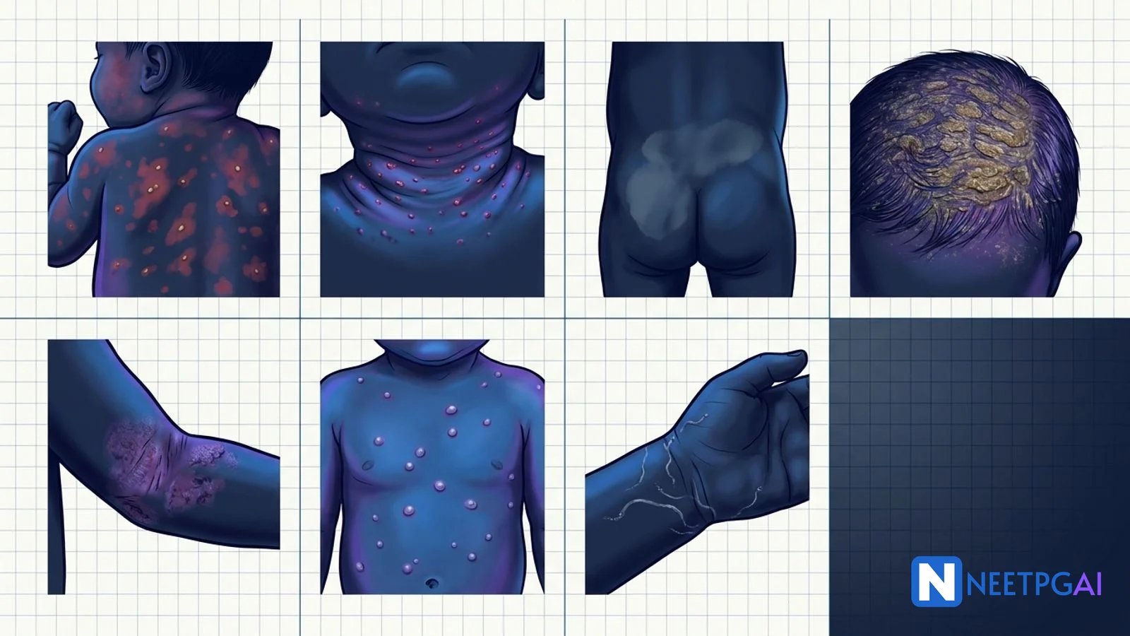
Every image MCQ in this set follows the exam-shape: a clinical scenario and a prompt-image description, four options, correct answer and detailed explanation with the high-yield one-liner you will carry into the exam hall.
MCQ 1 - Erythema Toxicum Neonatorum
Clinical scenario: A 2-day-old term baby boy born by normal vaginal delivery is noted by the paediatric resident to have a rash. On examination there are 2-3 mm erythematous macules with central yellow-white pustules scattered over the trunk, face and proximal limbs. Palms and soles are spared. The baby is feeding well, afebrile and alert.
Which stain of pustule contents will confirm the diagnosis?
A. Gram stain showing cocci in clusters
B. Wright stain showing eosinophils
C. Tzanck smear showing multinucleate giant cells
D. KOH preparation showing hyphae
Correct answer: B. Wright stain showing eosinophils.
Explanation: Erythema toxicum neonatorum (ETN) is the commonest benign neonatal rash, affecting 40-70 percent of term newborns, typically appearing on days 1-2 and resolving spontaneously by 7-14 days. The pustule contains sterile eosinophilic infiltrate - Wright stain demonstrating eosinophils is the diagnostic finding. Gram stain positive cocci would suggest neonatal staphylococcal pustulosis or bullous impetigo. Tzanck smear with multinucleate giant cells identifies herpes simplex. KOH hyphae indicates candida or dermatophyte. ETN is a diagnosis of inspection - no treatment, no workup needed if the baby is otherwise well.
One-liner: Flea-bite rash in first 48 hours + sterile eosinophilic pustules = erythema toxicum neonatorum. Observe only.
MCQ 2 - Miliaria Rubra (Prickly Heat)
Clinical scenario: A 5-week-old infant presents in June in Chennai (hot humid climate) with multiple 1-2 mm erythematous papulovesicles over the forehead, neck, upper chest and groin skin folds. The baby is restless, sweating and the mother reports swaddling with multiple layers.
What is the pathogenesis?
A. Blocked eccrine sweat ducts with sweat extravasation into the epidermis
B. Blocked apocrine ducts producing follicular pustules
C. Superantigen-mediated epidermal cleavage
D. IgE-mediated mast cell degranulation
Correct answer: A. Blocked eccrine sweat ducts with sweat extravasation into the epidermis.
Explanation: Miliaria rubra (prickly heat) is caused by obstruction of eccrine sweat ducts at the level of the stratum corneum, leading to sweat leakage into the surrounding epidermis and dermis with resultant inflammation. Four variants by depth of obstruction: miliaria crystallina (most superficial, clear vesicles like dewdrops, blocked at stratum corneum surface, no inflammation); miliaria rubra (intermediate, erythematous papulovesicles with pruritus and burning, blocked at deeper epidermis); miliaria pustulosa (secondary pustulation); miliaria profunda (deepest, blocked at dermal-epidermal junction, flesh-coloured papules).
Treatment is cooling - reduce ambient temperature, lighter clothing, air circulation, lukewarm baths, and avoid heavy occlusive emollients. Topical calamine or 1 percent hydrocortisone briefly for pruritus. Lesions resolve within days of cooling.
One-liner: Hot climate + swaddled infant + 1-2 mm red papulovesicles in sweat distribution = miliaria rubra from blocked eccrine ducts.
MCQ 3 - Mongolian Spot (Dermal Melanocytosis)
Clinical scenario: A 1-day-old baby girl of Indian descent has large flat bluish-grey patches over the lumbosacral region extending to the buttocks, measuring 8 cm by 10 cm. The surface is smooth without induration. The paediatrician must counsel the parents and document the finding.
Which of the following is TRUE about this lesion?
A. It is a precursor to melanoma and requires excision
B. It is dermal melanocytosis that typically resolves by school age
C. It is a sign of child abuse (bruising) and should be reported
D. It indicates spinal dysraphism and requires MRI
Correct answer: B. It is dermal melanocytosis that typically resolves by school age.
Explanation: Mongolian spot (now called congenital dermal melanocytosis) is a benign birthmark caused by melanocytes trapped in the dermis during their migration from the neural crest to the epidermis in utero. The Tyndall effect scatters shorter blue wavelengths and gives the characteristic bluish-grey colour. Prevalence varies by ethnicity: 80-90 percent in East Asian, Native American and sub-Saharan African infants; 60-70 percent in Indian infants; 10 percent in Caucasian infants.
Most Mongolian spots are over the lumbosacral region and buttocks, less commonly on shoulders and backs. They fade and typically resolve by age 3-6 years, but larger extrasacral or atypical lesions may persist and warrant evaluation for GM1 gangliosidosis, Hurler syndrome and other lysosomal storage disorders. No treatment is required for typical lesions.
Documentation is critical because Mongolian spots are frequently mistaken for bruising in child abuse evaluations - always photograph, measure and record at birth to avoid false abuse accusations later. MRI is NOT indicated unless there is additional midline anomaly (hair tuft, dimple, haemangioma over spine) suggesting dysraphism.
One-liner: Bluish-grey flat patch over lumbosacral region in Indian/Asian/African infant = Mongolian spot (dermal melanocytosis); benign, document to prevent false abuse report.
MCQ 4 - Infantile Seborrheic Dermatitis (Cradle Cap)
Clinical scenario: A 6-week-old infant presents with greasy yellow adherent scales covering the scalp vertex and extending onto the forehead and behind the ears. There are also mild salmon-coloured patches in the axillae and diaper area. The infant is not itching and feeding well.
What is the organism implicated and the first-line treatment?
A. Candida albicans - topical nystatin
B. Malassezia furfur - gentle shampoos and emollient softening
C. Staphylococcus aureus - oral cephalexin
D. Sarcoptes scabiei - permethrin 5 percent cream
Correct answer: B. Malassezia furfur - gentle shampoos and emollient softening.
Explanation: Infantile seborrheic dermatitis (cradle cap on scalp) is a self-limiting benign dermatosis of the first 3-6 months of life, driven by proliferation of Malassezia (Pityrosporum) yeast on sebum-rich sites in combination with maternal androgen stimulation of sebaceous glands. Lesions are greasy yellow adherent scales on scalp vertex, forehead, retroauricular folds, eyebrows and nasolabial folds; salmon-coloured poorly demarcated patches in flexures (axillae, groin, diaper area). Pruritus is minimal to absent - a key distinguishing feature from atopic dermatitis which is intensely pruritic.
Treatment is gentle - emollient soaking with mineral oil or petrolatum applied for 30-60 minutes to soften scale, followed by gentle brushing with a soft baby brush and shampooing with a mild baby shampoo or ketoconazole 2 percent shampoo twice weekly. Topical 1 percent hydrocortisone briefly for marked inflammation. Avoid vigorous scrubbing which traumatises skin. Lesions resolve by 6-8 months.
The classic exam distinction from infantile atopic dermatitis: cradle cap is in the first 3 months, affects scalp and flexures, is greasy yellow, non-pruritic; infantile atopic dermatitis typically starts after 2 months, affects face cheeks and extensor limbs, is dry scaly erythematous, intensely pruritic with sleep disturbance.
One-liner: Greasy yellow scalp scales in a 6-week-old + axillary/diaper salmon patches + no itching = infantile seborrheic dermatitis, Malassezia-driven, resolves spontaneously.
MCQ 5 - Atopic Dermatitis Age Distribution
Clinical scenario: A 7-year-old boy with a history of childhood asthma presents with chronically pruritic lichenified plaques in both antecubital and popliteal fossae, worse at night. Secondary excoriations are present. His father has allergic rhinitis.
At which stage of childhood does atopic dermatitis classically affect FLEXURAL surfaces?
A. Infantile phase (2 months to 2 years)
B. Childhood phase (2 to 12 years)
C. Adolescent phase (over 12 years)
D. Senile phase (over 60 years)
Correct answer: B. Childhood phase (2 to 12 years).
Explanation: Atopic dermatitis has a textbook age-specific distribution that is the single most tested fact in pediatric dermatology MCQs:
| Age | Classic Distribution | Morphology |
|---|
| Infantile (2 months to 2 years) | Cheeks, forehead, scalp, extensor surfaces of arms/legs; sparing central face and diaper | Acute, weeping, erythematous |
| Childhood (2 to 12 years) | Flexures - antecubital and popliteal fossae, wrist, ankle, neck | Lichenified, excoriated, chronic |
| Adolescent/adult (over 12 years) | Hands, feet, face (eyelids, periorbital), flexures | Lichenified, dry, chronic |
Diagnosis uses Hanifin and Rajka criteria: 3 or more major features (pruritus, typical morphology and distribution for age, chronic/relapsing course, personal or family atopy) plus 3 or more of 23 minor features (xerosis, keratosis pilaris, Dennie-Morgan infraorbital fold, allergic shiners, atopic pleats, white dermatographism, elevated IgE, eosinophilia).
Treatment: emollient saturation 2-4 times daily (ceramide-based preferred); topical corticosteroid graded by site (hydrocortisone 1 percent on face only, triamcinolone 0.1 percent on body); topical calcineurin inhibitors (tacrolimus 0.03/0.1 percent, pimecrolimus) for maintenance and face; bleach baths (1/2 cup bleach in full tub twice weekly) for Staph aureus colonisation; dupilumab (anti IL-4 receptor alpha) for moderate to severe refractory disease aged 6 months and above. Second-line systemic: cyclosporine, methotrexate, azathioprine. Avoid long-term oral steroids.
One-liner: Age 2-12 + flexural lichenified pruritic plaques + atopic history = childhood atopic dermatitis.
MCQ 6 - Molluscum Contagiosum
Clinical scenario: A 5-year-old boy who swims at a public pool presents with 15 small 3-4 mm firm pearly dome-shaped papules on the trunk and axilla. On close inspection with a magnifying lens there is a central dimple on each papule.
What is the causative agent and recommended management?
A. Human papillomavirus (HPV) - cryotherapy
B. Molluscum contagiosum virus (poxvirus) - watchful waiting
C. Varicella zoster virus - oral acyclovir
D. Herpes simplex virus - topical acyclovir
Correct answer: B. Molluscum contagiosum virus (poxvirus) - watchful waiting.
Explanation: Molluscum contagiosum is caused by Molluscum contagiosum virus (MCV), a large double-stranded DNA poxvirus with four subtypes (MCV1-4). It is highly contagious, spreading via direct skin contact, fomites (towels, shared swimming pools) and autoinoculation (scratching one lesion deposits virus on fingers). The pathognomonic lesion is a 2-5 mm firm pearly dome-shaped papule with central umbilication (central dell), visible on dermoscopy with surrounding vascular pattern.
Common in school-age children, typically on trunk, limbs, axillae and face. In sexually active adults, lesions in the genital region are a sexually transmitted infection; multiple widespread atypical facial lesions in an adult should prompt HIV testing and immunosuppression evaluation. The BOTE (Beginning Of The End) sign - a surrounding dermatitis around resolving lesions - signals immune clearance and should not be aggressively treated.
Management in healthy children is watchful waiting - lesions resolve spontaneously in 6-18 months, and most treatments (cryotherapy, cantharidin, curettage) are more traumatic than the disease itself. Treatment is indicated for painful, cosmetically distressing, genital, extensive, or immunocompromised cases. Options: cantharidin applied in-clinic, cryotherapy with liquid nitrogen (risk hypopigmentation in dark skin), curettage, podophyllotoxin or imiquimod for genital lesions. Prevention: avoid sharing towels, cover lesions, treat atopic dermatitis vigorously (atopy predisposes).
One-liner: Pearly dome-shaped umbilicated papules in a school-age child = molluscum contagiosum (poxvirus); watchful waiting first-line.
MCQ 7 - Scabies
Clinical scenario: A 4-year-old girl presents with severe nocturnal itching for 3 weeks. Examination shows multiple 2-5 mm serpiginous grey-brown linear tracts in the finger web spaces, flexor wrists and axillae, with widespread excoriations. Her younger brother and mother also complain of itching.
What is the first-line treatment?
A. Oral ivermectin single dose
B. Topical permethrin 5 percent cream applied neck down overnight, repeated after 7 days
C. Topical hydrocortisone 1 percent cream twice daily
D. Topical mupirocin 2 percent cream TID for 7 days
Correct answer: B. Topical permethrin 5 percent cream applied neck down overnight, repeated after 7 days.
Explanation: Scabies is caused by Sarcoptes scabiei var. hominis, a mite that burrows into the stratum corneum where the female lays eggs. The pathognomonic clinical finding is the burrow - a 2-10 mm serpiginous or S-shaped grey-brown linear tract most visible in the finger web spaces, flexor wrists, elbows, axillae, areolae (females), waistline, scrotum/penis (males), and buttocks. In infants and young children, involvement extends to palms, soles, face and scalp - unlike adults who classically spare the head.
The hallmark symptom is severe nocturnal pruritus, worst after a hot bath, out of proportion to visible lesions. Secondary excoriations, impetiginisation and eczematous changes are common. Nodular scabies (chronic firm pruritic nodules on scrotum, penis and axillae) and crusted (Norwegian) scabies (massive mite burden in immunocompromised hosts) are important variants.
Diagnosis is clinical, confirmed by mineral oil preparation showing mites, eggs or scybala (fecal pellets); dermoscopy shows jet-with-contrail appearance. Treatment:
- First-line: Permethrin 5 percent cream applied neck down (and including scalp/face in infants under 2 years) overnight for 8-14 hours, repeated after 7 days. Safe from 2 months of age.
- Second-line / failed topical: Oral ivermectin 200 microgram/kg single dose, repeated at 7 days. For patients over 15 kg.
- Crusted scabies: Combine oral ivermectin plus topical permethrin or benzyl benzoate for multiple cycles.
Treat all household contacts simultaneously regardless of symptoms - a classic board point. Wash all clothes, bedding and towels used in the past 72 hours at over 60 degrees Celsius or seal in plastic bags for 72 hours. Pruritus can persist for 2-4 weeks after successful treatment (post-scabetic itch) - counsel the patient and use topical steroid plus antihistamine, not a second scabicide.
One-liner: Nocturnal itching + interdigital burrows + family contact itch = scabies; permethrin 5 percent cream neck down, repeat at 7 days, treat ALL contacts.
High-Yield One-Liners
- Erythema toxicum neonatorum: eosinophils on Wright stain; first 48 hours; benign.
- Transient neonatal pustular melanosis: neutrophils on Wright stain; dark-skinned infants; residual hyperpigmentation.
- Miliaria rubra: blocked eccrine ducts; hot humid climate.
- Milia: retained keratin cysts; white papules; spontaneous resolution.
- Mongolian spot: dermal melanocytosis; benign; document to avoid child abuse false flag.
- Cradle cap (infantile seborrheic dermatitis): Malassezia-driven; non-pruritic; emollient and gentle shampoo.
- Atopic dermatitis infantile phase: face cheeks + extensor limbs; sparing diaper area.
- Atopic dermatitis childhood phase: flexural (antecubital, popliteal).
- Atopic dermatitis adolescent/adult phase: hands, feet, face eyelids, flexures.
- Hanifin and Rajka criteria: 3 major + 3 of 23 minor for atopic dermatitis.
- Dennie-Morgan fold, allergic shiners, white dermatographism: atopic markers.
- Molluscum contagiosum: poxvirus; pearly umbilicated papules; watchful waiting first-line.
- BOTE sign: surrounding dermatitis = beginning of immune clearance (molluscum).
- Impetigo non-bullous: honey crust; S. aureus greater than Strep pyogenes; mupirocin topical.
- Impetigo bullous: S. aureus exfoliative toxin cleaves desmoglein 1.
- Scabies burrow: interdigital web space, flexor wrist, axilla; nocturnal itch; involves face/palms in infants.
- Permethrin 5 percent: first-line scabicide; safe from 2 months.
- Oral ivermectin 200 mcg/kg: alternative scabies; over 15 kg; repeat at 7 days.
- Treat all household contacts of scabies simultaneously - mandatory.
- Post-scabetic itch: persists 2-4 weeks after successful treatment; use steroid, not another scabicide.
Common Mistakes
- Mistaking Mongolian spot for bruising - always document in birth records; failure to do so has created false child-abuse accusations in paediatric practice.
- Treating molluscum aggressively in healthy children - watchful waiting is first-line; cryotherapy and curettage are traumatic for a self-resolving condition.
- Treating only the index case in scabies - mandatory to treat all household contacts simultaneously or recurrence is certain.
- Giving topical fluorinated corticosteroid on infant face - risks atrophy, striae, perioral dermatitis and HPA axis suppression. Only 1 percent hydrocortisone on face in infants.
- Starting a second scabicide for post-scabetic itch - post-scabetic itch persists 2-4 weeks after successful treatment and reflects immune reaction to dead mites, not treatment failure. Treat with topical steroid and antihistamine, not another course of permethrin or ivermectin.
Related Topics on NEETPGAI
Practice This on NEETPGAI
Pediatric dermatology image MCQs are a reliable 2-3 question yield on NEET PG and INI-CET every cycle. NEETPGAI's free 28,000+ MCQ bank includes real previous year image questions with step-by-step explanations and spaced-repetition revision. Sign up free and build your visual-diagnosis instinct with a daily set.