Version 1.0 — Published October 2026
Quick Answer
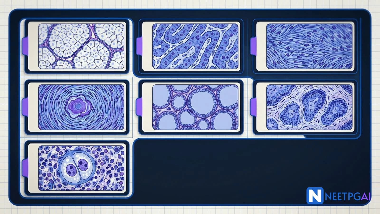
Seven classic pathology slide MCQs with full options, answers and detailed explanations — clear cell RCC, HCC trabecular, GIST spindle, meningioma whorls with psammoma, follicular thyroid carcinoma, basal cell carcinoma peripheral palisading, and Hodgkin Reed-Sternberg cells. Each slide is tested routinely on INI-CET and NEET PG. Train pattern recognition, not just facts.
Why This Matters for NEET PG & INI-CET
Image-based pathology MCQs are the single highest-yield block for recognition-speed training. On INI-CET you can expect 6 to 10 pathology image questions per paper; on NEET PG, 3 to 5. Each is worth a straight +1 (INI-CET) or +4 (NEET PG) if recognised in under 20 seconds — which frees up precious time for the longer clinical vignettes elsewhere.
The seven slides below are perennial classics. Every one of them has appeared in the AIIMS/INI-CET/NEET PG previous-year archives at least three times across 2017-2025. The pattern is predictable: identify the lesion, name the diagnostic feature, pick the correct molecular association or the correct management. Train with the full question-and-explanation workflow below — your goal is to recognise each slide in under 15 seconds and answer the follow-up in under 30.
Pathology image training also sharpens your approach to radiology, dermatology and clinical photographs — the mental habit of "name the lesion before reading the stem" generalises across all image MCQs.
How to Use This Article
Each MCQ below is formatted as:
- Image prompt (a short visual description — you will see the real slide on NEETPGAI)
- Clinical stem
- Four options
- Correct answer
- Detailed explanation
- One-liner takeaway
Set a 60-second timer per MCQ on first pass. Mark the ones you missed and revisit after reading the explanations. Do all seven, then re-do them from scratch in 10 minutes total.
MCQ 1: Clear Cell Renal Cell Carcinoma
Stem: A 62-year-old smoker presents with left flank pain and intermittent painless hematuria. CT abdomen shows an 8 cm enhancing mass arising from the upper pole of the left kidney with areas of necrosis. Nephrectomy specimen histology shows nests of polygonal cells with abundant clear cytoplasm separated by a delicate branching capillary network. Which molecular alteration is most likely driving this tumor?
A. MET gene mutation on chromosome 7
B. VHL gene inactivation on chromosome 3p25
C. Loss of chromosomes 1 and multiple others including 2, 6, 10, 13, 17, 21
D. ETV6-NTRK3 fusion
Answer: B
Explanation: The slide shows classical clear cell RCC — chicken-wire vascular network and polygonal cells with glycogen-and-lipid-rich clear cytoplasm. The molecular hallmark is VHL gene inactivation on chromosome 3p25, present in approximately 90 percent of sporadic clear cell RCC and essentially 100 percent of VHL-syndrome-associated cases. VHL loss uncouples HIF-1 alpha degradation, driving VEGF and PDGF upregulation and the hypervascular tumor phenotype — which is why anti-VEGF therapy (sunitinib, pazopanib, cabozantinib) works. MET mutations drive papillary type 1 RCC. Chromosomal losses listed in option C describe chromophobe RCC. ETV6-NTRK3 is translocation-associated RCC (pediatric).
One-liner: Chicken-wire vasculature + clear cytoplasm + VHL loss = clear cell RCC.
MCQ 2: Hepatocellular Carcinoma
Stem: A 58-year-old man with chronic hepatitis B infection and known cirrhosis presents with right upper quadrant discomfort and 10 kg weight loss over three months. Serum AFP is 2,400 ng/mL. Biopsy of a 6 cm liver lesion shows thick trabecular cords of eosinophilic tumor cells more than three cells thick with occasional bile pigment and loss of reticulin framework on silver stain. Which single immunohistochemical marker is most specific for confirming the diagnosis in a well-differentiated case where HepPar-1 is weak?
A. CK7
B. Glypican-3
C. CDX2
D. TTF-1
Answer: B
Explanation: The slide is HCC — thick trabeculae, loss of reticulin, bile within tumor cells. HepPar-1 is the standard hepatocellular marker but can be patchy in poorly differentiated or well-differentiated nodules. Glypican-3 is more specific for malignant hepatocellular lesions (positive in 70-90 percent of HCC, negative in cirrhosis and dysplastic nodules), making it the single best confirmatory IHC in diagnostically difficult cases. Arginase-1 is also highly sensitive and specific and may be used alongside. CK7 is a bile duct marker (cholangiocarcinoma). CDX2 is intestinal; TTF-1 is lung and thyroid.
One-liner: Thick trabeculae + reticulin loss + glypican-3 positive = HCC.
MCQ 3: Gastrointestinal Stromal Tumor (GIST)
Stem: A 58-year-old woman presents with vague epigastric discomfort and iron deficiency anemia. Upper GI endoscopy shows a 4 cm submucosal mass in the gastric body with central umbilication. Biopsy shows interlacing fascicles of elongated spindle cells with pale eosinophilic cytoplasm and perinuclear vacuoles. Which immunohistochemistry panel best confirms the diagnosis and predicts response to imatinib?
A. CD117 positive, DOG1 positive, SMA negative, S100 negative; KIT exon 11 mutation on sequencing
B. CD117 negative, S100 strongly positive, SMA negative; no actionable mutation
C. SMA strongly positive, desmin positive, CD117 negative; no actionable mutation
D. CD117 positive, DOG1 negative, SMA positive, S100 positive; PDGFRA D842V mutation
Answer: A
Explanation: The spindle cell morphology with perinuclear vacuoles in the muscularis propria is classic GIST. CD117 (c-KIT) positivity is diagnostic in 95 percent, and DOG1 is the most sensitive and specific marker (97 percent). Negative SMA/desmin rules out leiomyoma and leiomyosarcoma; negative S100 rules out schwannoma. KIT exon 11 mutation is the most common (50-60 percent of GIST) and confers the strongest imatinib sensitivity — median progression-free survival exceeds 2 years on 400 mg daily. Exon 9 mutations respond to 800 mg daily. PDGFRA D842V (option D) is imatinib-resistant and uses avapritinib. Option B describes schwannoma. Option C describes leiomyoma.
One-liner: Spindle cells + CD117 + DOG1 + KIT exon 11 = imatinib-sensitive GIST.
MCQ 4: Meningioma with Psammoma Bodies
Stem: A 54-year-old woman presents with slowly progressive right hemiparesis and new-onset focal seizures. MRI brain shows a 3 cm dural-based extra-axial lesion over the left frontal convexity with homogeneous enhancement and a dural tail sign. Resection specimen shows meningothelial cells arranged in syncytial whorls with intranuclear pseudoinclusions and multiple concentrically laminated calcifications. Which WHO 2021 CNS tumor grade is this lesion by default, and which immunohistochemistry marker is most useful?
A. WHO grade 1; EMA positive and SSTR2A positive
B. WHO grade 2; GFAP positive and SSTR2A negative
C. WHO grade 3; Ki-67 greater than 20 percent and chordoid features
D. WHO grade 4; MYCN amplification required
Answer: A
Explanation: The histology is classical meningioma — whorls, intranuclear pseudoinclusions, psammoma bodies. Classical meningioma without atypia defaults to WHO grade 1. EMA (epithelial membrane antigen) is positive in meningioma — a defining IHC marker. SSTR2A (somatostatin receptor 2A) is highly sensitive and specific for meningioma and is now a routine confirmatory marker; it also enables Ga-68 DOTATATE PET imaging and peptide receptor radionuclide therapy for inoperable cases. Grade 2 (atypical) requires 4-19 mitoses per 10 HPF or specific morphological criteria; grade 3 (anaplastic/malignant) requires over 20 mitoses or papillary/rhabdoid morphology. Grade 4 does not exist in the WHO 2021 meningioma classification but H3K27M-mutant diffuse midline glioma and some pediatric embryonal tumors are grade 4.
One-liner: Whorls + psammoma bodies + EMA/SSTR2A positive = meningioma WHO grade 1.
MCQ 5: Follicular Carcinoma of Thyroid
Stem: A 48-year-old woman has a single palpable thyroid nodule for 2 years. FNAC shows microfollicular architecture, nuclei without grooves or pseudoinclusions, and is reported as follicular neoplasm (Bethesda IV). Lobectomy specimen shows microfollicles with capsular invasion through the full thickness of the fibrous capsule with a mushrooming tumor thrust into the surrounding thyroid. Which molecular alteration is most frequently associated with this tumor?
A. BRAF V600E mutation
B. PAX8-PPARG fusion
C. RET-PTC rearrangement
D. CTNNB1 (beta-catenin) mutation
Answer: B
Explanation: Follicular carcinoma is distinguished from follicular adenoma solely by capsular and/or vascular invasion — nuclear features are NOT follicular carcinoma specific (BRAF V600E, grooves and pseudoinclusions describe papillary carcinoma, including follicular variant). Molecular hallmarks of follicular carcinoma are PAX8-PPARG fusion (30-35 percent) and RAS family mutations (NRAS, HRAS, KRAS — 40-50 percent). RET-PTC rearrangements are in papillary thyroid carcinoma. CTNNB1 is poorly differentiated or anaplastic. Clinical note: follicular carcinoma spreads hematogenously (lung and bone metastases) more than papillary (which goes to lymph nodes). Minimally invasive follicular carcinoma has excellent prognosis; widely invasive has worse outcomes.
One-liner: Capsular invasion + microfollicles + PAX8-PPARG = follicular carcinoma.
MCQ 6: Basal Cell Carcinoma
Stem: A 68-year-old farmer presents with a 1.2 cm pearly papule with telangiectasia and a central non-healing ulcer on the right ala of the nose, present for 8 months. Biopsy shows nests of basaloid cells with peripheral palisading of nuclei, retraction clefting between tumor nests and the surrounding myxoid stroma, and connection to the overlying epidermis. Which molecular pathway is most commonly dysregulated in this lesion?
A. Hedgehog pathway (PTCH1 loss or SMO activation)
B. BRAF V600E activation
C. HPV E6/E7 oncogene expression
D. BCR-ABL fusion
Answer: A
Explanation: The slide is classical basal cell carcinoma — basaloid nests with peripheral palisading and stromal retraction. The driver pathway is the Hedgehog signaling pathway. PTCH1 (patched-1) is a tumor suppressor normally inhibiting SMO (smoothened). Loss of PTCH1 or activating mutations in SMO result in constitutive hedgehog signaling and downstream GLI1-driven transcription. This is the molecular basis of Gorlin syndrome (nevoid BCC syndrome) and the pharmacological target of vismodegib and sonidegib — oral SMO inhibitors used in locally advanced or metastatic BCC. BRAF drives melanoma. HPV drives cervical cancer, oropharyngeal SCC, and some anogenital tumors. BCR-ABL is CML. BCC rarely metastasises (less than 0.1 percent) but is locally destructive — hence the term rodent ulcer.
One-liner: Peripheral palisading + retraction cleft + Hedgehog pathway = BCC.
MCQ 7: Hodgkin Lymphoma Reed-Sternberg Cells
Stem: A 24-year-old woman presents with painless right cervical lymphadenopathy for 4 weeks, night sweats, 6 kg weight loss and intermittent fever. Excisional biopsy shows a mixed inflammatory background with scattered large binucleate cells containing two mirror-image nuclei, each with a prominent eosinophilic nucleolus and surrounding clear halo. Which immunohistochemistry panel is most consistent with the diagnosis?
A. CD30 positive, CD15 positive, CD20 negative, CD45 negative, PAX-5 weak positive
B. CD30 negative, CD20 strongly positive, CD45 positive, BCL-2 positive
C. CD20 strongly positive, CD45 positive, CD15 negative, CD30 negative, Ki-67 near 100 percent
D. CD3 positive, CD5 positive, cyclin D1 positive, CD23 negative
Answer: A
Explanation: Classical Reed-Sternberg cell appearance — binucleate with owl-eye macronucleoli. In classical Hodgkin lymphoma, the RS cells are CD30 positive (membrane and Golgi), CD15 positive, CD20 usually negative (though a small subset can be weakly positive), CD45 negative (critical to distinguish from NHL where lymphoid cells are CD45 positive), and PAX-5 weakly positive (confirms B-cell origin despite loss of most B-cell markers). Option B describes DLBCL. Option C describes Burkitt lymphoma (Ki-67 ~100 percent). Option D describes mantle cell lymphoma (cyclin D1, CD5 positive). Subtypes of classical Hodgkin include nodular sclerosis (most common, young women, mediastinal), mixed cellularity (EBV-associated, older), lymphocyte-rich and lymphocyte-depleted. Note: NLPHL (nodular lymphocyte-predominant Hodgkin) is a distinct entity — the tumor cells are popcorn/LP cells which are CD20+ CD45+ CD15- CD30- and behave more like indolent B-cell lymphoma.
One-liner: Owl-eye binucleate + CD30/CD15 positive, CD45 negative = classical Hodgkin RS cell.
High-Yield One-Liners
- Clear cell RCC shows chicken-wire vasculature and VHL inactivation on 3p25.
- HCC shows thick trabeculae over 3 cells with reticulin loss; glypican-3 confirms malignancy.
- GIST is CD117 and DOG1 positive; KIT exon 11 mutation drives imatinib response.
- PDGFRA D842V GIST is imatinib-resistant; uses avapritinib.
- Meningioma whorls plus psammoma bodies plus EMA/SSTR2A positivity = WHO grade 1.
- SSTR2A positivity enables Ga-68 DOTATATE PET imaging in meningioma.
- Follicular thyroid carcinoma is defined by capsular or vascular invasion; molecular driver is PAX8-PPARG or RAS.
- Papillary thyroid shows Orphan Annie nuclei, grooves, pseudoinclusions and BRAF V600E in 60 percent.
- BCC shows peripheral palisading, retraction clefts, and Hedgehog pathway (PTCH1 loss or SMO activation).
- Vismodegib and sonidegib are SMO inhibitors for advanced BCC.
- Classical Reed-Sternberg cells are CD30+/CD15+/CD20-/CD45-; PAX-5 weak positive.
- NLPHL popcorn cells are CD20+/CD45+/CD30-/CD15- — different disease entity.
- Psammoma bodies: Papillary thyroid, Serous ovarian, Mesothelioma, Meningioma, Prolactinoma (PSaMMoma mnemonic).
- Nodular sclerosis Hodgkin shows lacunar cells and bands of fibrosis; most common subtype in young women.
- EBV-LMP1 positivity is highest in mixed cellularity Hodgkin subtype.
Common Mistakes
- Confusing follicular variant papillary thyroid with follicular carcinoma. The nuclear features (grooves, pseudoinclusions, Orphan Annie) are papillary — presence of these in a follicular architecture still means papillary carcinoma, not follicular carcinoma. Molecular signature differs (BRAF/RAS vs PAX8-PPARG).
- Calling any binucleate cell a Reed-Sternberg cell. Reactive immunoblasts and some NHL cells can appear large and binucleate. CD30/CD15 positive AND CD45 negative with a mixed inflammatory background is required.
- Missing SSTR2A for meningioma. EMA alone can be positive in some carcinomas and ependymomas. SSTR2A is the modern specific marker and is now routinely included.
- Assuming all GIST responds to imatinib. PDGFRA D842V exon 18 is resistant; SDH-deficient wild-type GIST (young patients, Carney triad) also responds poorly — knows this trap.
- Over-relying on AFP for HCC. AFP is elevated in only 60-70 percent of HCC and can rise in germ cell tumors and some hepatic conditions. Reticulin loss plus glypican-3 is the diagnostic combo.
Related Topics on NEETPGAI
Practice This on NEETPGAI
Train pattern recognition with our 2,400+ image-based MCQs across pathology, radiology, dermatology, ophthalmology and ENT. Each MCQ carries a full explanation with molecular associations and WHO classification references. Our analytics track your image-recognition speed and surface the slides you consistently miss.
Start free on NEETPGAI and build the image reflex that scores on exam day.