Version 1.0 - Published October 2026
Quick Answer
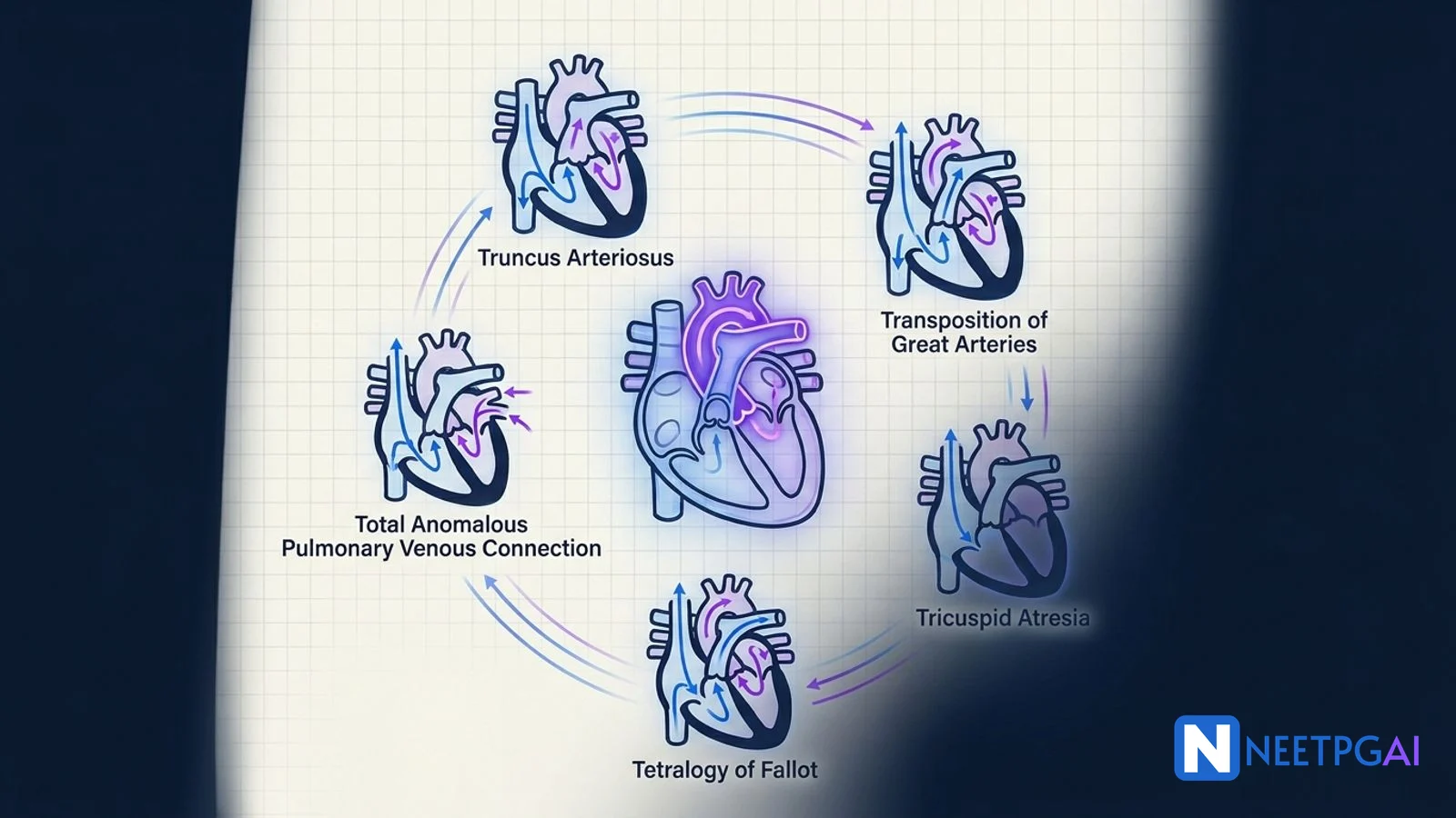
Pediatric congenital heart disease contributes 3-5 questions per NEET PG paper and typically 2-3 at INI-CET. The clinical reasoning tested is almost always: (1) is this cyanotic or acyanotic, (2) is it ductal-dependent, (3) when does it need surgery, and (4) what is the palliation strategy. Memorise the five Ts of cyanotic CHD (Tetralogy of Fallot, Transposition of great arteries, Truncus arteriosus, TAPVC, Tricuspid atresia) and the age at presentation each produces - TGA within the first week, TOF between 3 and 6 months with spells, truncus and TAPVC within the first month with congestive heart failure.
The three load-bearing concepts: IV prostaglandin E1 at 0.05-0.1 microgram per kg per minute keeps the ductus open and buys time to transfer any cyanotic newborn to a cardiac centre; the arterial switch operation for TGA must happen within the first 2 weeks before the left ventricle de-conditions; and large VSDs must be closed by 1-2 years to prevent Eisenmenger syndrome (6 months in Down syndrome because pulmonary vascular disease progresses faster).
Why This Matters for NEET PG & INI-CET
NEET PG and INI-CET love congenital heart disease because the questions follow a predictable clinical reasoning chain that distinguishes a prepared candidate from a rote memoriser. The exam does not ask you to recite the anatomy of truncus arteriosus. It gives you a day-3 cyanotic newborn with a single S2, no murmur, SpO2 60 percent that does not improve with 100 percent oxygen (hyperoxia test negative), and asks what to do next. The answer is PGE1 and urgent cardiology referral - and the stem is testing TGA, not truncus. The images on the exam are increasingly a chest X-ray (egg-on-side cardiac silhouette for TGA, boot-shaped for TOF, snowman for supracardiac TAPVC) paired with a hyperoxia test result and a 2-D echo clip.
INI-CET goes further and asks surgical timing in multi-step vignettes: a 4-month-old with a large VSD, failure to thrive, and bidirectional shunt on echo - what is the next step? Here the answer is urgent VSD closure before pulmonary vascular disease fixes. A 6-week-old with Down syndrome and a complete AV canal is referred for repair at 3 months because trisomy 21 accelerates pulmonary vascular disease. Learn the timing table once and these questions become reflex.
Beyond the exam, this is the one chapter in pediatrics where a 30-minute delay in starting PGE1 kills a newborn and a correctly scheduled Fontan completion gives a child with HLHS a 70 percent chance of reaching adulthood. The clinical stakes are immediate and visible.
The cyanotic versus acyanotic framework
The first branch point in every CHD question is cyanotic versus acyanotic.
| Feature | Cyanotic CHD | Acyanotic CHD |
|---|
| Direction of shunt | Right-to-left (deoxygenated blood bypasses the lungs) | Left-to-right (oxygenated blood recirculates to the lungs) |
| SpO2 at presentation | Less than 85 percent, does not improve with 100 percent oxygen (hyperoxia test negative - PaO2 less than 150 mmHg on 100 percent FiO2) | Normal (greater than 95 percent) early; falls late if Eisenmenger develops |
| Classic examples | Five Ts - TOF, TGA, Truncus arteriosus, TAPVC, Tricuspid atresia; plus HLHS, Ebstein anomaly, pulmonary atresia | VSD, ASD, PDA, coarctation, AV canal defect, aortic stenosis |
| Timing of presentation | First days to weeks (ductal-dependent) or 3-6 months (TOF with spells) | Later - heart failure in infancy (large VSD, large PDA) or asymptomatic until school age (ASD, small VSD) |
| Pulmonary blood flow on CXR | Decreased (TOF, tricuspid atresia, pulmonary atresia) or increased (TGA, truncus, TAPVC) | Increased (left-to-right shunt) |
| Growth | Usually poor (chronic hypoxemia) | Failure to thrive if large shunt (VSD, PDA, AV canal) |
The hyperoxia test is a bedside differential: give 100 percent oxygen for 10 minutes and measure arterial PaO2 from a right radial sample. PaO2 greater than 250 mmHg suggests pulmonary cause; PaO2 less than 150 mmHg strongly suggests cardiac right-to-left shunt (cyanotic CHD).
The five Ts of cyanotic congenital heart disease
Memorise the mnemonic and the age at presentation for each.
| Lesion | Mnemonic position | Age at presentation | Classic CXR | Treatment |
|---|
| Truncus arteriosus | 1 vessel (T1) | First month, CHF with mild cyanosis | Cardiomegaly, increased pulmonary vascular markings | Rastelli-type repair at 4-6 weeks |
| Transposition of great arteries (d-TGA) | 2 vessels transposed (T2) | Day 1-7, severe cyanosis within hours of birth | Egg on side (narrow mediastinum, oval cardiac shadow) | PGE1, Rashkind septostomy, arterial switch (Jatene) within 2 weeks |
| Tricuspid atresia | 3 (T3) - no 3rd leaflet | Depends on associated lesions; cyanotic if restrictive VSD | Decreased pulmonary vascularity | BT shunt if ductal-dependent, then Glenn 4-6 months, Fontan 2-4 years |
| Tetralogy of Fallot | 4 features (T4) | 3-6 months with hypercyanotic spells | Boot-shaped (coeur-en-sabot) with decreased pulmonary vascularity | Complete repair at 3-6 months; BT shunt if critical neonate |
| TAPVC (Total Anomalous Pulmonary Venous Connection) | 5 (all 4 pulmonary veins + atrial communication = 5 openings) | First month; obstructed infracardiac type presents hours after birth with severe cyanosis and respiratory distress | Snowman (figure-of-8) in supracardiac type | Immediate surgical repair - re-implant pulmonary veins to left atrium |
Age at presentation - the diagnostic shortcut
| Age | Most likely lesion if cyanotic | Why |
|---|
| First 24 hours | d-TGA, obstructed TAPVC, HLHS, severe Ebstein | Parallel circuits (TGA) or severe outflow obstruction |
| Day 1-7 | d-TGA, ductal-dependent pulmonary lesions (critical PS, pulmonary atresia), ductal-dependent systemic lesions (HLHS, critical AS, interrupted arch, severe coarctation) | Ductus closes between day 1 and day 14 |
| First month | Truncus arteriosus, TAPVC, complete AV canal | Pulmonary vascular resistance falls, left-to-right shunt increases, CHF develops |
| 3-6 months | TOF (hypercyanotic spells) | Right ventricular outflow obstruction progresses with infundibular hypertrophy |
| School age | ASD, small VSD, mild coarctation | Incidental murmur or exercise intolerance; late presentation of acyanotic lesion |
| Adolescent / adult | Eisenmenger syndrome from untreated large shunt | Pulmonary vascular disease has fixed |
The ductal-dependent lesions and PGE1
Three physiological groups of ductal-dependent lesions.
| Group | Lesions | Role of duct | Presentation on day 1-7 |
|---|
| Ductal-dependent pulmonary circulation | Critical pulmonary stenosis, pulmonary atresia, tricuspid atresia with PS, severe TOF, Ebstein | Supplies pulmonary blood flow | Severe cyanosis, SpO2 less than 70, metabolic acidosis |
| Ductal-dependent systemic circulation | HLHS, critical aortic stenosis, interrupted aortic arch, severe coarctation | Supplies systemic flow to lower body | Shock, pale-grey appearance, absent femoral pulses, differential pulse oximetry (upper greater than lower), lactic acidosis, renal failure, necrotising enterocolitis |
| Ductal-dependent mixing | d-TGA | Allows atrial and ductal-level mixing between parallel circuits | Severe cyanosis within hours; no response to oxygen |
PGE1 (alprostadil) protocol:
- Start at 0.05-0.1 microgram per kg per minute IV infusion
- Target: increase SpO2 to 75-85 percent (cyanotic lesions) or restore femoral pulses and perfusion (systemic lesions)
- Known side effects: apnoea (keep intubation ready - prophylactic intubation for transfer), fever, flushing, hypotension at high doses, cortical hyperostosis with prolonged use
- Once stable, taper to lowest effective dose (0.01-0.025 microgram per kg per minute)
- Continue until definitive surgery or catheter intervention
The acyanotic shunts - VSD, ASD, PDA
VSD (most common CHD overall, 20-30 percent)
| Size | Clinical features | Natural history | Management |
|---|
| Small (less than 3 mm, restrictive) | Loud harsh pansystolic murmur, no symptoms, normal growth | 70 percent close spontaneously by age 5 | Observe with serial echo; endocarditis prophylaxis only for high-risk situations |
| Moderate (3-6 mm) | Pansystolic murmur, mild tachypnoea, mild failure to thrive | 30-40 percent close spontaneously | Observe through infancy; close by age 2-5 if persistent large shunt |
| Large (greater than 6 mm, non-restrictive) | Soft or absent murmur (equal ventricular pressures), heart failure 6-8 weeks (as PVR falls), failure to thrive, repeated LRTI, hepatomegaly | Pulmonary vascular disease by 1-2 years | Diuretics, ACE inhibitor, high-calorie feeds, surgical closure by 6-12 months |
Eisenmenger prevention window: close large VSDs before 1-2 years; the window is 6 months in Down syndrome because pulmonary vascular disease progresses faster.
ASD (10 percent of CHD; secundum most common, 70 percent)
| Type | Location | Features | Management |
|---|
| Secundum | Mid-septum (fossa ovalis) | Fixed splitting of S2, soft pulmonary flow murmur, right ventricular lift | Device closure percutaneously at age 3-5 years if right ventricular dilatation or Qp:Qs greater than 1.5 |
| Primum | Lower septum, part of AV canal defect | Associated with cleft mitral valve, left-axis deviation on ECG | Surgical closure with mitral repair |
| Sinus venosus | High septum near SVC-right atrium junction | Associated with partial anomalous pulmonary venous return (right upper pulmonary vein to SVC) | Surgical repair - rerouting of pulmonary vein and ASD closure |
Secundum ASD rarely causes heart failure in childhood but produces atrial arrhythmia, paradoxical embolism (stroke in young adults), and right heart failure by adulthood. Eisenmenger develops but typically only in the 4th-5th decade.
PDA (5-10 percent of CHD; higher in preterm infants)
- Continuous machinery murmur (Gibson murmur) at left infraclavicular area
- Wide pulse pressure, bounding peripheral pulses
- Preterm infants: trial of medical closure with indomethacin, ibuprofen, or paracetamol (success 60-80 percent); surgical ligation if medical fails and the duct is hemodynamically significant
- Term infants and children: device closure percutaneously at 6-12 months
- Eisenmenger prevention: close by 2 years for large PDA
Hypercyanotic (Tet) spells - the acute management ladder
A hypercyanotic spell is a sudden decrease in pulmonary blood flow caused by infundibular spasm, dropping systemic vascular resistance, or dehydration - producing severe cyanosis, hyperpnea, agitation, and loss of consciousness in severe spells. Peak incidence: 3-24 months.
| Step | Intervention | Mechanism |
|---|
| 1 | Knee-chest position (child flexes hips, mother squats, holds child to chest) | Increases systemic vascular resistance - reduces right-to-left shunt |
| 2 | 100 percent oxygen via mask or hood | Pulmonary vasodilation; corrects hypoxemia |
| 3 | Morphine 0.1-0.2 mg per kg IV or IM | Sedates, reduces hyperpnea, breaks the cycle |
| 4 | IV crystalloid bolus 10-20 mL per kg | Raises preload, pushes blood through right ventricular outflow tract |
| 5 | IV propranolol 0.1-0.2 mg per kg or esmolol | Relieves infundibular spasm (negative inotropy of RVOT) |
| 6 | Sodium bicarbonate 1-2 mEq per kg if pH less than 7.2 | Corrects metabolic acidosis; reduces pulmonary vascular resistance |
| 7 | Phenylephrine 5-20 microgram per kg IV (or norepinephrine) | Raises SVR when all above fails |
| 8 | Emergency surgical referral - advance repair | Recurrent spells are an indication to operate sooner |
Surgical timing - the master table
| Lesion | Palliation (if needed) | Definitive repair | Why that age |
|---|
| d-TGA | Rashkind balloon septostomy in neonate if restrictive foramen ovale | Arterial switch (Jatene) day 3-14 | LV de-conditions after 2 weeks on pulmonary pressures |
| TOF (classic) | BT shunt in neonate if critical cyanosis | Complete repair 3-6 months | Balances technical feasibility and prevention of RV hypertrophy and polycythaemia |
| TOF with pulmonary atresia or MAPCAs | BT shunt or unifocalisation of MAPCAs | Staged repair, final completion 1-2 years | Pulmonary arteries need to grow |
| Truncus arteriosus | None (do not delay) | Rastelli-type repair 4-6 weeks | Prevents pulmonary vascular disease (which develops fast) |
| TAPVC obstructed | None - emergency | Immediate surgery within hours | Life-threatening pulmonary edema |
| Tricuspid atresia / HLHS | Norwood or BT shunt in neonate | Staged - Glenn 4-6 months, Fontan 2-4 years | Allows pulmonary artery growth and lung maturation |
| Large VSD | None if not critical | Primary closure 6-12 months (symptomatic) or 1-2 years (asymptomatic) | Eisenmenger prevention |
| Complete AV canal | None | Repair 3-6 months (especially Down syndrome) | PVD develops faster in trisomy 21 |
| Secundum ASD | None | Device closure 3-5 years | Elective; prevents late atrial arrhythmia and RV dilatation |
| PDA (term) | None | Device closure 6-12 months | Prevents RVH and endocarditis |
| PDA (preterm) | Medical closure (indomethacin, ibuprofen, paracetamol) | Surgical ligation if medical fails | Preterm duct has prostaglandin dependency |
| Coarctation of aorta | PGE1 if critical neonate; balloon dilatation in older children | End-to-end anastomosis or subclavian flap aortoplasty, neonate | Prevents LVH, systemic hypertension, cerebral aneurysm |
The modified Blalock-Taussig-Thomas shunt and other palliations
The modified BT shunt is the workhorse neonatal palliation when definitive repair is impossible. It is a Gore-Tex tube (3-4 mm) connecting the right or left subclavian artery to the ipsilateral pulmonary artery, providing a controlled source of pulmonary blood flow in lesions with inadequate pulmonary circulation.
Indications:
- Severe TOF or pulmonary atresia in the newborn before complete repair
- Tricuspid atresia with restrictive VSD before Glenn
- HLHS as part of the Norwood procedure
- Pulmonary atresia with intact ventricular septum
Historical BT shunt (classic Blalock-Taussig): direct anastomosis of subclavian artery to pulmonary artery; abandoned for the modified version to preserve subclavian flow to the ipsilateral arm.
Pulmonary artery banding: opposite physiological intent - a surgical band is placed around the main pulmonary artery to reduce excessive pulmonary blood flow in lesions like large VSD with pulmonary over-circulation, buying time until definitive repair. Rarely used now that primary VSD closure is feasible in infancy.
Signs of congestive heart failure in infants
NEET PG asks you to recognise the picture: a 6-week-old with a large VSD or complete AV canal in failure.
| System | Signs |
|---|
| General | Failure to thrive, poor feeding, sweating during feeds (sympathetic stress), low weight-for-age |
| Respiratory | Tachypnoea (RR greater than 60 awake), subcostal and intercostal retractions, grunting, recurrent LRTI |
| Cardiovascular | Tachycardia (HR greater than 160), gallop rhythm (S3), displaced apex, soft or absent murmur in large VSD (equal pressures) |
| Hepatomegaly | Liver edge 2-3 cm below costal margin - the most reliable sign of right heart failure in infants (JVP is unreliable) |
| Peripheral | Cool extremities with poor perfusion (cardiogenic shock in critical lesions); delayed capillary refill |
Medical management of CHF in infancy: diuretics (furosemide 1 mg per kg BID), ACE inhibitor (captopril 0.5 mg per kg TID titrated), high-calorie formula (24-30 kcal per oz), nasogastric feeds if feeding fatigue, treat intercurrent infections aggressively. Digoxin is used less commonly now; typical dose 10 microgram per kg per day in two divided doses, with careful monitoring for toxicity. The real treatment is timely surgery; medical therapy is a bridge.
Eisenmenger syndrome - the fate of untreated shunts
Chronic high pulmonary blood flow and pressure from an uncorrected left-to-right shunt (large VSD, large PDA, truncus, AV canal defect, less commonly ASD) causes:
- Phase 1 (hyperkinetic pulmonary hypertension): reversible, PVR not yet elevated; closing the defect normalises hemodynamics
- Phase 2 (reactive pulmonary vascular disease): medial hypertrophy of pulmonary arterioles, PVR rising but still partially reversible with vasodilators
- Phase 3 (fixed pulmonary vascular disease): intimal fibrosis, plexiform lesions, PVR greater than 10 Wood units irreversible; shunt reverses to right-to-left (Eisenmenger)
Once Eisenmenger establishes:
- Cyanosis, polycythaemia (compensated erythrocytosis, haematocrit greater than 55 percent), clubbing
- Hemoptysis (fragile pulmonary vasculature), heart failure, paradoxical embolism, cerebral abscess
- Pulmonary vasodilators (sildenafil, bosentan, prostacyclin analogues) reduce symptoms but do not cure
- Pregnancy is contraindicated (maternal mortality 30-50 percent)
- Definitive treatment: heart-lung transplantation
Prevention is everything. Close large VSD by 1-2 years (6 months in Down syndrome). Close AV canal defect by 3-6 months. Close large PDA by 1-2 years. Secundum ASD by 3-5 years. The exam question that reads "a 25-year-old with a known VSD since childhood now presents with cyanosis" is testing whether you understand the window has closed.
High-Yield One-Liners
- Hyperoxia test negative (PaO2 less than 150 on 100 percent oxygen) equals cyanotic CHD
- PGE1 0.05-0.1 microgram per kg per minute keeps the duct open; apnoea is the key side effect
- TGA presents day 1-7 with severe cyanosis and single S2; CXR shows egg-on-side
- Arterial switch (Jatene) within 2 weeks before LV de-conditions
- TOF presents 3-6 months with hypercyanotic spells; CXR shows boot-shaped heart
- Hypercyanotic spell management: knee-chest, oxygen, morphine, fluids, propranolol, bicarb, phenylephrine
- Truncus arteriosus: Rastelli repair at 4-6 weeks to prevent fast pulmonary vascular disease
- TAPVC obstructed is a surgical emergency within hours
- HLHS: Norwood day 1-7, Glenn 4-6 months, Fontan 2-4 years
- BT shunt is a Gore-Tex tube from subclavian to pulmonary artery
- Large VSD: close by 1-2 years to prevent Eisenmenger (6 months in Down syndrome)
- Secundum ASD: device closure at 3-5 years for RV dilatation or Qp:Qs greater than 1.5
- PDA (preterm): try indomethacin, ibuprofen, or paracetamol first; surgical ligation if medical fails
- Eisenmenger: PVR greater than 10 Wood units; irreversible; heart-lung transplant is the only cure
- Hepatomegaly 2-3 cm below costal margin is the most reliable sign of right heart failure in infants
- Fixed splitting of S2 equals ASD; continuous machinery murmur equals PDA; harsh pansystolic equals VSD; boot-shaped heart equals TOF
Common Mistakes
- Giving oxygen to a cyanotic newborn without starting PGE1. The hyperoxia test takes 10 minutes to interpret; PGE1 can be started empirically if cyanotic CHD is clinically suspected. Oxygen does not save a ductal-dependent newborn.
- Delaying arterial switch beyond 2 weeks in d-TGA. The left ventricle de-conditions on pulmonary pressures; after 2 weeks it cannot acutely take systemic afterload. Delayed presenters need pulmonary artery banding first (LV training) before deferred arterial switch at 2-4 months.
- Diagnosing hypercyanotic spell and only giving oxygen. Oxygen alone fails because the obstruction is at the right ventricular outflow tract, not at the lung. Knee-chest position and morphine do the heavy lifting.
- Closing a VSD in a 25-year-old with established Eisenmenger. Fixed pulmonary vascular resistance means the right ventricle depends on the shunt to decompress. Closing the VSD causes acute right heart failure and death. Operability is tested by acute pulmonary vasodilator challenge (nitric oxide, oxygen) with pre- and post-PVR measurement.
- Treating a large VSD with Down syndrome in the same timeline as a non-syndromic child. Pulmonary vascular disease develops faster in trisomy 21; close by 6 months, not 1-2 years.
Related Topics on NEETPGAI
Practice This on NEETPGAI
Congenital heart disease is a reasoning chain, not a fact list. Train the chain on 20 CHD MCQs per week and the pattern recognition becomes automatic. Sign up free and start practicing: NEETPGAI's 28,000+ MCQ bank is free for every registered user, with the full pediatric cardiology set, instant AI explanations of surgical timing, and spaced-repetition review of your mistakes. The AI Deep Dive can produce a labeled diagram of any CHD lesion you ask about - truncus with VSD and shared great artery, TGA with parallel circuits, tricuspid atresia with hypoplastic right ventricle - so the anatomy and the physiology become inseparable in your memory.
This content is for educational purposes for NEET PG exam preparation. It is not a substitute for professional medical advice, diagnosis, or treatment. Clinical information has been reviewed by qualified medical professionals.
Written by: NEETPGAI Editorial Team
Reviewed by: Pending SME Review
Last reviewed: October 2026