Version 1.0 - Published October 2026
Quick Answer
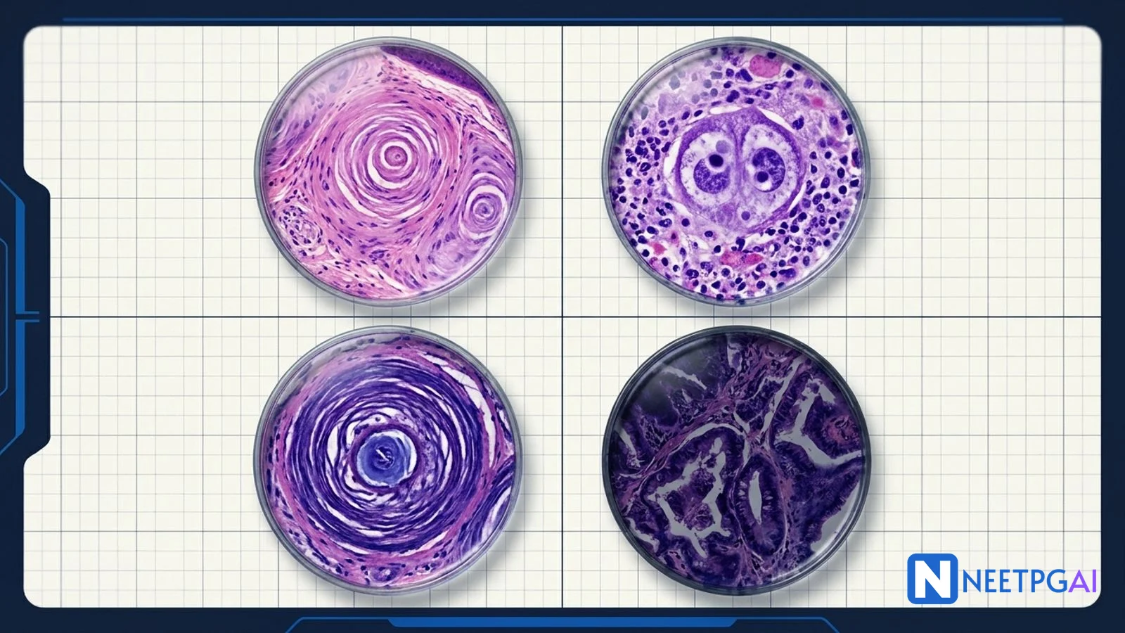
Histopathology slide identification contributes 3-5 questions per NEET PG paper and typically 2-3 at INI-CET, with the question invariably paired with a classic microscopic image and 4 options that include at least two convincing mimics. Eight tumor patterns recur across every recent exam cycle: adenocarcinoma (glands and mucin), squamous cell carcinoma (keratin pearls and intercellular bridges), GIST (uniform spindle cells, CD117 and DOG1 positive), meningioma (whorls and psammoma bodies), follicular carcinoma of thyroid (capsular or vascular invasion - the clinching feature versus adenoma), hepatocellular carcinoma (trabecular pattern, sinusoidal vasculature, lost reticulin), basal cell carcinoma (peripheral palisading with retraction artefact), and Hodgkin lymphoma (Reed-Sternberg owl-eye binucleate cells).
The three load-bearing distinctions: glands plus mucin equals adenocarcinoma; keratin pearls equal squamous cell carcinoma; owl-eye binucleate RS cell equals Hodgkin lymphoma. The IHC panel ties them to the tissue of origin.
Why This Matters for NEET PG & INI-CET
Histopathology image MCQs are the single highest-yield category of pathology questions on NEET PG, and the slides tested are the same 15-20 patterns that medical pathology has recognised for a century. The exam is not asking you to be a histopathologist. It is asking you to recognise the Reed-Sternberg cell, the keratin pearl, the whorl of meningothelial cells, and the trabecular pattern of HCC the moment you see them - because these pattern recognitions are the backbone of clinical oncology.
INI-CET pushes further by pairing the slide with an IHC panel or a clinical vignette and asking you to interpret both: a 35-year-old woman with a mediastinal mass and a slide showing lacunar cells with sclerotic bands equals nodular sclerosis Hodgkin lymphoma, which drives ABVD chemotherapy. A thyroid lobectomy specimen showing follicles with capsular invasion equals follicular carcinoma, which drives completion thyroidectomy plus radioactive iodine. The slide identification is only step one; the exam rewards the candidate who can run the full decision chain in 90 seconds.
The eight MCQs below cover almost every histopathology image MCQ asked in NEET PG 2019-2024. Learn the pattern once, drill the IHC panel beside it, and you will not miss them in the exam or on the ward when a biopsy report lands on your desk.
MCQ 1: 65-year-old man with a 3 cm colonic mass; biopsy under H&E
Image description: [High-power H&E photomicrograph showing malignant epithelial cells forming irregular glandular structures (tubules) with central lumens. The gland lumens contain basophilic mucinous material. Cells are columnar with pseudostratified elongated hyperchromatic nuclei and prominent nucleoli. The glands invade through a desmoplastic stromal response with dense fibrosis. Mitotic figures are frequent. Alcian blue stain positive in cytoplasm and lumens.]
Clinical vignette: A 65-year-old man presents with 3 months of change in bowel habit (alternating diarrhoea and constipation), weight loss of 6 kg, and intermittent rectal bleeding. Colonoscopy shows a 3 cm ulcerated mass at the hepatic flexure. Hemoglobin 9.8, CEA elevated at 42 ng per mL. Biopsy taken.
Options:
- (a) Squamous cell carcinoma
- (b) Adenocarcinoma, moderately differentiated
- (c) Carcinoid (neuroendocrine) tumor
- (d) Gastrointestinal stromal tumor (GIST)
Correct answer: (b) Adenocarcinoma, moderately differentiated
Reasoning: The hallmark of adenocarcinoma is gland formation - tumor cells arrange into tubules or acini with central lumens, and produce mucin stainable with PAS-diastase, mucicarmine, and alcian blue. Moderate differentiation retains gland formation (50-95 percent glandular architecture) with moderate nuclear atypia and desmoplastic response. SCC would show keratin pearls and intercellular bridges without glands - squamous epithelium is not native to colon mucosa so SCC of colon is extraordinarily rare (anal canal is the exception). Carcinoid shows uniform nests of monomorphic cells with salt-and-pepper nuclear chromatin, synaptophysin and chromogranin positive. GIST arises from the muscularis propria, shows uniform spindle or epithelioid cells, and is CD117/DOG1 positive.
IHC panel for colonic adenocarcinoma: CK20 positive, CDX2 positive, CK7 usually negative (classical colon pattern), villin positive, CDX2 strongly positive. MSI status (microsatellite instability) is now mandatory - MMR IHC panel (MLH1, MSH2, MSH6, PMS2) identifies Lynch syndrome and predicts response to immune checkpoint inhibitors.
Teaching pearl: Grading of adenocarcinoma:
| Grade | Gland formation | Mitotic activity | Nuclear atypia |
|---|
| Well-differentiated (G1) | Greater than 95 percent | Low | Mild |
| Moderately differentiated (G2) | 50-95 percent | Moderate | Moderate |
| Poorly differentiated (G3) | Less than 50 percent (solid sheets with rare glands) | High | Marked |
| Undifferentiated (G4) | No glands | High | Marked |
MCQ 2: 68-year-old smoker with lung mass; CT-guided biopsy
Image description: [High-power H&E photomicrograph showing nests of polygonal eosinophilic tumor cells with abundant pink cytoplasm, large vesicular nuclei, and prominent nucleoli. In the centre of several nests are concentric lamellated whorls of eosinophilic keratin (keratin pearls). At higher magnification, delicate linear bridges connect adjacent cells (intercellular bridges). No gland formation is seen. Individual cells show cytoplasmic keratinisation (dyskeratotic cells).]
Clinical vignette: A 68-year-old male smoker (50 pack-years) presents with a 3-month history of cough, hemoptysis, and 5 kg weight loss. CT chest shows a 4 cm central mass in the right upper lobe with ipsilateral hilar lymphadenopathy. CT-guided biopsy is taken.
Options:
- (a) Small cell lung carcinoma
- (b) Adenocarcinoma of lung
- (c) Squamous cell carcinoma of lung
- (d) Large cell neuroendocrine carcinoma
Correct answer: (c) Squamous cell carcinoma of lung
Reasoning: Keratin pearls (concentric lamellated whorls of keratin) and intercellular bridges are the two pathognomonic features of well- and moderately-differentiated squamous cell carcinoma. Central location in a long-term smoker is classical for SCC of lung. Small cell lung carcinoma shows small round blue cells with scant cytoplasm, nuclear moulding, salt-and-pepper chromatin, abundant mitoses, and crush artefact - not keratin pearls. Lung adenocarcinoma shows glandular formation, acinar or papillary patterns, often with mucin production, and is typically peripheral in location. Large cell neuroendocrine carcinoma shows large cells in organoid or rosette patterns with neuroendocrine markers (synaptophysin, chromogranin).
IHC panel for lung SCC: p40 positive (highly specific), p63 positive, CK5/6 positive, CK7 variable, TTF-1 negative, Napsin A negative. The p40 stain has largely replaced p63 for distinguishing SCC (p40 positive) from adenocarcinoma (p40 negative, TTF-1 positive, Napsin A positive) in poorly differentiated tumors.
Teaching pearl: Lung cancer classification by central versus peripheral location:
| Location | Common cell type | Smoking association |
|---|
| Central (hilar, mainstem bronchi) | Squamous cell carcinoma, small cell carcinoma | Strong (greater than 90 percent) |
| Peripheral | Adenocarcinoma, large cell carcinoma | Variable (adenocarcinoma includes non-smokers, EGFR-mutated) |
Adenocarcinoma is now the most common lung cancer worldwide, including in non-smokers (women, Asian populations with EGFR mutations). SCC and small cell remain strongly smoking-related.
MCQ 3: 55-year-old with gastric antral mass; biopsy under H&E
Image description: [High-power H&E photomicrograph showing a tumor composed of relatively uniform spindle-shaped cells with elongated eosinophilic cytoplasm and oval nuclei with blunt ends. The cells are arranged in interlacing fascicles and short bundles. Nuclear atypia is minimal; mitotic figures 3 per 50 high-power fields. No glands, no keratinisation. IHC panel: CD117 strongly positive in cytoplasm and membrane, DOG1 strongly positive, CD34 positive, SMA focally positive, S100 negative, desmin negative.]
Clinical vignette: A 55-year-old man presents with epigastric discomfort and occasional melena. OGD shows a 4 cm submucosal mass in the gastric antrum with a central ulceration. The overlying mucosa is intact. EUS-FNA biopsy is taken.
Options:
- (a) Leiomyoma
- (b) Gastrointestinal stromal tumor (GIST)
- (c) Schwannoma
- (d) Gastric adenocarcinoma
Correct answer: (b) Gastrointestinal stromal tumor (GIST)
Reasoning: The combination of spindle cell morphology, submucosal location, and CD117 plus DOG1 positive IHC is diagnostic of GIST. GIST arises from the interstitial cells of Cajal (pacemaker cells of the gut) and is driven by activating mutations in KIT (exons 9, 11, 13, 17) or PDGFRA (exon 18). Leiomyoma shows cigar-shaped nuclei with blunt ends and perinuclear vacuoles, is SMA and desmin positive, CD117 negative. Schwannoma shows nuclear palisading (Antoni A and B areas, Verocay bodies), is S100 positive, CD117 negative. Gastric adenocarcinoma shows glands and arises from the mucosa, not from submucosa.
IHC panel for GIST: CD117 positive 95 percent, DOG1 positive nearly 100 percent (critical when CD117 is negative), CD34 positive 70 percent, SMA focally positive 30 percent, desmin negative, S100 negative.
Teaching pearl: GIST risk stratification (Fletcher-Miettinen criteria) uses size, mitotic count, and location:
| Risk | Features |
|---|
| Very low | Less than 2 cm and less than 5 mitoses per 50 HPF |
| Low | 2-5 cm and less than 5 mitoses per 50 HPF |
| Intermediate | 5-10 cm and less than 5 mitoses per 50 HPF; or less than 5 cm and 6-10 mitoses |
| High | Greater than 10 cm, or greater than 10 mitoses per 50 HPF, or greater than 5 cm with greater than 5 mitoses, or non-gastric location greater than 5 cm |
Imatinib 400 mg per day is standard adjuvant for 3 years in high-risk resected cases; neoadjuvant imatinib for marginally resectable or metastatic disease. KIT exon 11 mutation has the best response; PDGFRA D842V is imatinib-resistant.
MCQ 4: 48-year-old woman with frontal parasagittal brain tumor; craniotomy specimen
Image description: [Low-power H&E photomicrograph showing a well-circumscribed tumor composed of sheets and whorls of plump polygonal cells with pale pink cytoplasm and oval nuclei with delicate chromatin and small nucleoli. The whorls are concentric swirls of cells with cells at the periphery wrapping around a central group. Scattered within the whorls are concentric lamellated calcified structures (psammoma bodies). No necrosis, mild atypia, mitoses less than 1 per 10 HPF. Dural attachment is present.]
Clinical vignette: A 48-year-old woman presents with 6 months of progressive headaches and left leg weakness. MRI shows a dural-based parasagittal mass with homogeneous contrast enhancement and a dural tail. Gross total resection is performed.
Options:
- (a) Glioblastoma
- (b) Meningioma, WHO grade I
- (c) Metastatic breast carcinoma to dura
- (d) Oligodendroglioma
Correct answer: (b) Meningioma, WHO grade I
Reasoning: The classical triad of whorls, psammoma bodies, and dural attachment is diagnostic of meningioma. Whorls are concentric arrangements of meningothelial cells; psammoma bodies are concentric lamellated calcifications also seen in papillary thyroid carcinoma, papillary serous carcinoma of ovary, and some mesotheliomas. Meningioma is the most common primary brain tumor in adults (not glioma; gliomas are the most common primary malignant CNS tumor but meningiomas are predominantly benign). Glioblastoma shows pseudopalisading necrosis, microvascular proliferation, and dense cellularity with marked atypia. Metastatic breast carcinoma shows glandular or ductal structures with pleomorphic cells, often ER positive and GATA3 positive. Oligodendroglioma shows the fried-egg appearance (perinuclear halo, round nuclei, chicken-wire vasculature) and the 1p/19q codeletion.
IHC panel for meningioma: EMA positive, vimentin positive, progesterone receptor positive in 70 percent, S100 variable, SSTR2a positive (useful for octreotide imaging in recurrent cases).
Teaching pearl: WHO grading of meningioma:
| Grade | Features | 5-year recurrence |
|---|
| Grade I (benign, 80-90 percent) | Classical whorls, psammoma bodies, mild atypia, less than 4 mitoses per 10 HPF | 10-15 percent |
| Grade II (atypical, 5-15 percent) | 4-19 mitoses per 10 HPF, OR brain invasion, OR 3 of 5 features (hypercellularity, small cells, prominent nucleoli, sheeting, necrosis) | 30-40 percent |
| Grade III (anaplastic, 1-3 percent) | 20 or more mitoses per 10 HPF, or frank anaplasia (resembling carcinoma or sarcoma) | 50-80 percent, poor survival |
Classical subtypes (meningothelial, fibrous, transitional, psammomatous) are all WHO Grade I.
MCQ 5: 42-year-old woman thyroid lobectomy for solitary nodule; capsule section
Image description: [Low-power H&E photomicrograph showing a well-encapsulated thyroid tumor composed of microfollicles lined by cuboidal cells with monomorphic round nuclei, mildly enlarged. Follicles contain scant colloid. At the capsule, tumor cells are seen penetrating through the full thickness of the fibrous capsule into the adjacent thyroid parenchyma - the tumor-capsule interface is irregular with tumor extending into and beyond the capsule. In an adjacent high-power field, tumor cells are seen inside a vascular channel within the capsule, attached to the endothelium with associated thrombus.]
Clinical vignette: A 42-year-old woman presents with a solitary 2.5 cm thyroid nodule. FNAC reports Bethesda category IV (follicular neoplasm) - the cytology shows a monomorphic follicular pattern but cannot distinguish adenoma from carcinoma. She undergoes hemithyroidectomy. Final histopathology awaited.
Options:
- (a) Follicular adenoma
- (b) Follicular carcinoma
- (c) Papillary thyroid carcinoma, follicular variant
- (d) Hurthle cell adenoma
Correct answer: (b) Follicular carcinoma
Reasoning: The two diagnostic features of follicular carcinoma - capsular invasion (full-thickness penetration of the fibrous capsule) and vascular invasion (tumor inside a vascular channel in the capsule, attached to the vessel wall with thrombus) - distinguish it from follicular adenoma. Both features are visible only on the surgical resection specimen - FNAC can diagnose follicular neoplasm but not distinguish benign from malignant, which is why Bethesda IV always leads to lobectomy. Follicular adenoma shows the same monomorphic follicular pattern but with an intact capsule and no vascular invasion. Papillary thyroid carcinoma, follicular variant, shows the nuclear features of PTC (Orphan Annie nuclei, nuclear grooves, intranuclear pseudoinclusions) despite the follicular architecture - the nuclei here are not Orphan Annie. Hurthle cell adenoma shows cells with abundant granular eosinophilic cytoplasm (mitochondria-rich oncocytes).
IHC panel favouring malignancy in a follicular lesion: HBME-1 positive, galectin-3 positive, CK19 positive, CD56 negative. None are definitive - histopathology of capsule remains the gold standard.
Teaching pearl: Thyroid cancer summary:
| Type | Frequency | Spread | Treatment |
|---|
| Papillary (PTC) | 70-80 percent | Lymphatic, often multifocal, bilateral | Total thyroidectomy + central neck dissection + RAI; prognosis excellent (10-year survival greater than 95 percent) |
| Follicular (FTC) | 10-15 percent | Hematogenous (lung, bone), rarely nodal | Total thyroidectomy + RAI; prognosis good |
| Hurthle cell | 3-5 percent | Hematogenous and lymphatic | Total thyroidectomy; less RAI-avid |
| Medullary (MTC) | 3-5 percent | Lymphatic + hematogenous; parafollicular C cells; calcitonin positive | Total thyroidectomy + bilateral central and lateral neck dissection; genetic testing for RET mutation (MEN 2A, 2B, familial MTC) |
| Anaplastic | Less than 2 percent | Rapid invasion; mortality 100 percent at 1 year | Palliative; occasionally lenvatinib or dabrafenib-trametinib if BRAF mutated |
MCQ 6: 60-year-old cirrhotic patient with liver mass; biopsy
Image description: [Medium-power H&E photomicrograph showing a tumor composed of plates of large polygonal eosinophilic hepatocyte-like cells arranged in trabeculae that are 3-5 cells thick. The trabeculae are separated by thin sinusoidal spaces lined directly by tumor cells, with no reticulin framework between tumor cells and sinusoids. Several intracytoplasmic hyaline eosinophilic globules are seen (Mallory-Denk bodies). Bile pigment is present in a few canaliculi within the tumor. Nuclear atypia is moderate, mitotic figures occasional.]
Clinical vignette: A 60-year-old man with Child-Pugh A cirrhosis from chronic hepatitis B presents on surveillance ultrasound with a new 3.2 cm hepatic lesion. AFP is elevated at 480 ng per mL. Triphasic CT shows arterial hyperenhancement and portal venous washout - LI-RADS 5. Core biopsy is taken for atypical features.
Options:
- (a) Hepatocellular carcinoma
- (b) Metastatic colonic adenocarcinoma
- (c) Hepatic adenoma
- (d) Cholangiocarcinoma
Correct answer: (a) Hepatocellular carcinoma
Reasoning: HCC classically shows the trabecular pattern (tumor hepatocytes arranged in plates 3 or more cells thick, versus normal liver which is 1-2 cells thick), sinusoidal vascular pattern (endothelial cells line the trabeculae directly without a reticulin framework - lost reticulin staining is a classic finding), bile production (pathognomonic when present as it indicates hepatocellular origin), and Mallory-Denk bodies (eosinophilic hyaline intracytoplasmic inclusions, also seen in alcoholic hepatitis). Clinical context of cirrhosis plus elevated AFP plus LI-RADS 5 lesion is diagnostic even without biopsy (AASLD guidelines: biopsy is not required when imaging is diagnostic). Metastatic adenocarcinoma shows glands and is CDX2 positive, CK20 positive (colonic). Hepatic adenoma lacks nuclear atypia and the trabecular pattern is normal; it is seen in young women on OCPs. Cholangiocarcinoma shows glands with abundant desmoplastic stroma and is CK7 and CK19 positive.
IHC panel for HCC: HepPar-1 positive, arginase-1 positive (highly specific), glypican-3 positive, AFP positive, polyclonal CEA shows canalicular pattern (specific), CD34 highlights sinusoidal pattern.
Teaching pearl: HCC variants and prognosis:
| Variant | Setting | Features | Prognosis |
|---|
| Classical HCC | Cirrhosis (HBV, HCV, alcohol, NASH) | Trabecular, elevated AFP | BCLC-staged; curative options (resection, transplant, ablation) for early stage |
| Fibrolamellar HCC | Young adults, non-cirrhotic liver | Large eosinophilic hepatocytes with lamellar fibrous bands; normal AFP; DNAJB1-PRKACA fusion | Better than classical HCC |
| Combined hepatocellular-cholangiocarcinoma | Cirrhosis | Mixed hepatocellular and biliary differentiation | Worse than pure HCC |
Treatment is BCLC-staged - resection and transplant for very early and early stage; TACE for intermediate; sorafenib, lenvatinib, or atezolizumab plus bevacizumab for advanced.
MCQ 7: 72-year-old man with nodular facial lesion; excisional biopsy
Image description: [Low-power H&E photomicrograph showing nests and islands of basaloid cells with uniform small round dark nuclei and scant basophilic cytoplasm, extending from the overlying epidermis into the dermis. At the periphery of each nest, the outermost row of cells is aligned like a fence with nuclei perpendicular to the basement membrane (peripheral palisading). Thin clefts or spaces separate the tumor nests from the surrounding stroma (retraction artefact). The surface shows ulceration. No keratin pearls, no gland formation.]
Clinical vignette: A 72-year-old farmer presents with a 1.5 cm pearly nodule with central ulceration on the left ala of the nose, slowly growing over 8 months with occasional bleeding. He has significant long-term sun exposure. Excisional biopsy is taken with 3 mm margins.
Options:
- (a) Squamous cell carcinoma
- (b) Nodular basal cell carcinoma
- (c) Nodular melanoma, amelanotic
- (d) Merkel cell carcinoma
Correct answer: (b) Nodular basal cell carcinoma
Reasoning: The classical features of BCC are peripheral palisading (fence-like alignment of nuclei at the periphery of tumor nests, oriented perpendicular to the basement membrane) and retraction artefact (clefts between the tumor and the stroma - a fixation artefact but highly characteristic of BCC). The basaloid cells resemble the basal layer of the epidermis - uniform, small, dark, with scant cytoplasm. SCC shows keratin pearls, intercellular bridges, and larger eosinophilic cells. Nodular melanoma, even amelanotic, shows spindle or epithelioid cells with marked pleomorphism, prominent nucleoli, and would be S100, Melan-A, HMB-45, and SOX10 positive. Merkel cell carcinoma shows small round blue cells with salt-and-pepper chromatin, nuclear moulding, and is CK20 positive (classic paranuclear dot pattern) and neuroendocrine markers positive.
IHC panel for BCC: BerEP4 positive, CK5/6 positive, p63 positive, EMA negative (versus SCC which is EMA positive), CK20 negative.
Teaching pearl: BCC subtypes by prognosis:
| Subtype | Features | Prognosis |
|---|
| Nodular (most common) | Pearly papule with telangiectasia, central ulceration; well-demarcated | Low recurrence with excision |
| Superficial | Erythematous scaly patch; often on trunk | Low metastatic potential but recurs locally |
| Pigmented | Confused clinically with melanoma; dermoscopy shows blue-grey ovoid nests | Same as nodular |
| Morpheaform / Infiltrative | Scar-like plaque with ill-defined margins; infiltrative growth deep into dermis | Highest recurrence (10-20 percent after conventional excision); Mohs surgery preferred |
| Basosquamous | Features of both BCC and SCC; higher metastatic potential | Treat as high-risk BCC |
Treatment: standard excision with 4 mm margins for low-risk; Mohs micrographic surgery for high-risk BCC (face, infiltrative subtype, recurrent lesions). Vismodegib and sonidegib (hedgehog pathway inhibitors) for locally advanced or metastatic BCC. Metastasis is rare (less than 0.1 percent); local destruction is the main concern.
MCQ 8: 28-year-old woman with mediastinal mass; cervical lymph node biopsy
Image description: [Low-power H&E photomicrograph showing a lymph node with partially effaced architecture by broad fibrous bands dividing the node into nodules. Within the nodules, scattered large cells with abundant eosinophilic cytoplasm and clear retraction halos around the cell (lacunar cells) are seen in a mixed inflammatory background of small lymphocytes, eosinophils, plasma cells, and histiocytes. One high-power field shows a classic binucleate cell with two mirror-image nuclei, each with a prominent eosinophilic nucleolus the size of a small lymphocyte and thick nuclear membrane (owl-eye binucleate Reed-Sternberg cell). IHC: CD30 positive, CD15 positive, PAX5 weakly positive, CD45 (LCA) negative, CD20 negative.]
Clinical vignette: A 28-year-old woman presents with a 6-week history of painless neck swelling, drenching night sweats, 8 percent weight loss over 3 months, and intermittent fever. Chest X-ray shows a large anterior mediastinal mass. Cervical lymph node excisional biopsy is taken.
Options:
- (a) Diffuse large B-cell lymphoma
- (b) Nodular sclerosis classical Hodgkin lymphoma
- (c) Burkitt lymphoma
- (d) Castleman disease, hyaline vascular variant
Correct answer: (b) Nodular sclerosis classical Hodgkin lymphoma
Reasoning: The combination of Reed-Sternberg cells (owl-eye binucleate large cells with prominent nucleoli), lacunar cells (the retraction-halo variant typical of nodular sclerosis subtype), broad fibrous bands dividing the node into nodules, mixed inflammatory background (eosinophils, plasma cells, histiocytes, small lymphocytes), and the classic IHC profile (CD30+, CD15+, PAX5 weak+, CD45-, CD20 usually -) is diagnostic. Young adult female with mediastinal mass and B symptoms is the textbook presentation. DLBCL shows diffuse sheets of large B cells, CD20 strongly positive, CD30 variable, and lacks the mixed inflammatory background. Burkitt lymphoma shows medium-sized monotonous cells with starry-sky appearance (macrophages ingesting apoptotic bodies), MYC translocation, and Ki-67 nearly 100 percent. Castleman disease shows atretic germinal centres with onion-skin mantles and penetrating hyalinized vessels.
IHC panel for classical Hodgkin lymphoma: CD30 positive (nearly 100 percent), CD15 positive 75-85 percent, PAX5 weakly positive, CD45 (LCA) negative (opposite to NHL), CD20 usually negative (10-20 percent weak positive), EBV-LMP1 positive in mixed cellularity and lymphocyte-depleted (EBV-related).
Teaching pearl: Classical Hodgkin lymphoma subtypes:
| Subtype | Frequency | Classic patient | Microscopy | Prognosis |
|---|
| Nodular sclerosis | 70 percent | Young adult female, mediastinal mass | Lacunar cells, broad fibrous bands dividing nodes into nodules | Excellent (5-year OS greater than 90 percent) |
| Mixed cellularity | 20 percent | Older, EBV-associated, abdominal nodes, HIV | Classic RS cells in mixed inflammatory background without sclerosis | Good |
| Lymphocyte-rich | 5 percent | Older adult | Few RS cells in lymphocyte-rich background | Best |
| Lymphocyte-depleted | 5 percent | Elderly, HIV, developing countries | Abundant RS cells with few lymphocytes; sarcomatoid | Worst |
Nodular lymphocyte predominant Hodgkin lymphoma (NLPHL) is a separate entity (not classical HL) with popcorn cells (L&H cells, multilobated nucleus), CD20 positive, CD45 positive, CD15 and CD30 negative - treated more like indolent B-cell lymphoma. Treatment of classical HL: ABVD (adriamycin, bleomycin, vinblastine, dacarbazine) is standard first-line; brentuximab-vedotin (anti-CD30) and PD-1 inhibitors (nivolumab, pembrolizumab) for relapsed-refractory.
High-Yield One-Liners
- Glands plus mucin equals adenocarcinoma; CK7/CK20 pattern localises site
- Keratin pearls plus intercellular bridges equal squamous cell carcinoma; p40 and p63 confirm
- GIST equals CD117 and DOG1 positive; imatinib target; KIT exon 11 is the most common mutation and best responder
- Whorls plus psammoma bodies equal meningioma (WHO I in 80-90 percent); EMA positive
- Follicular carcinoma of thyroid is diagnosed only on capsule section - capsular or vascular invasion
- Papillary thyroid carcinoma has Orphan Annie nuclei, nuclear grooves, intranuclear pseudoinclusions
- HCC shows trabecular pattern plus sinusoidal vessels plus lost reticulin plus bile production
- Fibrolamellar HCC in young non-cirrhotic patient; DNAJB1-PRKACA fusion
- BCC equals peripheral palisading plus retraction artefact; Mohs for infiltrative face lesions
- Owl-eye binucleate equals Reed-Sternberg equals Hodgkin lymphoma
- Lacunar cells plus broad fibrous bands equal nodular sclerosis Hodgkin
- Classical HL IHC: CD30+/CD15+/CD45- (opposite of NHL)
- Starry-sky equals Burkitt (macrophages ingesting apoptotic bodies); MYC translocation
- Fried-egg appearance plus 1p/19q codeletion equals oligodendroglioma
- Pseudopalisading necrosis plus microvascular proliferation equal glioblastoma
- Psammoma bodies in: papillary thyroid Ca, papillary serous Ca ovary, meningioma, some mesotheliomas
Common Mistakes
- Calling a biopsy adenocarcinoma without seeing glands. True adenocarcinoma must show gland formation on at least one high-power field, or produce mucin, or be IHC-positive for adenocarcinoma markers (CDX2, TTF-1, Napsin A). Solid sheets of malignant cells without differentiation are poorly differentiated carcinoma - the lineage requires IHC.
- Diagnosing follicular carcinoma from FNAC. FNAC can only report follicular neoplasm (Bethesda IV); the carcinoma versus adenoma distinction requires the capsule, which is removed with the lobectomy. Any Bethesda IV result mandates lobectomy for histopathology.
- Confusing psammoma bodies with calcification. Psammoma bodies are concentric lamellated calcified structures from the degeneration of individual tumor cells - they have a specific onion-ring appearance. Random dystrophic calcification in necrotic tissue is not psammoma.
- Missing the owl-eye RS cell in a lymph node with reactive hyperplasia. The RS cell is rare in Hodgkin lymphoma (1-2 percent of total cellularity). Scan every lymph node biopsy with suspicion for mediastinal mass or B symptoms carefully; CD30 and CD15 IHC resolves doubt.
- Diagnosing SCC of lung based on p63 alone. The newer standard is p40 (deltaN p63), which is more specific for SCC. p63 can also mark urothelial carcinoma, basal cells of prostate, myoepithelial cells, and some lymphomas. Pair p40 with TTF-1/Napsin A (adenocarcinoma) and synaptophysin/chromogranin (neuroendocrine) to resolve lung lineage.
Related Topics on NEETPGAI
Practice This on NEETPGAI
Histopathology slide recognition is a pattern-matching reflex that only drill-based practice builds. Drill 15 slide MCQs per day for two weeks and your pathology image accuracy on NEET PG will jump from 55 percent to 90 percent. Sign up free and start practicing: NEETPGAI's 28,000+ MCQ bank is free for every registered user, with the full histopathology image set covering 20+ tumor patterns, instant AI explanations of IHC panels, and spaced-repetition review of your mistakes. The AI Deep Dive feature can generate a labelled annotation of any slide you flag - cycle the keratin pearl, the lacunar cell, the whorl, and the trabecula into muscle memory in two weeks and the exam MCQs become fast marks.
This content is for educational purposes for NEET PG exam preparation. It is not a substitute for professional medical advice, diagnosis, or treatment. Clinical information has been reviewed by qualified medical professionals.
Written by: NEETPGAI Editorial Team
Reviewed by: Pending SME Review
Last reviewed: October 2026