Quick Answer
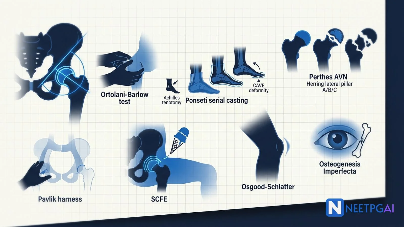
Pediatric orthopedics is a 3 to 4 question topic per NEET PG paper — largely covered under Orthopedics and Pediatrics. Lock these:
- DDH screening — Ortolani (reduction) + Barlow (provocation) in newborn; Galeazzi + skin folds after 3 months.
- DDH imaging — USG Graf method under 6 months (alpha angle over 60 degrees normal); X-ray after 4-6 months (Hilgenreiner, Perkin, Shenton, acetabular index).
- DDH treatment ladder — Pavlik under 6 months, closed reduction + spica 6-18 months, open reduction + osteotomy over 18 months.
- Club foot CAVE — Cavus + Adductus + Varus + Equinus.
- Ponseti method — serial weekly casts then percutaneous Achilles tenotomy + foot abduction brace; 90 percent success.
- Legg-Calve-Perthes — idiopathic AVN age 4-8 years, boys, painless limp, Herring lateral pillar A/B/C.
- SCFE — obese adolescent, obligate external rotation, Klein line, in situ pinning; do NOT reduce.
- Osgood-Schlatter — tibial tubercle apophysitis; adolescent athletes; activity modification.
- Osteogenesis imperfecta — COL1A1/A2; blue sclera + hearing loss + easy fracture; bisphosphonates.
- Achondroplasia — FGFR3 mutation; short-limb dwarfism; foramen magnum stenosis in infancy.
- Cerebral palsy — non-progressive, spastic diplegia commonest; primitive reflex persistence; multidisciplinary care.
- India context — RBSK screening, PMDDK deformity correction, low-cost Ponseti coverage.
Pediatric orthopedic disorders concentrate on the hip (DDH, Perthes, SCFE), the foot (club foot), the growth plate (SCFE, Osgood-Schlatter), and the systemic bone disease (osteogenesis imperfecta, achondroplasia) — with cerebral palsy overlapping neurology and rehabilitation. NEET PG tests each entity across orthopedics, pediatrics, and community medicine (RBSK, screening).
This NEETPGAI deep dive walks entity by entity through etiology → screening and clinical features → imaging → treatment ladder → India programmatic context. Pair this with a solid grasp of fracture healing and the Salter-Harris classification before your exam.
Developmental dysplasia of the hip (DDH)
Risk factors
Screening picks up 90 percent of DDH if you know the risk set:
- Breech presentation in the third trimester (largest single risk — 1 in 25)
- First-born
- Female (4-6 times more common than male)
- Family history in a first-degree relative
- Oligohydramnios
- Congenital torticollis or metatarsus adductus (packaging disorders share aetiology)
- Cultural swaddling in extension (Native American papoose, some traditional Indian swaddling — hip should ride in flexion and abduction)
Clinical signs by age
| Age | Sign |
|---|
| Newborn to 3 months | Ortolani (reduction) and Barlow (provocation) — palpable clunks |
| 3 to 12 months | Asymmetric skin creases, limited hip abduction, Galeazzi sign (unequal knee heights with hips flexed) |
| Walking child | Trendelenburg gait, apparent limb shortening, waddling if bilateral |
Imaging
- Under 6 months — hip ultrasound by the Graf method — alpha angle over 60 degrees is normal, 43-60 is dysplastic, under 43 is dislocated. Use for high-risk infants and any clinical suspicion.
- After 4-6 months — plain AP pelvic X-ray once the ossific nucleus of the femoral head appears. Assess:
- Hilgenreiner line — horizontal through the triradiate cartilages
- Perkin line — vertical through the lateral acetabular margin; normal femoral head sits in the inferomedial quadrant
- Shenton line — smooth arc from the medial femoral neck to the superior obturator foramen; broken in dislocation
- Acetabular index — angle between Hilgenreiner line and the acetabular roof; over 30 degrees at 6 months suggests dysplasia
Treatment ladder
| Age at diagnosis | Treatment |
|---|
| Under 6 months | Pavlik harness (hip flexion and abduction; maintained 3-6 months; avoid over-abduction — femoral nerve palsy or AVN) |
| 6 to 18 months | Closed reduction under GA + adductor tenotomy if needed + hip spica cast in human position (100 degrees flexion, 40-60 abduction) |
| 18 months to 8 years | Open reduction + femoral shortening + acetabular osteotomy (Salter, Pemberton, or Dega) |
| Older child / adolescent salvage | Chiari osteotomy, Salter innominate osteotomy, periacetabular osteotomy; total hip replacement in the adult with secondary OA |
Club foot (congenital talipes equinovarus, CTEV)
The deformity (CAVE)
- Cavus of the midfoot
- Adductus of the forefoot
- Varus of the hindfoot
- Equinus of the ankle
Types
- Idiopathic (postural) — commonest; supple; excellent response to Ponseti
- Syndromic — arthrogryposis, spina bifida (myelomeningocele), distal arthrogryposis; rigid; often need soft-tissue release
Assessment
- Dimeglio classification — grades severity of each CAVE component
- Pirani score — 6-point scoring of hindfoot and midfoot contracture; used to track Ponseti progression
Ponseti method (standard of care)
- Correct cavus first — supinate the forefoot to line up with the hindfoot
- Serial long-leg plaster casts changed weekly for 5-7 weeks; abduct the foot around the fixed talus (never pronate)
- Percutaneous Achilles tenotomy in about 90 percent to correct residual equinus once forefoot is abducted 60-70 degrees; final cast for 3 weeks
- Foot abduction brace (Denis Browne bar) — 23 hours per day for 3 months, then nightly to age 4-5 years — brace non-compliance is the main cause of relapse
- Anterior tibialis transfer at age 2-4 years for dynamic supination relapse
Success
- Idiopathic CTEV — over 90 percent correction with Ponseti
- Syndromic — poorer; may need staged posteromedial release
Legg-Calve-Perthes disease
Epidemiology and clinical
- Idiopathic avascular necrosis of the femoral capital epiphysis
- Age 4-8 years (range 2-14); boys 4:1
- Painless limp; hip pain referred to knee or thigh; loss of internal rotation and abduction
Stages (Waldenstrom)
- Initial (ischaemic) — subchondral crescent sign, sclerosis, joint space widening
- Fragmentation — femoral head collapses and fragments
- Reossification — new bone deposition
- Residual (healed) — final shape determines long-term outcome
Herring lateral pillar classification (predictor)
- A — full lateral pillar height retained; good outcome
- B — up to 50 percent loss; intermediate
- C — over 50 percent loss; poor outcome
Treatment principle — containment
Keep the softened femoral head deep in the acetabulum during reossification so it heals spherical.
| Age / severity | Treatment |
|---|
| Under 6 years | Observation, activity modification, physiotherapy |
| Over 6 years + Herring B or B/C | Containment surgery — femoral varus derotation osteotomy or Salter innominate osteotomy |
| Herring C in older child | Poor outcome regardless |
| Adult deformed hip | Total hip replacement |
Slipped capital femoral epiphysis (SCFE)
Clinical picture
- Salter-Harris type I fracture through the proximal femoral physis
- Obese adolescent aged 10-16 years during peri-pubertal growth spurt
- Endocrine screen if atypical (very young, very thin, bilateral, short stature) — hypothyroidism, growth hormone deficiency, hypogonadism, renal osteodystrophy
- Painful limp; leg held in external rotation; obligate external rotation on hip flexion; loss of internal rotation
- Referred knee pain classic — always X-ray the hip in adolescent knee pain
Imaging
- Bilateral AP and frog-leg lateral pelvic X-ray
- Klein line — line along superior femoral neck; should intersect the epiphysis on normal AP; failure to intersect suggests SCFE
- Frog-leg lateral — classic ice-cream slipping off the cone appearance
Loder classification and management
- Stable — patient can weight-bear (with or without crutches); low AVN risk
- Unstable — cannot weight-bear; up to 50 percent AVN risk
- Definitive management — urgent in situ percutaneous single screw fixation across the physis to prevent further slip
- Do NOT attempt acute reduction of a chronic slip — dramatically raises AVN risk
- Prophylactic contralateral pinning — increasingly recommended for endocrinopathy, very young onset, and open triradiate cartilage (bilaterality reaches 60 percent in these groups)
Osgood-Schlatter disease
- Traction apophysitis at the tibial tubercle from repetitive quadriceps pull
- Adolescent athletes 10-15 years (jumping and kicking sports)
- Painful bony prominence at the tibial tubercle, tender to palpation
- Diagnosis clinical; X-ray shows tibial tubercle irregularity or a small avulsion fragment
- Treatment — activity modification, ice, NSAIDs, quadriceps stretching; self-limiting with skeletal maturity; rarely persistent ossicle needs excision
Osteogenesis imperfecta (OI)
- Autosomal dominant defect of type I collagen — COL1A1 or COL1A2 mutation
- Sillence classification I-IV; Type II is perinatal lethal; Type I is the mildest and commonest
- Type I features — blue sclera, easy fracturing (multiple long-bone fractures with minor trauma), conductive-then-sensorineural hearing loss, dentinogenesis imperfecta, ligamentous laxity, normal or near-normal intellect
- Investigations — clinical + skeletal survey (multiple fractures at different stages of healing — differential includes non-accidental injury); genetic confirmation
- Management — bisphosphonates (pamidronate IV, alendronate PO) reduce fracture rate and bone pain; intramedullary rods (Fassier-Duval telescoping) for long-bone deformity; multidisciplinary rehabilitation; audiology
Achondroplasia
- Autosomal dominant activating mutation of FGFR3 (most sporadic — new mutations paternal age effect)
- Short-limb dwarfism (rhizomelic — proximal segments proportionally shorter), macrocephaly with frontal bossing, midface hypoplasia, trident hand, normal trunk length, normal intelligence
- Complications — foramen magnum stenosis in infancy (sudden death, cervicomedullary compression), thoracolumbar kyphosis, spinal stenosis in adulthood, obstructive sleep apnoea, otitis media
- Management — surveillance MRI cervicomedullary junction, decompression if compression; growth hormone modestly effective; vosoritide (CNP analogue, approved 2021) increases growth velocity
Cerebral palsy (CP)
Definition and classification
- Non-progressive disorder of movement and posture from a fixed brain lesion sustained before, during, or in the first 2 years of life
- Motor pattern — spastic (about 70 percent), dyskinetic (kernicterus classic), ataxic, mixed
- Topographic — hemiplegic, diplegic, quadriplegic
- GMFCS I-V — grades functional mobility; guides prognostic counselling and hip surveillance
Etiology
- Antenatal — intrauterine infection (TORCH), genetic, congenital brain malformations
- Perinatal — hypoxic-ischaemic encephalopathy, kernicterus, preterm periventricular leukomalacia
- Postnatal (first 2 years) — meningitis, encephalitis, head injury, near-drowning
Diagnosis and comorbidities
- Delayed motor milestones, persistence of primitive reflexes past normal ages (Moro past 6 months, ATNR past 6 months), abnormal tone, asymmetry
- MRI brain identifies underlying lesion and rules out progressive mimics
- Comorbidities — epilepsy, intellectual disability, sensorineural hearing loss, cortical visual impairment, dysphagia, GORD, poor growth, hip subluxation/dislocation, progressive scoliosis, osteoporosis
Management
- Multidisciplinary — physical therapy, occupational therapy, speech and language therapy
- Orthotics — AFOs for equinus, resting splints
- Spasticity — focal (botulinum toxin injections), generalised (oral baclofen, tizanidine, diazepam), severe (intrathecal baclofen pump, selective dorsal rhizotomy)
- Orthopaedic surgery — soft-tissue lengthening and bony realignment once contractures established
- Hip surveillance — X-rays every 6-12 months in GMFCS III-V children to catch progressive subluxation
- Nutritional support, seizure management, communication aids
Common Salter-Harris pearls (SCFE cross-over)
| Type | Description | Example |
|---|
| I | Fracture through physis only | SCFE (chronic), birth injuries |
| II | Physis + metaphysis (Thurston-Holland fragment) | Commonest; distal radius |
| III | Physis + epiphysis (intra-articular) | Tillaux fracture (distal tibia) |
| IV | Physis + metaphysis + epiphysis (intra-articular) | Distal humerus |
| V | Crush of physis | Poor prognosis; growth arrest |
India-specific context
- Rashtriya Bal Swasthya Karyakram (RBSK) — under National Health Mission; screens 0-18 year-olds at anganwadis and schools for the 4Ds (Defects at birth, Deficiencies, Diseases, Development delays including disability); referral pathway for DDH, club foot, cerebral palsy
- Rashtriya Bal Suraksha Karyakram and district early intervention centres (DEICs) provide multidisciplinary follow-up
- Ponseti coverage — India is a global success story; the technique needs only plaster and a Denis Browne bar and is delivered through district hospitals and Miracle Feet partner clinics
- Pradhan Mantri Divyang Kalyan and district disability schemes provide surgical correction for congenital limb and spine deformities
- Ayushman Bharat PM-JAY — covers pediatric orthopedic surgery including DDH, CTEV, spine, and cerebral palsy interventions in empanelled hospitals
NEET PG MCQ traps
- Ortolani = reduction; Barlow = provocation — palpable clunks in the newborn.
- DDH risk factors — breech, first-born, female, family history, oligohydramnios, torticollis.
- Under 6 months — USG Graf method (alpha angle over 60 normal).
- After 4-6 months — X-ray with Hilgenreiner, Perkin, Shenton, acetabular index.
- DDH treatment ladder — Pavlik under 6, closed reduction + spica 6-18 months, open reduction + osteotomy over 18 months.
- Pavlik complications — femoral nerve palsy, AVN with over-abduction.
- CAVE — Cavus + Adductus + Varus + Equinus of club foot.
- Ponseti — supinate to correct cavus, abduct around talus (never pronate), Achilles tenotomy in 90 percent, brace to age 4-5.
- Perthes age 4-8, boys 4:1 — painless limp.
- Perthes Herring A/B/C — lateral pillar height at fragmentation predicts outcome.
- Perthes containment — Salter or femoral varus osteotomy over 6 years with Herring B.
- SCFE — obese adolescent 10-16 years — external rotation deformity.
- Klein line — should intersect epiphysis on normal AP; failure suggests SCFE.
- SCFE — in situ pinning — do NOT reduce chronic slip (AVN risk).
- Prophylactic contralateral pinning — endocrinopathy, very young, open triradiate cartilage.
- Osgood-Schlatter — tibial tubercle apophysitis; adolescent jumping sports; self-limiting.
- Osteogenesis imperfecta Type I — blue sclera + hearing loss + fractures; COL1A1/A2.
- Achondroplasia — FGFR3; foramen magnum stenosis in infancy; vosoritide.
- Cerebral palsy spastic diplegia — commonest; often preterm; periventricular leukomalacia on MRI.
- Kernicterus — dyskinetic (choreoathetotic) CP; basal ganglia injury.
- GMFCS I-V — grades functional mobility in CP.
- Hip surveillance — GMFCS III-V children need 6-12 monthly X-rays.
- Salter-Harris I — SCFE and birth injuries; through physis only.
- Salter-Harris V — crush of physis; worst prognosis, growth arrest.
- Referred knee pain in adolescent — always X-ray hips (SCFE trap).
- RBSK — screens 0-18 year-olds for 4Ds; feeds district early intervention centres.
Recent updates and Indian context
- Vosoritide (CNP analogue) approved 2021 for achondroplasia — increases growth velocity; access limited in India by cost
- Bisphosphonates in OI — standard of care; pamidronate IV cycles + oral alendronate; new anti-sclerostin (romosozumab) under trial for OI
- Selective dorsal rhizotomy — increasing role in ambulant GMFCS II-III spastic diplegia; long-term data now supportive
- Miracle Feet + district Ponseti clinics — scaling clubfoot correction in low-resource India; RBSK referral pathway central
- PMJAY orthopedic packages — cover DDH open reduction, CTEV soft-tissue release, hip surveillance surgery in CP
- Neonatal hip ultrasound screening — universal in some Indian tertiary centres; selective (risk-based) is national practice
Frequently asked questions
How do Ortolani and Barlow tests differ and when should DDH be screened?
Ortolani and Barlow are the paired neonatal screening manoeuvres for developmental dysplasia of the hip (DDH) and are complementary — Barlow is a provocation test that attempts to dislocate a reducible hip and Ortolani is a reduction test that relocates an already-dislocated hip. In Barlow the examiner flexes and adducts the neonatal hip and applies gentle posterior pressure — a palpable clunk as the femoral head slips out of the acetabulum is positive. In Ortolani the examiner flexes and abducts the hip while lifting the greater trochanter forward — a palpable clunk as a dislocated head slips back into the acetabulum is positive. Both are most reliable in the first 3 months while ligamentous laxity permits reduction; after 3 months the tests become negative even in true DDH because soft tissues contract, and clinical signs shift to asymmetric skin creases, limited hip abduction, apparent limb-length discrepancy (Galeazzi sign — knee heights differ when both hips are flexed 90 degrees with the child supine), and eventually a Trendelenburg gait once walking begins. Universal clinical screening of all newborns at every well-baby visit for the first year is standard; selective ultrasound (Graf method) is added for high-risk infants — breech presentation in the third trimester, positive family history, first-born female, oligohydramnios, torticollis, or metatarsus adductus. X-ray replaces ultrasound after 4-6 months once the ossific nucleus appears.
What is the Ponseti method for club foot and why has it replaced surgical release?
The Ponseti method is a sequenced non-operative correction of idiopathic congenital talipes equinovarus (CTEV) that has replaced primary surgical soft-tissue release as the standard of care worldwide because it delivers 90 percent or better correction with less stiffness, less scar, and better long-term function than surgery. The deformity is remembered by the mnemonic CAVE — Cavus of the midfoot, Adductus of the forefoot, Varus of the hindfoot, and Equinus of the ankle. Ponseti correction begins in the first weeks of life and follows a strict order: (1) correct cavus first by supinating the forefoot to line up the metatarsals with the hindfoot, (2) correct adductus and varus together by abducting the foot around the fixed talus (never pronate the forefoot — that locks the calcaneus in varus), (3) apply a long-leg plaster cast in the corrected position and change it weekly for 5 to 7 weeks, (4) perform a percutaneous Achilles tenotomy in about 90 percent of cases once the forefoot is abducted 60-70 degrees to correct the residual equinus, then a final cast for 3 weeks, and (5) maintain correction with a foot abduction brace (Denis Browne bar or Mitchell brace) worn 23 hours per day for 3 months and then nightly to age 4-5 years — the brace phase is where compliance failure is the commonest cause of recurrence. Syndromic club foot (arthrogryposis, spina bifida, distal arthrogryposis) is stiffer and has poorer results; some still need soft-tissue release. India has excellent Ponseti coverage because the technique needs only plaster and simple bracing and is delivered through the Rashtriya Bal Swasthya Karyakram (RBSK) screening programme.
How do you stage and treat Legg-Calve-Perthes disease?
Legg-Calve-Perthes disease is idiopathic avascular necrosis of the proximal femoral capital epiphysis in a child aged 4-8 years, four times more common in boys, presenting as a painless limp with hip or referred knee pain and loss of hip internal rotation and abduction. The natural history moves through four radiographic stages — (1) initial or ischaemic — subchondral crescent sign, sclerosis of the femoral head, joint space widening; (2) fragmentation — the femoral head collapses and appears fragmented as revascularisation begins; (3) reossification — new bone deposition; (4) residual or healed — remodelling with a residual head shape that determines long-term outcome. The Herring lateral pillar classification (A, B, C) staged at the fragmentation phase predicts outcome — pillar A retains full lateral height and does well; C loses over 50 percent and does poorly; B is intermediate. Two other predictors matter more than any treatment — age at onset (under 6 years does well irrespective of treatment; over 8 years does poorly regardless of treatment) and lateral pillar class. The treatment principle is containment — keeping the softened femoral head deep in the acetabulum during the reossification phase so it heals as a sphere rather than a mushroom. Under 6 years — observation, activity modification, physiotherapy to maintain range. Over 6 years with Herring B or borderline B/C — containment surgery with a femoral varus derotation osteotomy or a Salter innominate osteotomy of the pelvis. Herring C in an older child — poor outcome regardless. Salvage in the deformed adult hip is total hip replacement. NEET PG most commonly tests the age group, the radiographic stages, the Herring classification, and the containment principle.
What is the clinical picture, imaging and definitive management of slipped capital femoral epiphysis?
Slipped capital femoral epiphysis (SCFE) is a Salter-Harris type I fracture through the proximal femoral physis with posterior and inferior displacement of the epiphysis relative to the metaphysis, occurring in obese adolescents aged 10-16 years during the peri-pubertal growth spurt. Risk factors — obesity is the biggest, and endocrine disorders (hypothyroidism, growth hormone deficiency, hypogonadism, renal osteodystrophy) should be actively looked for in an atypical child (very young, very thin, bilateral synchronous, or short stature). Presentation is a painful limp with the affected hip held in external rotation and reduced internal rotation; a classic examination finding is obligate external rotation of the hip when it is flexed passively (the child cannot maintain the leg neutral because the deformed head engages the acetabulum only in ER). Referred knee pain is common and misleading — always X-ray both hips in an adolescent with knee pain. Imaging is bilateral AP and frog-leg lateral pelvic X-rays — Klein line (a line drawn along the superior femoral neck) should intersect the epiphysis on a normal AP; if it fails to intersect the epiphysis, SCFE is present. Frog-leg lateral shows the classic ice-cream slipping off the cone appearance. Loder classification — stable (patient can weight-bear with or without crutches; low AVN risk) versus unstable (cannot weight-bear; up to 50 percent AVN risk). Definitive management is urgent in situ percutaneous screw fixation across the physis to prevent further slip; do not attempt to reduce the slip acutely as this dramatically raises AVN risk. Prophylactic contralateral pinning is increasingly recommended for endocrinopathy patients, very young onset, and open triradiate cartilage because bilaterality reaches 60 percent in these groups. Complications — avascular necrosis (worst in unstable slip and forced reduction) and chondrolysis.
What are the diagnostic pillars and management of cerebral palsy in children?
Cerebral palsy (CP) is a non-progressive disorder of movement and posture caused by a fixed lesion of the developing brain sustained before, during, or in the first 2 years of postnatal life. The lesion is fixed but the clinical picture evolves as the child grows and as musculoskeletal secondary changes appear. Classification is by motor pattern — spastic (about 70 percent — pyramidal tract injury, further split into hemiplegic, diplegic, quadriplegic), dyskinetic (extrapyramidal — choreoathetotic or dystonic; classically kernicterus damaging the basal ganglia), ataxic (cerebellar), and mixed — and by topographic distribution. The Gross Motor Function Classification System (GMFCS) grades functional mobility from level I (walks without limits) to level V (transported in a wheelchair, no independent mobility) and guides prognostic counselling and rehabilitation planning. Diagnosis is clinical — persistence of primitive reflexes (Moro, ATNR) past their normal disappearance age, delayed motor milestones, abnormal tone, hyperreflexia, and asymmetry; MRI brain identifies the underlying lesion (periventricular leukomalacia is the commonest finding in preterm survivors) and rules out progressive disorders that mimic CP. Comorbidities that shape management — epilepsy, intellectual disability, sensorineural hearing loss, cortical visual impairment, dysphagia, gastroesophageal reflux, poor growth, hip subluxation and progressive scoliosis in non-ambulant children, and osteoporosis. Management is multidisciplinary — physical therapy, occupational therapy, speech and language therapy, orthotics (AFOs for equinus, resting splints), botulinum toxin injections for focal spasticity, oral antispastics (baclofen, tizanidine, diazepam) for generalised spasticity, intrathecal baclofen pump and selective dorsal rhizotomy for severe spasticity in carefully selected patients, orthopaedic soft-tissue lengthening and bony realignment surgery once contractures are established, and hip surveillance X-rays every 6-12 months in GMFCS III-V children to catch progressive subluxation before it becomes a painful dislocated hip. In India CP is under-diagnosed and under-served; RBSK screening under the National Health Mission is the main early-detection pathway.
This content is for educational purposes for NEET PG exam preparation. It is not a substitute for professional medical advice, diagnosis, or treatment. Clinical information has been reviewed by qualified medical professionals.
Written by: NEETPGAI Editorial Team
Reviewed by: Pending SME Review
Last reviewed: July 2026