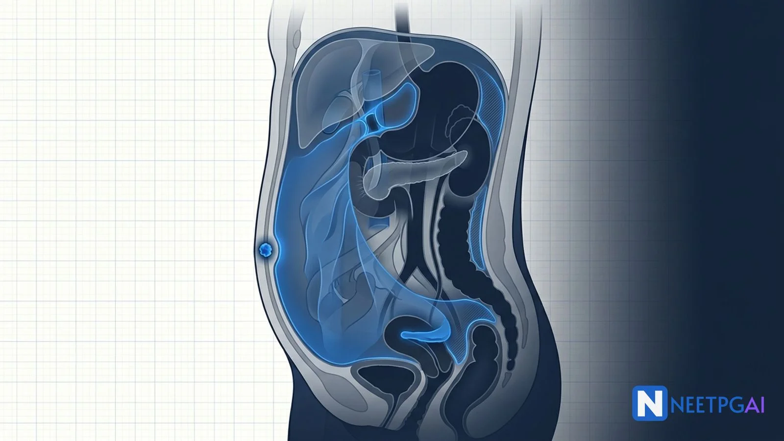
Master peritoneum, epiploic foramen, mesenteries, omenta, SAD PUCKER retroperitoneal organs, and ascites SAAG for NEET PG 2027 Anatomy MCQs with India context.
Dr. NEETPGAI Editorial TeamPublished 29 Aug 2026Updated 29 Sept 202611 min read
Quick Answer
Peritoneum and retroperitoneum anatomy is a 2 to 3 question topic in NEET PG Anatomy and Surgery. Lock these:
Epiploic foramen of Winslow — anterior lesser omentum (portal triad), posterior IVC, superior caudate lobe, inferior D1.
Portal triad — portal vein, hepatic artery proper, common bile duct in the free edge of the lesser omentum (hepatoduodenal ligament).
Morison's pouch — most common site for free fluid in a supine patient (FAST scan window).
Pouch of Douglas — most dependent recess in upright females.
SAAG greater than 1.1 — portal hypertension; SAAG less than 1.1 — malignancy/infection.
Sister Mary Joseph, Krukenberg, Blumer shelf — peritoneal spread of malignancy.
Peritoneum anatomy is high-yield because every surgeon and radiologist speaks the same language of recesses, ligaments, and mesenteries. NEET PG questions revolve around the epiploic foramen, the retroperitoneal-organ list, the pouch of Douglas, and the SAAG in ascites. India-specific context adds tuberculous peritonitis ("doughy abdomen") and CAPD peritonitis in the growing dialysis population. This NEETPGAI deep dive walks through the peritoneum layer by layer with the exam-relevant clinical correlations.
The peritoneum is a serous membrane of a single layer of mesothelium on a thin connective tissue base. Two continuous layers:
Parietal peritoneum — lines the abdominal wall, pelvic wall, and undersurface of the diaphragm. Pain sensitive, well innervated by somatic nerves — pain is sharp and precisely localised.
Visceral peritoneum — invests the abdominal organs. Innervated by visceral afferents travelling with the autonomic nerves — pain is dull, poorly localised, and referred to the midline dermatome of the corresponding embryological gut region.
The peritoneal cavity is a potential space between the two layers containing a thin film (about 100 mL in health) of serous fluid.
Greater and lesser sacs
The peritoneal cavity is divided into two communicating compartments:
Greater sac — the main peritoneal cavity, from the diaphragm to the pelvis.
Lesser sac (omental bursa) — a diverticulum behind the stomach and lesser omentum, in front of the pancreas. Created during embryological gut rotation.
Share this article
Ready to put this into practice?
Start practicing NEET PG MCQs with AI-powered explanations.
The two sacs communicate through the epiploic foramen (foramen of Winslow).
Epiploic foramen of Winslow — boundaries
Anterior — free right border of the lesser omentum (hepatoduodenal ligament) containing the portal triad: portal vein (posterior), hepatic artery proper (anterior-left), common bile duct (anterior-right).
Posterior — inferior vena cava covered by peritoneum.
Superior — caudate lobe of the liver.
Inferior — first part of the duodenum.
The Pringle manoeuvre places a vascular clamp across the free edge of the lesser omentum through the epiploic foramen to control liver haemorrhage — occluding the hepatic artery and portal vein.
Omenta and mesenteries
Lesser omentum
A double-layered peritoneal fold from the lesser curvature of the stomach and the first part of the duodenum to the porta hepatis and inferior surface of the liver. Two parts:
Hepatogastric ligament (broader medial part).
Hepatoduodenal ligament (free right edge) — contains the portal triad.
Greater omentum
A large four-layered peritoneal apron hanging from the greater curvature of the stomach in front of the small intestine. The "policeman of the abdomen" — walls off inflammatory foci (appendicitis, perforation). Components:
Gastrocolic ligament — greater curvature of stomach to transverse colon.
Gastrosplenic ligament — greater curvature to hilum of spleen; carries short gastric arteries.
Gastrophrenic ligament — greater curvature to diaphragm.
Mesenteries
Mesentery proper — of the small intestine (jejunum and ileum). Its root runs obliquely from the duodenojejunal flexure (left of L2) to the ileocaecal junction (right sacroiliac joint) — about 15 cm long. Contains superior mesenteric vessels, lymphatics, autonomic nerves.
Transverse mesocolon — from the anterior surface of the pancreas to the transverse colon. Divides the greater sac into supracolic and infracolic compartments.
Sigmoid mesocolon — inverted V-shaped attachment; apex over left ureter and left common iliac artery.
Mesoappendix — attached to the appendix, contains the appendicular artery.
Peritoneal ligaments
Ligament
Connects
Contents
Falciform ligament
Liver to anterior abdominal wall
Ligamentum teres (obliterated left umbilical vein) in its free edge
Peritoneal recesses are the "puddles" where free fluid, blood, pus, and metastases collect. High-yield sites:
Hepatorenal recess (Morison's pouch) — between the liver and right kidney. The most dependent portion of the supraumbilical peritoneal cavity in a supine patient — most common site for free fluid in supine trauma. FAST scan window.
Rectouterine pouch of Douglas — between the rectum and posterior vaginal fornix in females. Most dependent recess in an upright female — most common site for pelvic collections (ovarian metastases, pelvic abscess, blood from ruptured ectopic).
Rectovesical pouch — between the rectum and bladder in males. Most dependent recess in an upright male.
Paracolic gutters — right and left, lateral to the ascending and descending colon. Communicate the pelvic peritoneum with the subphrenic spaces; conduits for pus from a burst appendix to reach the subphrenic space.
Subphrenic spaces (right and left) — under each dome of the diaphragm; sites of subphrenic abscess after upper abdominal surgery.
Vesicouterine pouch — between the bladder and uterus in females; shallower than Douglas.
Retroperitoneal organs — SAD PUCKER
Retroperitoneal organs lie behind the parietal peritoneum with peritoneum covering only their anterior surface. Two categories:
Secondary retroperitoneal — start intraperitoneal in a mesentery, become fixed retroperitoneal during gut rotation (duodenum parts 2-4, pancreas except tail, ascending and descending colon).
SAD PUCKER mnemonic:
Suprarenal (adrenal) glands
Aorta and inferior vena cava
Duodenum (2nd, 3rd, 4th parts — 1st part is intraperitoneal)
Pancreas (head, neck, body; tail is intraperitoneal in the splenorenal ligament)
Ureters
Colon (ascending and descending; transverse and sigmoid are intraperitoneal)
Kidneys
Esophagus (thoracic — but "abdominal esophagus" is intraperitoneal)
Rectum (middle third; upper third intraperitoneal, lower third extraperitoneal)
Peritoneal spread of malignancy
Peritoneal carcinomatosis is a common late feature of gastrointestinal, ovarian, and pancreatic cancers. Named signs:
Sister Mary Joseph nodule — palpable metastatic nodule at the umbilicus; spread through the falciform ligament / round ligament. Primary is usually gastric, colonic, pancreatic, or ovarian.
Krukenberg tumour — bilateral ovarian metastasis from a signet-ring-cell primary (classically stomach, sometimes colon or breast). Transcoelomic and lymphatic spread.
Blumer's shelf — a palpable metastatic shelf in the rectovesical or rectouterine pouch on rectal examination; from disseminated peritoneal malignancy.
Virchow node (Troisier sign) — left supraclavicular lymph node metastasis; classical for gastric primary.
Irish node — left axillary lymphadenopathy; also from gastric primary.
Ascites and SAAG
Ascites is pathological accumulation of fluid in the peritoneal cavity. The serum-ascites albumin gradient (SAAG) classifies ascites better than the old transudate-exudate protein cut-off.
SAAG = serum albumin − ascitic-fluid albumin (same-day sample).
Spontaneous bacterial peritonitis (SBP) — cirrhotic ascitic-fluid infection with ascitic PMN count greater than 250 cells/µL. Empirical third-generation cephalosporin (ceftriaxone or cefotaxime); albumin infusion reduces mortality.
India-specific context
Tuberculous peritonitis — a common Indian cause of ascites. "Doughy abdomen" with matted mesentery and omental thickening on ultrasound / CT; SAAG less than 1.1; ADA elevated; culture is slow — start empirical ATT on high suspicion.
CAPD peritonitis — cloudy peritoneal dialysate with over 100 WBC/µL and over 50 percent PMN; commonest complication of continuous ambulatory peritoneal dialysis in the expanding Indian dialysis population.
Gastric cancer with Krukenberg and Sister Mary Joseph — India has significant gastric cancer burden, especially in the northeast and south. Presentation with peritoneal signs indicates advanced disease.
Ovarian cancer with omental caking — the "omental cake" on CT is a classic finding of transcoelomic spread; India lacks a national ovarian cancer screening programme.
NEET PG MCQ traps
Portal triad in the free edge of lesser omentum — portal vein, hepatic artery proper, CBD.
Sister Mary Joseph nodule — umbilical metastasis; GI/pelvic primary.
Krukenberg tumour — bilateral ovarian metastasis from signet-ring gastric primary.
Blumer shelf — pelvic peritoneal metastasis on PR.
Virchow node (Troisier) — left supraclavicular; gastric primary.
TB peritonitis — doughy abdomen, low SAAG, high ADA.
Falciform ligament — contains ligamentum teres (obliterated left umbilical vein).
Frequently asked questions
What are the boundaries of the epiploic foramen of Winslow?
The epiploic foramen (foramen of Winslow) connects the lesser sac to the greater sac of the peritoneal cavity. Boundaries: anteriorly — free right edge of lesser omentum containing the portal triad (portal vein, hepatic artery proper, common bile duct); posteriorly — inferior vena cava covered by peritoneum; superiorly — caudate lobe of the liver; inferiorly — first part of the duodenum. The Pringle manoeuvre is performed by clamping the hepatoduodenal ligament through this foramen to control liver bleeding.
What is the SAD PUCKER mnemonic for retroperitoneal organs?
SAD PUCKER lists the retroperitoneal organs — Suprarenal (adrenal) glands, Aorta and IVC, Duodenum (2nd, 3rd, 4th parts), Pancreas (except tail), Ureters, Colon (ascending and descending), Kidneys, Esophagus (thoracic), Rectum (mid). Primary retroperitoneal organs (kidneys, adrenals, ureters, aorta, IVC) develop and stay behind the peritoneum. Secondary retroperitoneal organs (duodenum 2-4, pancreas, ascending and descending colon) start intraperitoneal and become fixed behind the peritoneum during gut rotation.
Why is Morison's pouch the most common site for free intraperitoneal fluid in a supine patient?
Morison's pouch (hepatorenal recess) is the most dependent portion of the supraumbilical peritoneal cavity when the patient lies supine. Blood or fluid tracks by gravity into this recess between the liver and right kidney. Bedside FAST scan (Focused Assessment with Sonography in Trauma) evaluates Morison's pouch as one of its four windows because free fluid there in the setting of blunt abdominal trauma is highly sensitive for haemoperitoneum. In an upright female patient the most dependent recess is the rectouterine pouch of Douglas.
What does a SAAG greater than 1.1 mean in ascites?
The serum-ascites albumin gradient (SAAG) is calculated as serum albumin minus ascitic-fluid albumin, on the same day. SAAG greater than 1.1 g/dL indicates portal hypertension (transudate physiology) — cirrhosis, cardiac ascites, Budd-Chiari, portal vein thrombosis. SAAG less than 1.1 g/dL indicates non-portal-hypertensive causes (exudate physiology) — peritoneal malignancy, tuberculous peritonitis, pancreatic ascites, nephrotic syndrome. SAAG has replaced the old transudate-exudate protein cut-off for ascitic-fluid classification.
What are Sister Mary Joseph nodule and Krukenberg tumour?
Sister Mary Joseph nodule is a palpable metastatic deposit at the umbilicus — a sign of advanced intra-abdominal or pelvic malignancy that has spread through the falciform ligament and round ligament of the liver to the umbilical remnant. Common primaries are gastric, colonic, pancreatic, and ovarian cancer. Krukenberg tumour is bilateral ovarian metastasis from a signet-ring-cell gastric primary (occasionally colon or breast) that reaches the ovaries by transcoelomic or lymphatic spread. Both are grave prognostic signs indicating disseminated disease.
This content is for educational purposes for NEET PG exam preparation. It is not a substitute for professional medical advice, diagnosis, or treatment. Clinical information has been reviewed by qualified medical professionals.
Written by: NEETPGAI Editorial Team
Reviewed by: Pending SME Review
Last reviewed: July 2026