Quick Answer
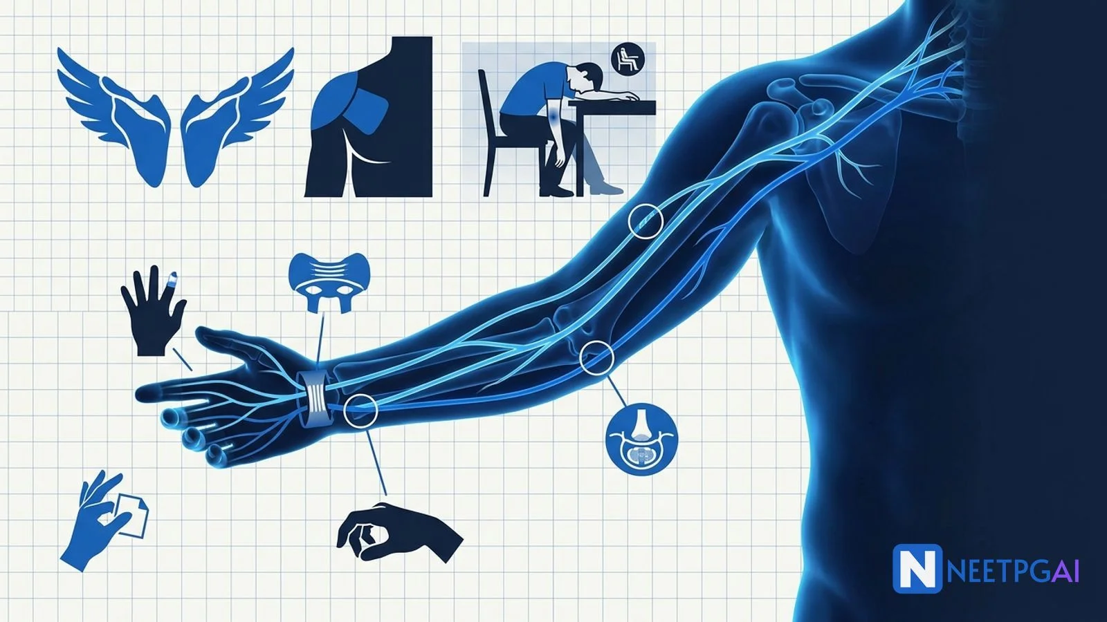
Upper limb peripheral nerve injuries are a 2 to 3 question topic in NEET PG Anatomy plus Orthopedics. Lock these:
- Median nerve — LOAF muscles, ape hand, hand of benediction (proximal), pointing index (anterior interosseous), carpal tunnel.
- Ulnar nerve — claw hand (distal worse — ulnar paradox), Froment sign, Wartenberg sign, cubital tunnel vs Guyon canal.
- Radial nerve — wrist drop (Saturday night palsy at radial groove), finger drop without wrist drop (posterior interosseous), sensory loss over dorsum of first web space.
- Axillary nerve — shoulder dislocation, regimental patch sensory loss, deltoid wasting.
- Long thoracic nerve — winged scapula (serratus anterior).
- Erb palsy (C5-C6) — waiter's tip; Klumpke palsy (C8-T1) — claw hand plus Horner syndrome.
Peripheral nerve injuries are a favourite NEET PG Anatomy topic because they map surface signs to precise root, plexus, or peripheral branch lesions. Indian practice adds specific injury patterns — motorcyclist Erb palsy from high-impact two-wheeler crashes, chronic auto-rickshaw driving carpal tunnel, and industrial hand injuries — that keep these questions clinically anchored. This NEETPGAI deep dive covers median, ulnar, radial, axillary, musculocutaneous, and long thoracic nerve palsies with the exam-relevant signs, deformities, and management points.
Pair this guide with the head and neck cancer ENT deep dive for cranial nerve and cervical anatomy overlap.
Median nerve
Course and root value
Median nerve arises from the lateral (C5, C6, C7) and medial (C8, T1) cords of the brachial plexus. It runs down the arm without branches, enters the cubital fossa medial to the brachial artery, passes between the two heads of pronator teres, gives off the anterior interosseous nerve, then travels through the carpal tunnel deep to the flexor retinaculum into the palm.
Motor supply
- Forearm — pronator teres, flexor carpi radialis, palmaris longus, flexor digitorum superficialis, lateral half of flexor digitorum profundus (index and middle), flexor pollicis longus, pronator quadratus.
- Hand (LOAF) — lateral 2 Lumbricals, Opponens pollicis, Abductor pollicis brevis, Flexor pollicis brevis (superficial head).
Sensory supply
Lateral 3.5 fingers on palmar aspect plus the corresponding nail beds and distal dorsal aspect. Palmar cutaneous branch (arises proximal to the flexor retinaculum) supplies the thenar eminence — this is spared in carpal tunnel syndrome and is a useful clinical discriminator.
Injury patterns
| Site | Cause | Key deficits |
|---|
| Supracondylar humerus fracture | Fall on outstretched hand (paediatric FOOSH) | Full LOAF loss, hand of benediction on fist, lateral 3.5 sensory loss |
| Anterior interosseous syndrome | Kiloh-Nevin; forearm trauma | Loss of pinch grip (FPL, FDP index) — cannot make an OK sign; pure motor, no sensory |
| Carpal tunnel (wrist) | Repetitive wrist flexion, pregnancy, hypothyroid, RA, acromegaly | Thenar wasting, ape hand, LOAF weakness; thenar sensation spared; Phalen and Tinel positive |
Clinical signs
- Ape hand — thumb lies adducted in the plane of the palm from opponens pollicis paralysis.
- Hand of benediction (fist-making sign) — inability to flex index and middle fingers when asked to make a fist; proximal median palsy only.
- Pointing index — anterior interosseous lesion; index stays extended when trying to make a fist because FDP (index) and FPL are paralysed.
- Phalen test — reproducing paraesthesia by holding wrists in maximal flexion for 60 seconds (carpal tunnel).
- Tinel sign — tapping over the flexor retinaculum reproduces paraesthesia in the median distribution.
Carpal tunnel syndrome — high-yield
Most common entrapment neuropathy globally. Female predominance. Nerve conduction studies (prolonged distal motor latency) confirm. Conservative — night splint, NSAIDs, steroid injection. Surgical — open or endoscopic carpal tunnel release for failed conservative management or thenar wasting.
Ulnar nerve
Course and root value
Ulnar nerve arises from the medial cord (C8, T1). It descends behind the medial epicondyle in the cubital tunnel (a common compression site), enters the forearm between the two heads of flexor carpi ulnaris, and crosses the wrist superficial to the flexor retinaculum through the Guyon canal (the second compression site) to enter the hand.
Motor supply
- Forearm — flexor carpi ulnaris, medial half of flexor digitorum profundus (ring and little).
- Hand (most intrinsic muscles) — hypothenar (abductor, opponens, flexor digiti minimi), medial 2 lumbricals, all interossei (dorsal and palmar), adductor pollicis, deep head of flexor pollicis brevis.
Sensory supply
Medial 1.5 fingers (little finger and medial half of ring) on both palmar and dorsal aspects.
Injury patterns and paradox
| Site | Cause | Sign |
|---|
| Medial epicondyle fracture, cubital tunnel | Elbow trauma, chronic flexion | Proximal ulnar palsy — FDP (ring/little) paralysed → less claw |
| Guyon canal at wrist | Handlebar palsy (cyclists), ganglia, pisiform trauma | Distal ulnar palsy — FDP intact → more claw (ulnar paradox) |
Clinical signs
- Claw hand (ulnar claw) — MCP hyperextension plus IP flexion of ring and little fingers due to loss of intrinsic muscle balance. Distal lesions produce a sharper claw because FDP is preserved.
- Froment sign — patient asked to pinch a paper between thumb and index side of hand flexes the thumb IP joint (using FPL, median-innervated) because adductor pollicis is paralysed.
- Wartenberg sign — persistent abduction of the little finger at rest because the third palmar interosseous (adductor) is paralysed while extensor digiti minimi is intact.
- Card test — inability to hold a card between adjacent fingers (interosseous weakness).
Note — hand of benediction is a median nerve sign (proximal palsy on fist-making), NOT ulnar. Ulnar produces a resting claw, not a benediction posture.
Radial nerve
Course and root value
Radial nerve arises from the posterior cord (C5, C6, C7, C8, T1). It runs in the spiral (radial) groove of the humerus behind the shaft — vulnerable to mid-shaft humeral fracture and to Saturday night palsy. It enters the forearm in front of the lateral epicondyle and divides into the superficial (sensory) branch and the posterior interosseous nerve (deep motor branch), which pierces supinator.
Motor supply
- Arm — triceps brachii, anconeus.
- Forearm (via posterior interosseous nerve) — brachioradialis, extensor carpi radialis longus and brevis (wrist extensors), supinator, extensor digitorum, extensor digiti minimi, extensor carpi ulnaris, abductor pollicis longus, extensor pollicis longus and brevis, extensor indicis.
Sensory supply
Dorsum of the lateral 3.5 fingers up to the DIP joints, dorsum of the hand on the radial side, and the dorsum of the first web space (a small autonomous zone — most reliable radial sensory territory).
Injury patterns
| Site | Cause | Deficits |
|---|
| Axilla | Crutch palsy, saturday night palsy at axilla | Triceps weakness, wrist drop, finger drop, sensory loss dorsum of arm and forearm |
| Radial (spiral) groove | Mid-shaft humerus fracture, Saturday night palsy at groove | Triceps spared; wrist drop, finger drop, sensory loss dorsum of first web space |
| Posterior interosseous (below elbow) | Supinator syndrome, forearm trauma | Finger drop without wrist drop (ECRL preserved); NO sensory loss (pure motor) |
| Superficial radial (Wartenberg syndrome) | Handcuff, tight watch | Pure sensory loss dorsum of first web space; no motor deficit |
Clinical pearls
- Wrist drop plus finger drop plus dorsum of first web space sensory loss — radial nerve at the groove.
- Finger drop only (wrist extension preserved with radial deviation) — posterior interosseous. Wrist is deviated radially because ECRL is intact but ECU is paralysed.
- Test radial nerve motor by asking the patient to extend the wrist against gravity and to extend the thumb (thumb-up sign).
Brachial plexus injuries — quick recap
| Palsy | Roots | Mechanism | Signs |
|---|
| Erb (upper trunk) | C5, C6 | Excess head-to-shoulder traction — obstetric, motorcyclist | Waiter's tip: shoulder adducted and internally rotated, elbow extended, forearm pronated, wrist flexed |
| Klumpke (lower trunk) | C8, T1 | Excess arm abduction — grabbing overhead on a fall | Total claw hand plus Horner syndrome (T1 sympathetic loss) — ptosis, miosis, anhidrosis |
Motorcyclist Erb palsy is common in Indian trauma centres because of unhelmeted two-wheeler crashes with head-shoulder impact stretching the upper trunk.
Axillary nerve
- Roots — C5, C6 (posterior cord).
- Course — winds around the surgical neck of the humerus with the posterior circumflex humeral artery through the quadrangular space.
- Motor — deltoid, teres minor.
- Sensory — upper lateral cutaneous nerve of arm (regimental badge / patch area over lower deltoid).
- Injury — anterior shoulder dislocation, surgical neck of humerus fracture, faulty crutch use.
- Signs — loss of shoulder abduction beyond 15 degrees (supraspinatus initiates), deltoid wasting, regimental patch sensory loss.
- Always test axillary function before and after reducing any shoulder dislocation.
Musculocutaneous nerve
- Roots — C5, C6, C7 (lateral cord).
- Motor — coracobrachialis, biceps brachii, brachialis.
- Sensory — lateral cutaneous nerve of forearm (lateral forearm sensation).
- Injury — penetrating axillary trauma; uncommon isolated injury.
- Signs — weak elbow flexion (brachioradialis, radial-innervated, still allows some flexion in mid-prone), loss of supination in flexed elbow, absent biceps reflex, lateral forearm numbness.
Long thoracic nerve
- Roots — C5, C6, C7 (branches directly from roots proximal to the plexus).
- Motor — serratus anterior only.
- Injury — mastectomy with axillary dissection, stab injury to lateral chest wall, backpack straps, direct blow to the shoulder.
- Sign — winged scapula — medial border of scapula lifts away from the chest wall when the patient pushes against a wall. Loss of scapular protraction and difficulty raising the arm above the shoulder.
NEET PG MCQ traps
- LOAF muscles — Lumbricals 1-2, Opponens pollicis, Abductor pollicis brevis, Flexor pollicis brevis (superficial); median nerve.
- Ape hand — median nerve; thumb in plane of palm.
- Hand of benediction — proximal median palsy on fist-making (index and middle fingers stay extended). NOT ulnar.
- Pointing index — anterior interosseous nerve palsy.
- Carpal tunnel — thenar sensation spared (palmar cutaneous branch arises proximal to flexor retinaculum).
- Phalen and Tinel — carpal tunnel bedside tests.
- Ulnar claw — MCP hyperextension plus IP flexion of ring and little.
- Ulnar paradox — distal lesion produces more claw than proximal.
- Froment sign — thumb IP flexion on pinch (adductor pollicis paralysis; ulnar).
- Wartenberg sign (ulnar) — persistent little finger abduction.
- Wrist drop — radial nerve.
- Saturday night palsy — radial at spiral groove; triceps spared; sensory loss dorsum of first web space.
- Posterior interosseous syndrome — finger drop without wrist drop; NO sensory loss.
- Regimental badge sensory loss — axillary nerve; shoulder dislocation.
- Winged scapula — long thoracic nerve; serratus anterior paralysis.
- Erb waiter's tip — C5, C6 upper trunk.
- Klumpke claw plus Horner — C8, T1 lower trunk.
- Musculocutaneous nerve — loss of biceps reflex, lateral forearm sensation.
- Mid-shaft humerus fracture — radial nerve at spiral groove.
- Medial epicondyle fracture — ulnar nerve at cubital tunnel.
India-specific injury patterns
- Motorcyclist Erb palsy — high-impact two-wheeler crashes without helmet; upper trunk stretched at cervicoshoulder junction.
- Auto-rickshaw / commercial driver carpal tunnel — prolonged wrist flexion and grip vibration.
- Construction and industrial injuries — supracondylar humerus fracture (paediatric FOOSH in India), open humeral shaft fracture from crush injury.
- Handcuff and ligature palsies — superficial radial nerve (Wartenberg syndrome).
- Traditional wrestling and akhara injuries — brachial plexus stretch and axillary nerve injuries.
- Delayed presentation — many Indian patients present weeks after injury with wasting and fixed deformity — physiotherapy, splinting, and tendon transfer play a central role.
Frequently asked questions
Why is the 'hand of benediction' a median nerve sign and not an ulnar nerve sign?
Hand of benediction appears when a patient with a proximal median nerve palsy tries to make a fist. The lateral two fingers (index and middle) fail to flex because flexor digitorum profundus (index and middle) and flexor digitorum superficialis are paralysed. The ring and little fingers still flex normally via the ulnar-innervated FDP. In contrast, ulnar claw is a resting deformity of the ring and little fingers due to intrinsic muscle paralysis. Benediction is a fist-making sign; claw is a rest sign.
What is the ulnar paradox?
Ulnar paradox describes the counter-intuitive observation that a distal ulnar nerve lesion (at the wrist, Guyon canal) produces a more prominent claw hand deformity than a proximal lesion (at the elbow, cubital tunnel). In a proximal lesion, the ulnar half of flexor digitorum profundus is also paralysed, so the ring and little fingers cannot flex at the DIP joints — reducing the claw. In a distal lesion, FDP is intact, MCP hyperextension by unopposed extensors combines with strong DIP/PIP flexion, producing a sharper claw.
What is Saturday night palsy and which nerve is affected?
Saturday night palsy is a compressive radial nerve injury in the spiral (radial) groove of the humerus caused by prolonged pressure on the arm — classically an intoxicated person falling asleep with the arm draped over a chair or bench. Presentation is wrist drop plus finger drop plus sensory loss on the dorsum of the first web space. Triceps is spared because its branches leave proximal to the groove. Recovery over 6 to 12 weeks with a cock-up splint is usual; nerve conduction studies confirm the lesion.
What are the LOAF muscles and why do they matter in carpal tunnel syndrome?
LOAF is a mnemonic for the four thenar and lateral hand muscles supplied by the recurrent motor branch of the median nerve: Lumbricals (1 and 2), Opponens pollicis, Abductor pollicis brevis, and Flexor pollicis brevis (superficial head). Carpal tunnel syndrome compresses the median nerve at the wrist, denervating these muscles — producing thenar wasting, weak thumb opposition and abduction, and the classic ape hand deformity where the thumb rests in the plane of the palm.
Which nerve is injured in a shoulder dislocation and what deficit results?
The axillary nerve (C5, C6) is most commonly injured in anterior shoulder dislocation and in surgical-neck-of-humerus fracture. It winds around the surgical neck and can be stretched or transected. The deficit is deltoid paralysis (loss of shoulder abduction beyond 15 degrees, which supraspinatus initiates) plus a small oval area of sensory loss over the lower deltoid — the so-called regimental badge or regimental patch area. Always test axillary nerve function before and after reduction of any dislocated shoulder.
This content is for educational purposes for NEET PG exam preparation. It is not a substitute for professional medical advice, diagnosis, or treatment. Clinical information has been reviewed by qualified medical professionals.
Written by: NEETPGAI Editorial Team
Reviewed by: Pending SME Review
Last reviewed: July 2026